吉林大学学报(医学版) ›› 2020, Vol. 46 ›› Issue (6): 1234-1240.doi: 10.13481/j.1671-587x.20200620

• 基础研究 • 上一篇    下一篇

BTg-3对甲状腺滤泡癌细胞增殖、侵袭和迁移及WNT/β-catenin信号通路的影响

杜静海,郭欣()   

  1. 河北医科大学附属唐山工人医院头颈外科,河北 唐山 063000
  • 收稿日期:2020-02-25 出版日期:2020-11-28 发布日期:2022-08-24
  • 通讯作者: 郭欣 E-mail:haijing592@163.com
  • 作者简介:杜静海(1982-),男,山东省招远市人,主治医师,医学硕士,主要从事甲状腺癌基础和临床方面的研究。
  • 基金资助:
    河北省卫计委医学科学研究项目资助课题(20181255)

Effects of BTg-3 on proliferation, invasion, migration and WNT / β-catenin signaling pathway of thyroid follicular carcinoma cells

Jinghai DU,Xin GUO()   

  1. Department of Head and Neck Surgery,Tangshan Workers’Hospital,Hebei Medical University,Tangshan 063000,China
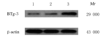
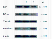
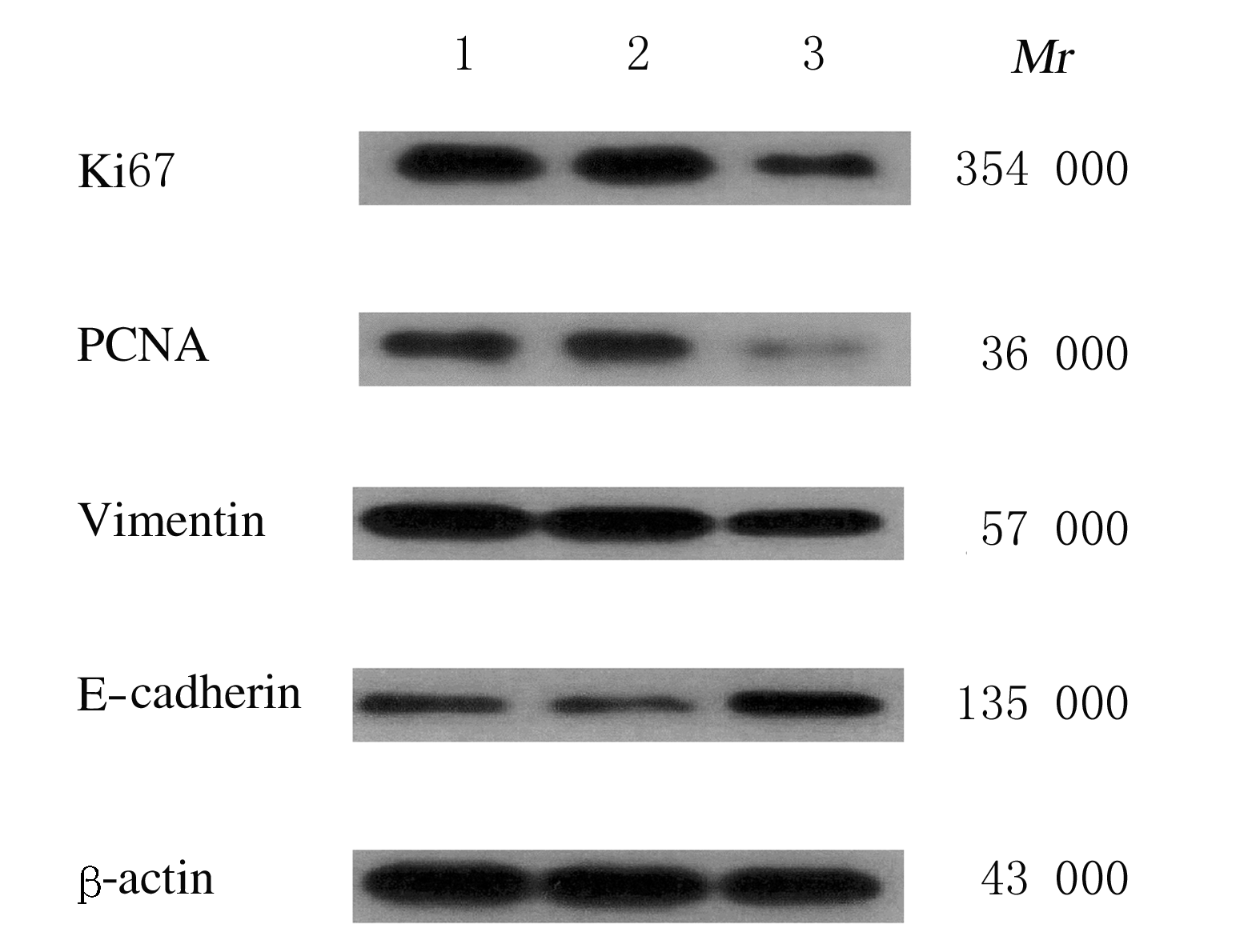
  • Received:2020-02-25 Online:2020-11-28 Published:2022-08-24
  • Contact: Xin GUO E-mail:haijing592@163.com

摘要: 目的

探讨B细胞转位基因3(BTg-3)在甲状腺滤泡癌组织和细胞中的表达及其对甲状腺滤泡癌细胞增殖、侵袭和迁移的影响,阐明BTg-3在甲状腺滤泡癌发生发展中的可能作用机制。

方法

选择经病理证实的甲状腺滤泡癌组织和甲状腺滤泡腺瘤组织标本各80例,免疫组织化学法检测甲状腺滤泡癌组织和甲状腺滤泡腺瘤组织中BTg-3表达情况,Western blotting法检测甲状腺滤泡症CGTHW-1和甲状腺滤泡上皮Nthy-ori细胞中BTg-3蛋白表达水平。将甲状腺滤泡癌CGTHW-1细胞随机分为空白对照组(不转染载体)、阴性对照组(转染阴性对照载体)和BTg-3过表达组(转染BTg-3过表达载体)。CCK-8法检测各组甲状腺滤泡癌CGTHW-1细胞增殖活性,平板克隆实验检测各组甲状腺滤泡癌CGTHW-1细胞克隆形成率,Transwell法检测各组甲状腺滤泡癌CGTHW-1细胞侵袭和迁移细胞数,Western blotting法检测各组甲状腺滤泡癌CGTHW-1细胞中Ki67、增殖细胞核抗原(PCNA)、波形蛋白(Vimentin)、上皮性钙黏蛋白(E-cadherin)、WNT1、β-连环蛋白(β-catenin)、糖原合成激酶3β(GSK3β)和磷酸化GSK3β(p-GSK3β)蛋白表达水平。

结果

甲状腺滤泡癌组织中BTg-3阳性表达率低于甲状腺滤泡腺瘤组织(χ2=62.864,P<0.01)。甲状腺滤泡癌CGTHW-1细胞中BTg-3蛋白表达水平低于甲状腺滤泡上皮Nthy-ori细胞(t=34.484,P<0.01)。与空白对照组和阴性对照组比较,BTg-3过表达组甲状腺滤泡癌CGTHW-1细胞中BTg-3蛋白表达水平升高(P<0.05),细胞增殖活性、克隆形成率、侵袭细胞数和迁移细胞数降低(P <0.05),甲状腺滤泡癌CGTHW-1细胞中Ki67、PCNA、Vimentin、WNT1、β-catenin和p-GSK3β蛋白表达水平降低(P<0.05),E-cadherin蛋白表达水平升高(P<0.05)。

结论

甲状腺滤泡癌组织和细胞中BTg-3表达水平降低,过表达BTg-3可抑制甲状腺滤泡癌细胞增殖、侵袭和迁移,抑制WNT/β-catenin信号通路激活。

关键词: BTg-3, 甲状腺滤泡癌, 细胞增殖, 细胞侵袭, 细胞迁移, WNT/β-catenin通路

Abstract:

Objective To investigate the expression of B-cell translocation gene-3 (BTg-3) in the thyroid follicular carcinoma tissue and cells and its effects on the proliferation, invasion and migration of thyroid follicular carcinoma cells, and to clarify its possible mechanism on the occurence and development of thyroid follicular carcinoma.

Methods

A total of 80 cases of thyroid follicular carcinoma tissue specimens and 80 cases of thyroid follicular adenoma tissue specimens confirmed by pathology were selected. Immunohistochemistry was used to detect the expressions of BTg-3 in thyroid follicular carcinoma and thyroid follicular adenoma tissues. Western blotting method was used to detect the expression levels of BTg-3 protein in thyroid follicular carcinoma CGTHW-1 cells and thyroid follicular adenoma Nthy-ori cells.The thyroid follicular carcinoma CGTHW-1 cells were randomly divided into blank control group (without transfection), negative control group(transfected with negative control vector),and BTg-3 over-expression group (transfected with BTg-3 over-expression vector).Plate clone experiment was used to detect the clone formation rates of the thyroid follicular carcinoma CGTHW-1 cells in various groups;Transwell chamber assay was used to determine the number of invasion and migration cells in various groups;Western blotting method was used to detect the expression levels of Ki67, proliferating cell nuclear antigen (PCNA), Vimentin, epithelial cadherin (E-cadherin), WNT1, β-catenin, glycogen synthesis kinase 3β (GSK3β) ,and phosphorylated GSK3β (p-GSK3β) proteins in the thyroid follicular carcinoma CGTHW-1 cells in various groups.

Results

The positive expression rate of BTg-3 in thyroid follicular carcinoma tissue was lower than that in thyroid follicular adenoma tissue (χ2 = 62.864, P<0.01).The expression level of BTg-3 protein in the thyroid follicular carcinoma CGTHW-1 cells was lower than that in thyroid follicular epithelium Nthy-ori cells (t = 34.484, P<0.01).Compared with blank control group and negative control group, the expression level of BTg-3 protein in the thyroid follicular carcinoma CGTHW-1 cells in BTg-3 over-expression group was increased (P<0.05);the clone formation rate, number of invation cells and number of migration cells in the thyroid follicular carcinoma CGTHW-1 cells in BTg-3 over-expression group were decreased (P<0.05);the expression levels of Ki67, PCNA, Vimentin, WNT1, β-catenin, and p-GSK3β proteins in the thyroid follicular carcinoma CGTHW-1 cells in BTg-3 over-expression group were decreased (P<0.05);the expression level of E-cadherin protein was increased (P<0.05).

Conclusion

The expression levels of BTg-3 in the thyroid follicular cancinoma tissue and cells are decreased. Over-expression of BTg-3 can inhibit the proliferation, invasion,and migration of thyroid follicular carcinoma cells, and inhibit the activation of WNT / β-catenin signaling pathway.

Key words: BTg-3, thyroid follicular carcinoma, cell proliferation, cell invasion, cell migration, WNT/β-catenin pathway

中图分类号: 

  • R736.1