吉林大学学报(医学版) ›› 2022, Vol. 48 ›› Issue (1): 9-17.doi: 10.13481/j.1671-587X.20220102

• 基础研究 • 上一篇    下一篇

鼻咽癌斑马鱼移植瘤模型的构建及姜黄素对CNE-2细胞的抑制作用

王泽泰1,娄丹丹1,彭燕1,朱道琦1,李爱武2,宫凤英2,吕英2,范钦1()   

  1. 1.南方医科大学中医药学院分子生物学教研室,广东 广州 510515
    2.南方医科大学南方医院 古中医科,广东 广州 510515
  • 收稿日期:2021-06-04 出版日期:2022-01-28 发布日期:2022-01-17
  • 通讯作者: 范钦 E-mail:fqin@163.com
  • 作者简介:王泽泰(1994-),男,广东省揭阳市人,在读硕士研究生,主要从事中医药抗肿瘤方面的研究。
  • 基金资助:
    国家自然科学基金项目(82074132);广东省中医药管理局科研项目(20213009);广东省广州市科技局科技 计划项目(202102080405)

Establishment of zebrafish xenograft model of nasopharyngeal carcinoma and inhibitory effect of curcumin on CNE-2 cells

Zetai WANG1,Dandan LOU1,Yan PENG1,Daoqi ZHU1,Aiwu LI2,Fengying GONG2,Ying LYU2,Qin FAN1()   

  1. 1.Department of Molecular Biology,School of Traditional Chinese Medicine,Southern Medical University,Guangzhou 510515,China
    2.Traditional Chinese Medicine,NanFang Hospital,Southern Medical University,Guangzhou 510515,China
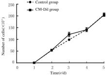
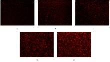
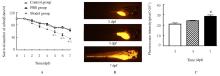
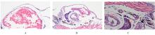
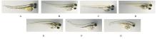
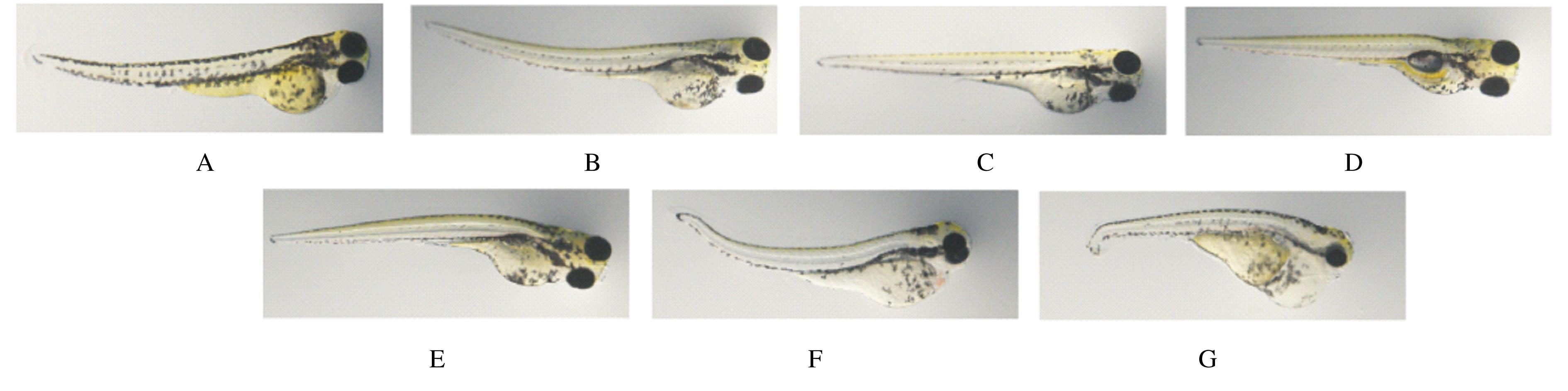
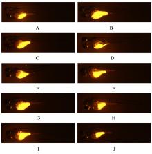
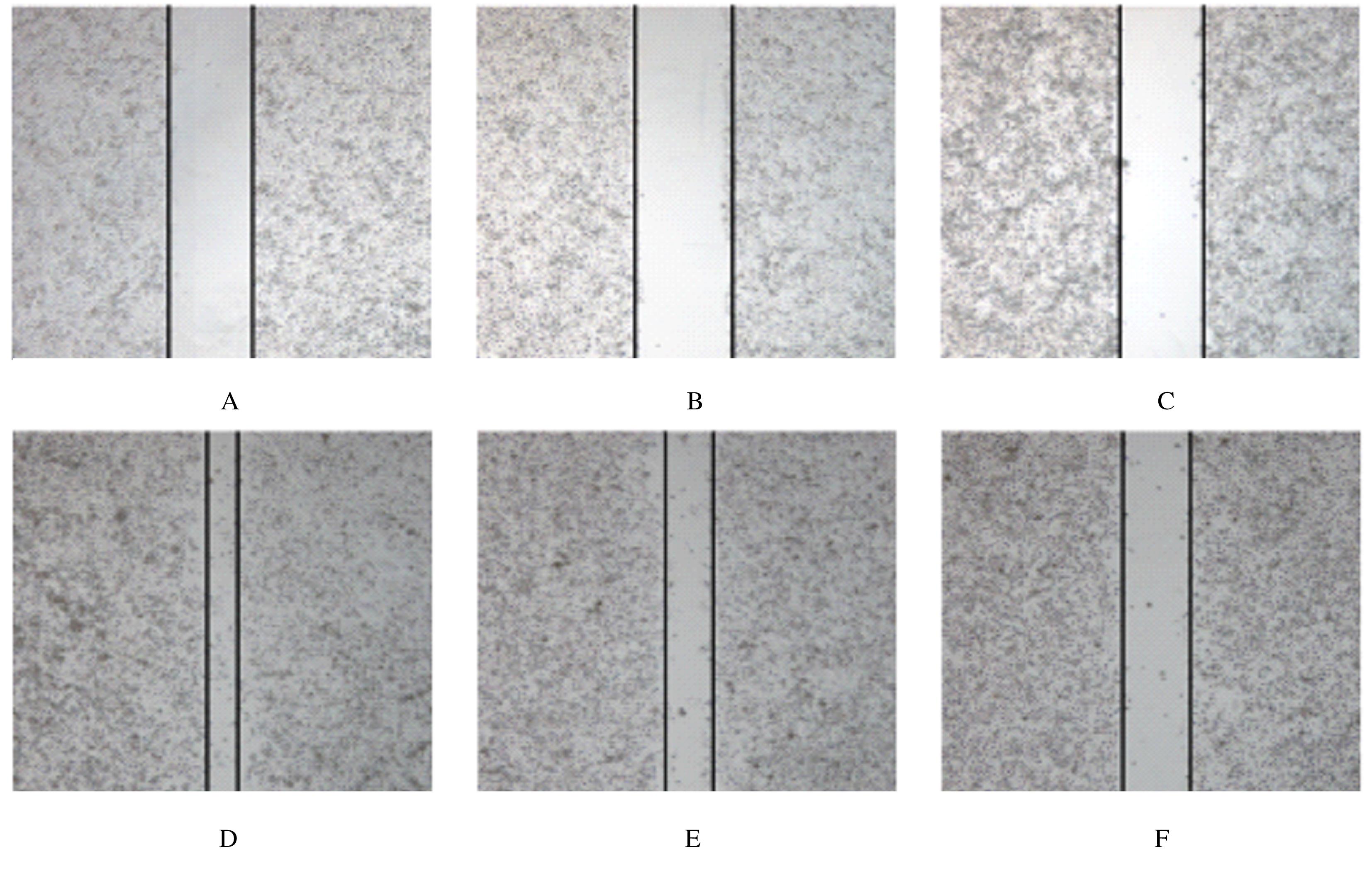
  • Received:2021-06-04 Online:2022-01-28 Published:2022-01-17
  • Contact: Qin FAN E-mail:fqin@163.com

摘要: 目的

建立人鼻咽癌斑马鱼异种移植瘤模型,探讨姜黄素在体内外对鼻咽癌的抑制作用,为阐明姜黄素抗鼻咽癌的分子机制提供依据。

方法

将鼻咽癌CNE-2细胞分为对照组和细胞膜红色荧光染色剂CM-Dil(CM-Dil)组,连续5 d进行细胞计数,并观察荧光强度变化。采用显微注射法构建鼻咽癌斑马鱼异种移植瘤模型,将斑马鱼胚胎分为对照组、PBS组和模型组,对照组斑马鱼胚胎不予处理,PBS组斑马鱼胚胎注射10 nL PBS缓冲液,模型组斑马鱼胚胎注射10 nL CNE-2细胞悬液,孵育培养后统计各组斑马鱼存活数,采用宏观体式显微镜观察并定量分析模型组斑马鱼体内移植瘤荧光强度,HE染色观察对照组和模型组斑马鱼移植瘤组织形态表现。将受精后天数(dpf)为3 的斑马鱼胚胎暴露于含不同浓度(0、0.625、1.250、2.500、5.000、7.500和10.000 μmol·L-1)姜黄素的饲养水中,观察斑马鱼死亡数和畸形数并计算斑马鱼死亡率,筛选姜黄素体内用药浓度。采用0、0.625、1.250、2.500和5.000 μmol·L-1 姜黄素作用于斑马鱼鼻咽癌模型48 h,检测移植瘤生长和转移情况。0、10和20 μmol·L-1姜黄素作用CNE-2细胞24 h,CCK-8法和划痕实验检测细胞增殖率和迁移率。

结果

与对照组比较,CM-Dil组CNE-2细胞数差异无统计学意义(P>0.05)。实验第5天,与对照组比较,PBS组斑马鱼存活数差异无统计学意义(P>0.05);与对照组和PBS组比较,模型组斑马鱼存活数明显减少(P<0.01)。模型组斑马鱼体内红色荧光随着时间的延长而增强;与注射后第1天比较,注射后第5天模型组斑马鱼体内红色荧光强度明显增强(P<0.05)。HE染色,模型组斑马鱼切片中的卵黄囊有CNE-2细胞,提示造模成功。浓度小于或等于5.000 μmol·L-1姜黄素组斑马鱼无死亡及明显毒性反应。与对照组(0 μmol·L-1 姜黄素组)比较, 2.500和5.000 μmol·L-1姜黄素组斑马鱼体内移植瘤荧光强度明显降低(P<0.05或P<0.01),5.000 μmol·L-1姜黄素组发生头尾部转移的斑马鱼数量明显减少(P<0.05)。CCK-8法检测和划痕实验,与对照组(0 μmol·L-1 姜黄素组)比较,10和20 μmol·L-1姜黄素组CNE-2细胞增殖率和迁移率明显降低(P<0.05)。

结论

人鼻咽癌CNE-2细胞可以成功植入斑马鱼体内并构建鼻咽癌斑马鱼模型,姜黄素在体内外均能抑制鼻咽癌细胞的增殖和转移,具有良好的抗肿瘤前景。

关键词: 姜黄素, 鼻咽肿瘤, 移植瘤, 斑马鱼, 细胞增殖率, 细胞迁移率

Abstract: Objective

To establish a zebrafish xenograft model of human nasopharyngeal carcinoma and explore the inhibitory effect of curcumin on nasopharyngeal carcinoma in vivo and in vitro, and to provide the basis for elucidating the molecular mechanism of anti-nasopharyngeal carcinoma of curcumin.

Methods

The nasopharyngeal carcinoma CNE-2 cells were divided into control group and CM-Dil group. The cells were counted for 5 consecutive days, and the fluorescence intensity changes were observed. The zebrafish xenograft model of nasopharyngeal carcinoma was established by microinjection. The zebrafishes were divided into control group, PBS group and model group. The zebrafishes in control group were not treated, the zebrafishes in PBS group were injected with 10 nL PBS buffer, and the zebrafishes in model group were injected with 10 nL cell suspension. The survival number of zebrafishes in each group was counted, and the fluorescence intensity of transplanted tumor of the zebrafishes in model group was observed and quantitatively analyzed by macro stereomicroscope. The morphology of zebrafish transplanted tumors in control group and model group were observed by HE staining. The zebrafishes with the days postfertilization(dpf) was 3 were exposed to feeding water containing different concentrations (0, 0.625, 1.250, 2.500, 5.000,and 7.500, 10.000 μmol·L-1) of curcumin, the death and deformity were observed;the death rates of zebrafishes were calculated,and the concentration of curcumin in vivo was selected. The zebrafish models were treated with differnent concentrations (0,0.625, 1.250, 2.500, and 5.000 μmol·L-1) of curcumin for 48 h, the growth and metastasis of the transplanted tumor were detected. The CNE-2 cells were treated with different concentrations(0, 10, and 20 μmol·L-1) of curcumin for 24 h, the proliferation rates and migration rates were detected by CCK-8 method and wound healing assay.

Results

Compared with control group, there was no significant difference in the number of CNE-2 cells in CM-Dil group (P>0.05). On the 5th day of the experiment, compared with control group, there was no significant difference in the survival number of zebrafishes in PBS group (P>0.05); compared with control group and PBS group, the survival number of zebrafishes in model group was decreased significantly (P<0.01). The red fluorescence intensity in zebrafishes in model group was increased with the prolongation of time. Compared with the first day after injection, the red fluorescence intensity of zebrafishes in model group was increased significantly on the fifth day after injection (P<0.05).The HE staining results showed that the CNE-2 cells were found in the yolk sac of the zebrafishes in model group, indicating that the models were successfully constructed. The zebrafishes in 0.625, 1.250, 2.500, and 5.000 μmol·L-1 concentrations of curcumin groups had no death and obvious toxicity. Compared with control group(0 μmol·L-1 cureumin group),the fluorescence intensities in the zebrafish xenograft tissue in 2.500 and 5.000 μmol·L-1 cureumin groups were signifricantly decreased(P<0.05 or P<0.01), and the number of zebrafishes with head and tail metastasis in 5.00 μmol·L-1 curcumin group was decreased (P<0.05). The results of CCK-8 method and wound healing assay showed that compared with control group (0 μmol·L-1 curcumin group),the proliferation rates and migration rates of CNE-2 cells in 10 and 20 μmol·L-1 curcumin groups were significantly decreased (P<0.05).

Conclusion

The CNE-2 cells can be successfully implanted in the zebrafishes and to estabish the zebrafish models of nasopharyngeal carcinoma. Curcumin can inhibit the proliferation and metastasis of nasopharyngeal carcinoma in vivo and in vitro, and it has a good prospect of cell anti-tumor.

Key words: Curcumin, Nasopharyngeal carcinoma, Xenograft model, Zebrafish, Cell poliferation rate, Cell migration rate

中图分类号: 

  • R285.5