吉林大学学报(医学版) ›› 2022, Vol. 48 ›› Issue (4): 962-970.doi: 10.13481/j.1671-587X.20220416

• 临床研究 • 上一篇    下一篇

m6A甲基化修饰结合蛋白YTHDF2在食管癌组织中的表达及其对食管癌细胞增殖和迁移的影响

马琰迪1,卢香云1,何尚峰1,俞雪燕1,胡云华1,高海霞1,陈云昭2,禹洁2,王文洁2,李锋1,3,崔晓宾1,4()   

  1. 1.石河子大学医学院病理学系,新疆 石河子 832002
    2.江苏省苏州市高新区人民医院病理科,江苏 苏州 215000
    3.首都医科大学附属北京朝阳医院病理科,北京 100200
    4.南京大学医学院附属鼓楼医院病理科,江苏 南京 210008
  • 收稿日期:2021-11-24 出版日期:2022-07-28 发布日期:2022-07-26
  • 通讯作者: 崔晓宾 E-mail:342029924@qq.com
  • 作者简介:马琰迪(1993-),女,河南省南阳市人,在读硕士研究生,主要从事食管癌基础方面的研究。
  • 基金资助:
    国家自然科学基金项目(82160542);新疆生产建设兵团科技局重点领域科技攻关计划项目(2020AB022);江苏省苏州市高新区医疗卫生科技计划项目(2018Q004);江苏省苏州市高新区人民医院科学创新基金项目(SGY2020A01);石河子大学科技项目(ZZZC202124)

Expression of m6A methylation binding protein YTHDF2 in esophageal carcinoma tissue and its effect on proliferation and migration of esophageal carcinoma cells

Yandi MA1,Xiangyun LU1,Shangfeng HE1,Xueyan YU1,Yunhua HU1,Haixia GAO1,Yunzhao CHEN2,Jie YU2,Wenjie WANG2,Feng LI1,3,Xiaobin CUI1,4()   

  1. 1.Department of Pathology, School of Medical Sciences, Shihezi University, Shihezi 832002, China
    2.Department of Pathology, Suzhou High Tech Zone People’s Hospital, Suzhou 215000, China
    3.Department of Pathology and Medical Research Center, Beijing Chaoyang Hospital, Capital Medical University, Beijing 100020, China
    4.Department of Pathology, Affiliated Nanjing Drum Tower Hospital, School of Medical Sciences, Nanjing University, Nanjing 210008, China
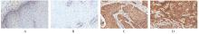
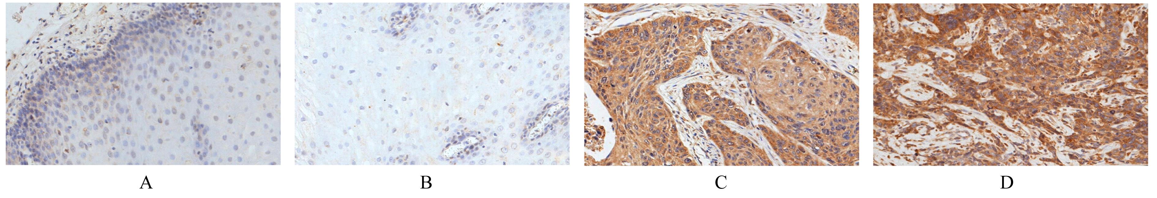
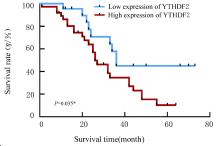
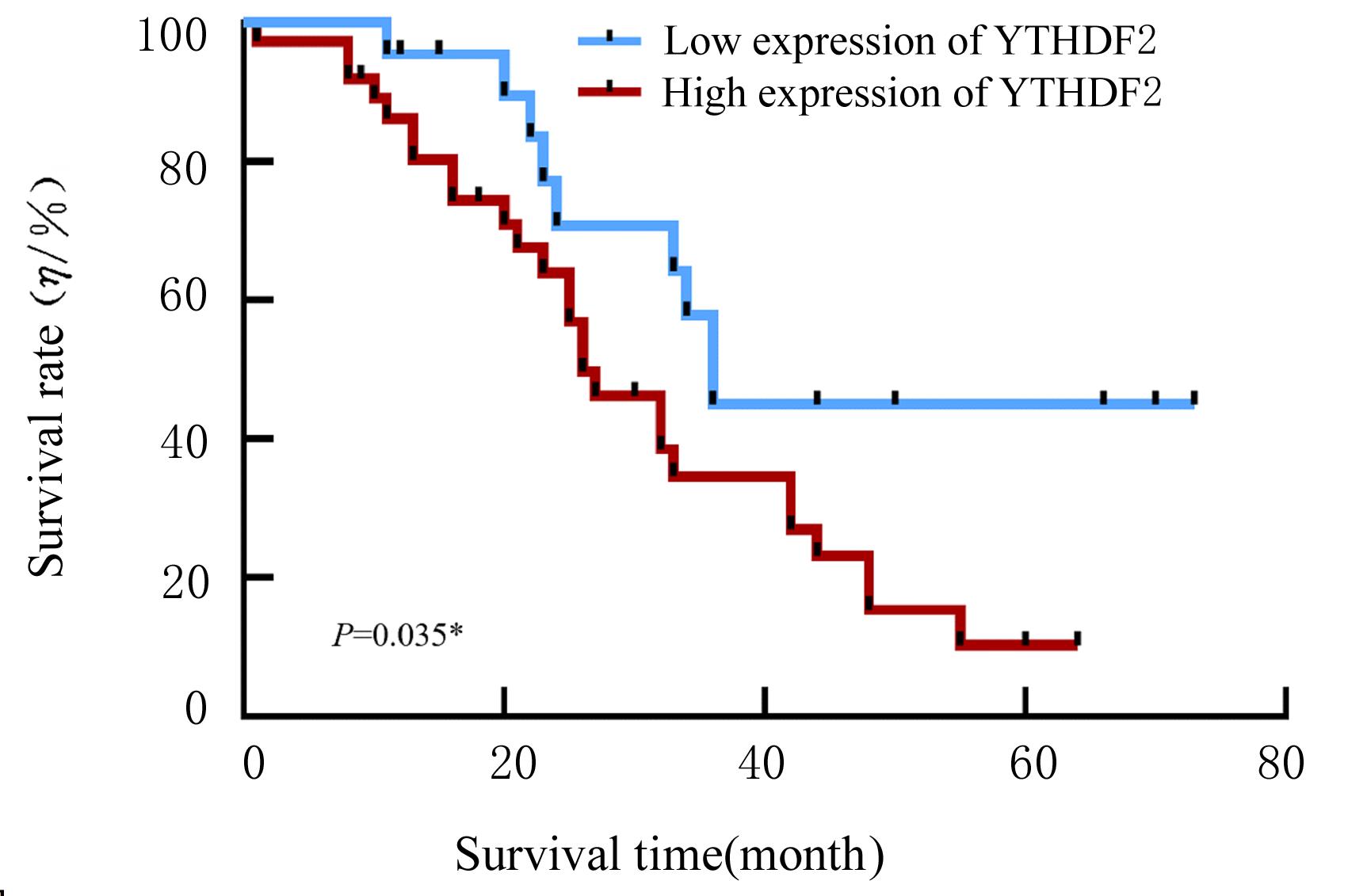
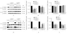
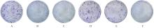
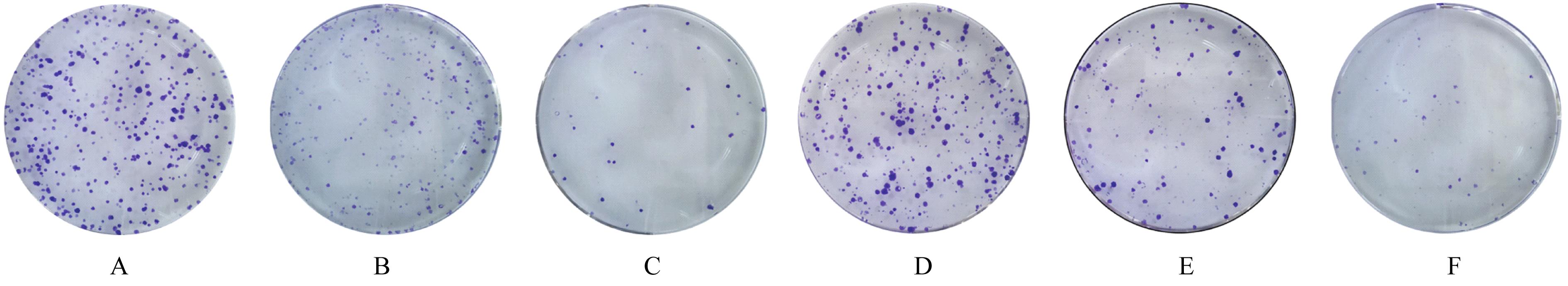
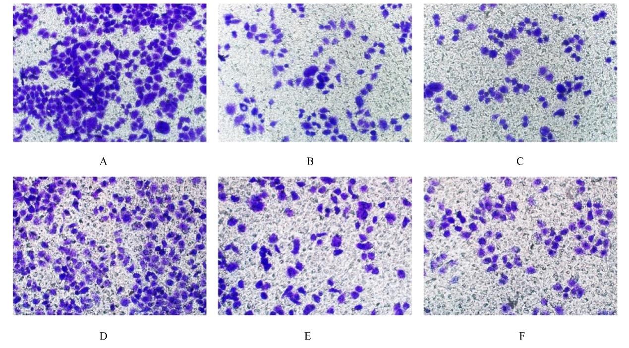
  • Received:2021-11-24 Online:2022-07-28 Published:2022-07-26
  • Contact: Xiaobin CUI E-mail:342029924@qq.com

摘要: 目的

探讨N6-甲基腺嘌呤(m6A)甲基化修饰结合蛋白YTH结构域连接蛋白2(YTHDF2)在食管鳞状细胞癌(ESCC)组织中表达及其对食管癌细胞生物学行为的影响,阐明其可能的作用机制。

方法

免疫组织化学法检测113例ESCC组织和95例癌旁正常食管上皮组织中YTHDF2蛋白表达情况,分析YTHDF2蛋白表达与ESCC患者临床病理特征和预后的关系。将体外培养的食管癌Eca109和EC9706细胞分为对照组(转染si-NC)和si-YTHDF2组(分别转染si-YTHDF2#1和si-YTHDF2#2)。Western blotting法检测各组细胞中YTHDF2蛋白表达水平,CCK-8法检测各组细胞增殖能力,平板克隆实验检测各组细胞克隆形成数,Transwell小室实验检测各组细胞中迁移细胞数。

结果

YTHDF2蛋白主要表达于ESCC细胞的细胞质,少量表达于细胞核,其在ESCC组织中的表达强度明显高于癌旁正常食管上皮组织(P<0.01)。不同发病年龄和 TNM分期ESCC患者间YTHDF2蛋白表达强度比较差异有统计学意义(P=0.008,P=0.041)。Kaplan-Meier 生存分析,与YTHDF2低表达组比较,YTHDF2高表达组ESCC患者生存率明显降低(P=0.035)。CCK-8法和平板克隆实验,与对照组比较,si-YTHDF2#1组和si-YTHDF2#2组食管癌细胞的增殖活性明显降低(P<0.05),克隆形成数明显减少(P<0.01)。Transwell 小室实验,与对照组比较,si-YTHDF2#1组和si-YTHDF2#2组食管癌细胞中迁移细胞数明显减少(P<0.01)。

结论

YTHDF2在ESCC组织中的表达强度明显高于正常食管上皮组织,敲低YTHDF2表达可抑制食管癌细胞的增殖、克隆形成和迁移。

关键词: 食管肿瘤, 鳞状细胞癌, YTH结构域连接蛋白2, 细胞增殖, 细胞迁移

Abstract:

Objective To investigate the expression of N6-methyladenosine(m6A) methylation binding protein YTH domain-containing family protein 2 (YTHDF2) in the esophageal squamous cell carcinoma (ESCC) tissue and its effect on the biological behavior of esophageal carcinoma cells,and to clarify its possible mechanism.

Methods

The expressions of YTHDF2 protein in 113 cases of ESCC tissue and 95 cases of normal tissue adjacent to esophageal carcinoma were detected by immunohistochemical method. The relationships between the expression of YTHDF2 protein and the clinicopathological features and prognosis of the ESCC patients were analyzed.The Eca109 and EC9706 cells cultured in vitro were divided into control group (transfected with si-NC) and si-YTHDF2 groups (transfected with si-YTHDF2#1 and si-YTHDF2#2). The expressions of YTHDF2 protein in the cells in various groups were detected by Western blotting method. The proliferation abilities of cells in various groups were detected by CCK-8 method, the number of colony formation in various groups was detected by colony formation experiment, and the number of migration cells in various groups were detected by Transwell chamber assay.

Results

The YTHDF2 protein mainly expressed in the cytoplasm of ESCC cells and a small amount of YTHDF2 protein expressed in the nucleus.The expression amount of YTHDF2 protein in ESCC tissue was significantly higher than that in paracancerous normal tissue (P<0.01). There were significant differences in the expression intensities of YTHDF2 protein in the ESCC patients with different ages of onset and TNM stages (P=0.008,P=0.041). The Kaplan-Meier survival analysis showed that compared with YTHDF2 low expression group,the survival time of the patients in YTHDF2 high expression group was significantly shortened(P<0.05). The CCK-8 method and colony formation experiment results showed that the proliferation activities and the number of colony formation of esophageal carcinoma cells in si-YTHDF2 transfection groups were significantly lower than those in control group (P<0.01). The Transwell chamber assay results showed that the number of migration esophageal carcinoma cells in si-YTHDF2 transfection groups was significantly lower than that in control group (P<0.01).

Conclusion

The expression of YTHDF2 in ESCC tissue is significantly higher than that in the normal tissue. YTHDF2 knockdown contributes to inhibiting the proliferation, colony formation and migration of esophageal carcinoma cells.

Key words: Esophageal neoplasms, Squamous cell carcinoma, YTH domain-containing family protein 2, Cell proliferation, Cell migration

中图分类号: 

  • R735.1