吉林大学学报(医学版) ›› 2023, Vol. 49 ›› Issue (4): 1034-1039.doi: 10.13481/j.1671-587X.20230426

• 临床医学 • 上一篇    

关节镜治疗膝关节滑膜内膜下血管瘤1例报告及文献复习

王庆帅,陈博,张海锐,唐雄风,高雪,李颖智()   

  1. 吉林大学第二医院运动医学关节镜科,吉林 长春 130041
  • 收稿日期:2022-08-22 出版日期:2023-07-28 发布日期:2023-07-26
  • 通讯作者: 李颖智 E-mail:dlyz2005@163.com
  • 作者简介:王庆帅(1998-),男,山东省临沂市人,医学硕士,主要从事运动医学关节镜治疗方面的研究。
  • 基金资助:
    吉林省科技厅自然科学基金项目(20200201536JC)

Arthroscopic treatment of subsynovial hemangioma of knee joint: A case report and literature review

Qingshuai WANG,Bo CHEN,Hairui ZHANG,Xiongfeng TANG,Xue GAO,Yingzhi LI()   

  1. Department of Sports Medicine Arthroscopy,Second Hospital,Jilin University,Changchun 130041,China
  • Received:2022-08-22 Online:2023-07-28 Published:2023-07-26
  • Contact: Yingzhi LI E-mail:dlyz2005@163.com

摘要:

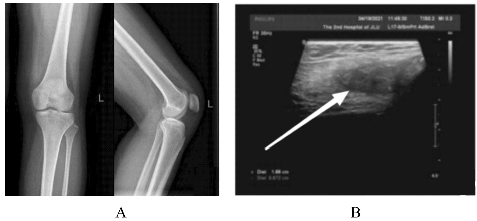
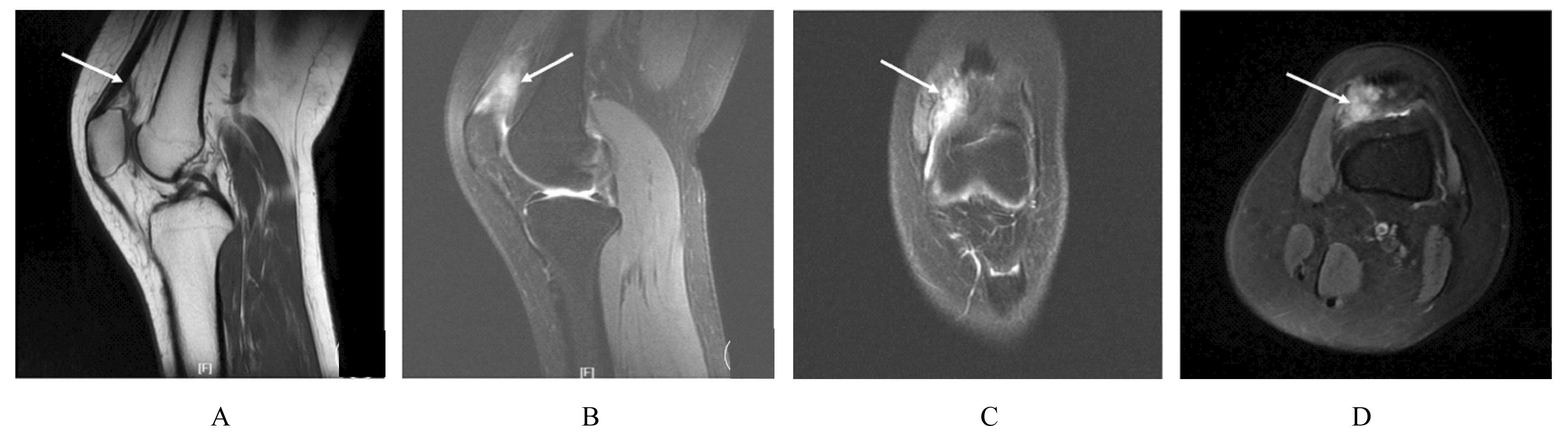
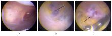
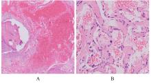
目的 分析1例膝关节滑膜内膜下血管瘤患者的临床表现和诊疗经过,以提高临床医生对该病的认识。 方法 回顾性分析1例膝关节滑膜内膜下血管瘤患者的临床资料、影像学资料、关节镜下表现和病理检查结果,并进行相关文献复习。 结果 患者,女性,22岁,7年内间断性左膝关节疼痛伴肿胀。查体,左膝关节内和外侧间隙压痛明显,左膝关节活动度降低(0°~90°)。超声检查,股内侧肌肌腱深层与滑膜组织间探及低回声光团;磁共振成像(MRI)检查,左膝关节内侧支持带深部靠近髌旁软组织肿胀,表现为T1加权序列上呈稍低信号,T2脂肪抑制序列上呈团片状稍高信号,滑膜组织内见高T2信号,边界欠清晰。初步诊断为左膝关节肿物。行关节镜下左膝关节病损切除术,镜下见内侧髌旁滑膜外观基本正常,清理表面滑膜组织内膜层后见肿物位于滑膜内膜下与关节囊之间,彻底切除肿物并送检,病理检查结果为滑膜内膜下血管瘤,术后患者左膝关节疼痛消失。 结论 膝关节滑膜内膜下血管瘤患者多有外伤病史,表现为不明原因的膝关节疼痛、肿胀和活动受限;结合影像学表现、关节镜和病理检查结果可明确诊断;关节镜手术治疗有助于改善患者预后,且具有损伤小、术后并发症少和恢复快等优势。

关键词: 关节镜, 滑膜内膜, 血管瘤, 膝关节, 病例报告

Abstract:

Objective To discuss the clinical manifestations and diagnosis and treatment processes of one patient with subintimal hemangioma of the knee joint, and to improve the clinicians’ understandings of the disease. Methods The clinical data, imagological data, arthroscopic manifestations, and pathological results of one patient with subsynovial hemangioma of the knee joint were retrospectively analyzed, and the related literatures were reviewed. Results A 22-year-old female patient presented with intermittent left knee joint pain and swelling for 7 years. The physical examination results showed obvious tenderness in the inner and outer spaces of the left knee joint, and the range of motion of the left knee joint was decreased (0°-90°).The ultrasound results showed that the hypoechoic light clusters were found between the deep layer of the vastus medialis tendon and the synovial tissue; the magnetic resonance imaging(MRI) results showed that the deep medial retinaculum of the left knee joint was swollen near the parapatellar soft tissue,which showed a slightly low signal on T1-weighted sequences and a patch-like signal with slightly higher intensity on T2 fat-suppressed sequences;the high T2 signal was seen in the synovial tissue, and the boundary was not clear. The preliminary consideration was left knee mass. The arthroscopic left knee joint lesion resection was performed; under the arthroscope, the appearance of the medial parapatellar synovium was basically normal,after removing the intimal layer of the superficial synovial tissue, the mass was located between the subintima of the synovium and the joint capsule, the mass was completely removed for submission, and the pathology results showed it was subsynovial hemangioma of the knee joint; the original pain in the left knee joint of the patient disappeared after operation. Conclusion Lots of patients with subsynovial hemangioma of the knee joint have medical history of trauma, manifesting as unexplained pain, swelling, and limited mobility of the knee joint; the diagnosis of the disease needs to be confirmed through the combination of imagological manifestations, arthroscope, and pathological examination results;the arthroscope surgery can significantly improve the prognosis of the patients, and it has some advantages,such as fewer surgical injuries, fewer postoperative complications, and faster recovery.

Key words: Arthroscopy, Subsynovial, Hemangioma, Knee joint, Case report

中图分类号: 

  • R686.7