吉林大学学报(医学版) ›› 2024, Vol. 50 ›› Issue (3): 620-627.doi: 10.13481/j.1671-587X.20240305

• 基础研究 • 上一篇    

高磷诱导下肢血管平滑肌细胞成骨分化关键差异表达基因筛选及验证

倪英群1,2,3(),杨矛1,杨迪1,郭呈林1,朱文君4,俞雅琴4,卢芹4,骆金芝4,吴春琴4,方朝晖1,2,3   

  1. 1.安徽中医药大学第一附属医院内分泌科,安徽 合肥 230031
    2.安徽省中医药科学院中医药防治糖尿病研究所,安徽 合肥 230031
    3.安徽中医药大学新安医学教育部重点实验室,安徽 合肥 230038
    4.安徽中医药大学研究生院,安徽 合肥 230038
  • 收稿日期:2023-06-30 出版日期:2024-05-28 发布日期:2024-07-01
  • 通讯作者: 倪英群 E-mail:Jessica8163@126.com
  • 作者简介:倪英群(1981-),女,安徽省芜湖市人,副主任医师,医学博士,主要从事中医药防治代谢内分泌疾病方面的研究。
  • 基金资助:
    国家自然科学基金项目(82274468);安徽省教育厅高校优秀人才支持计划项目(gxyqZD2021114);安徽中医药大学 新安医学教育部重点实验室开放项目(2022XAYX06);安徽中医药大学临床科研项目(2021yfylc07)

Screening of key differentially expressed genes involved in osteogenic differentiation of lower limb vascular smooth muscle cells and validation

Yingqun NI1,2,3(),Mao YANG1,Di YANG1,Chenglin GUO1,Wenjun ZHU4,Yaqin YU4,Qin LU4,Jinzhi LUO4,Chunqin WU4,Zhaohui FANG1,2,3   

  1. 1.Department of Endocrinology,First Affiliated Hospital,Anhui University of Chinese Medicine,Hefei 230031,China
    2.Institute of Traditional Chinese Medicine for Prevention and Treatment of Diabetes,Anhui Academy of Chinese Medicine,Hefei 230031,China
    3.Key Laboratory of Xin An Medicine,Ministry of Education,Anhui University of Chinese Medicine,Hefei 230038,China
    4.Graduate School,Anhui University of Chinese Medicine,Hefei 230038,China
  • Received:2023-06-30 Online:2024-05-28 Published:2024-07-01
  • Contact: Yingqun NI E-mail:Jessica8163@126.com

摘要:

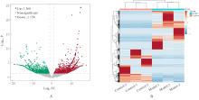
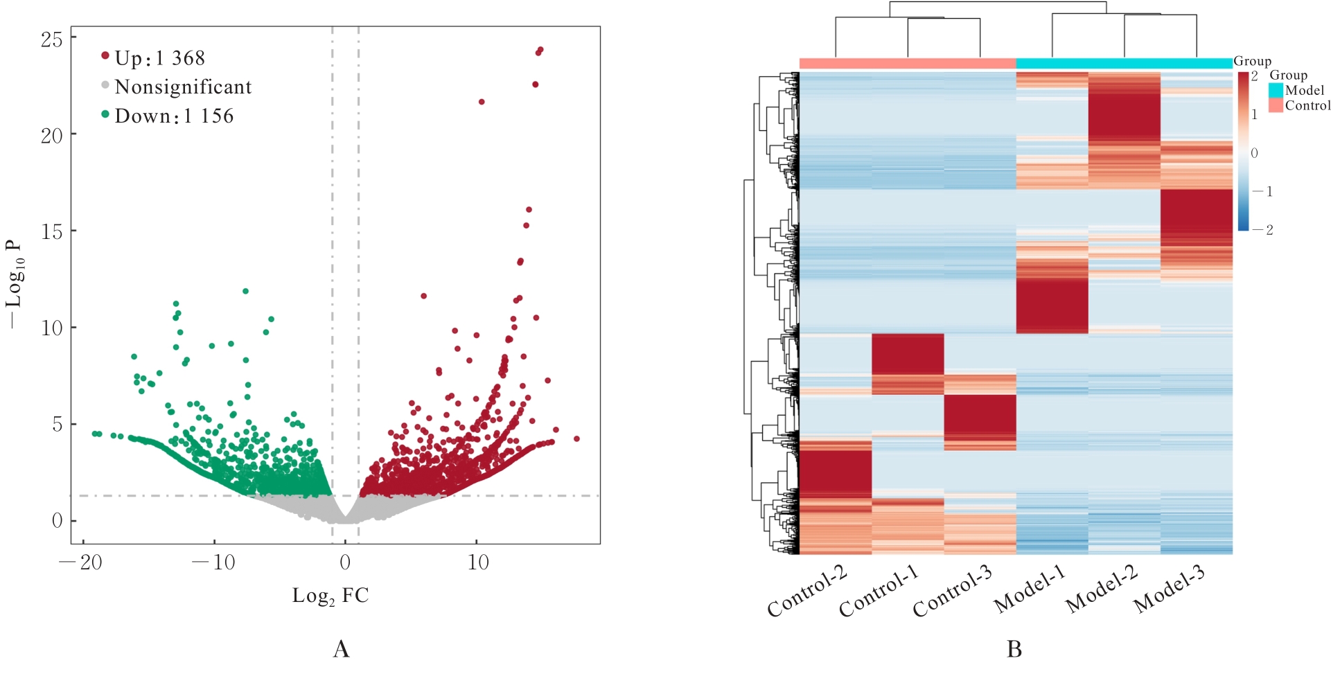
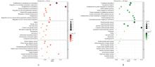
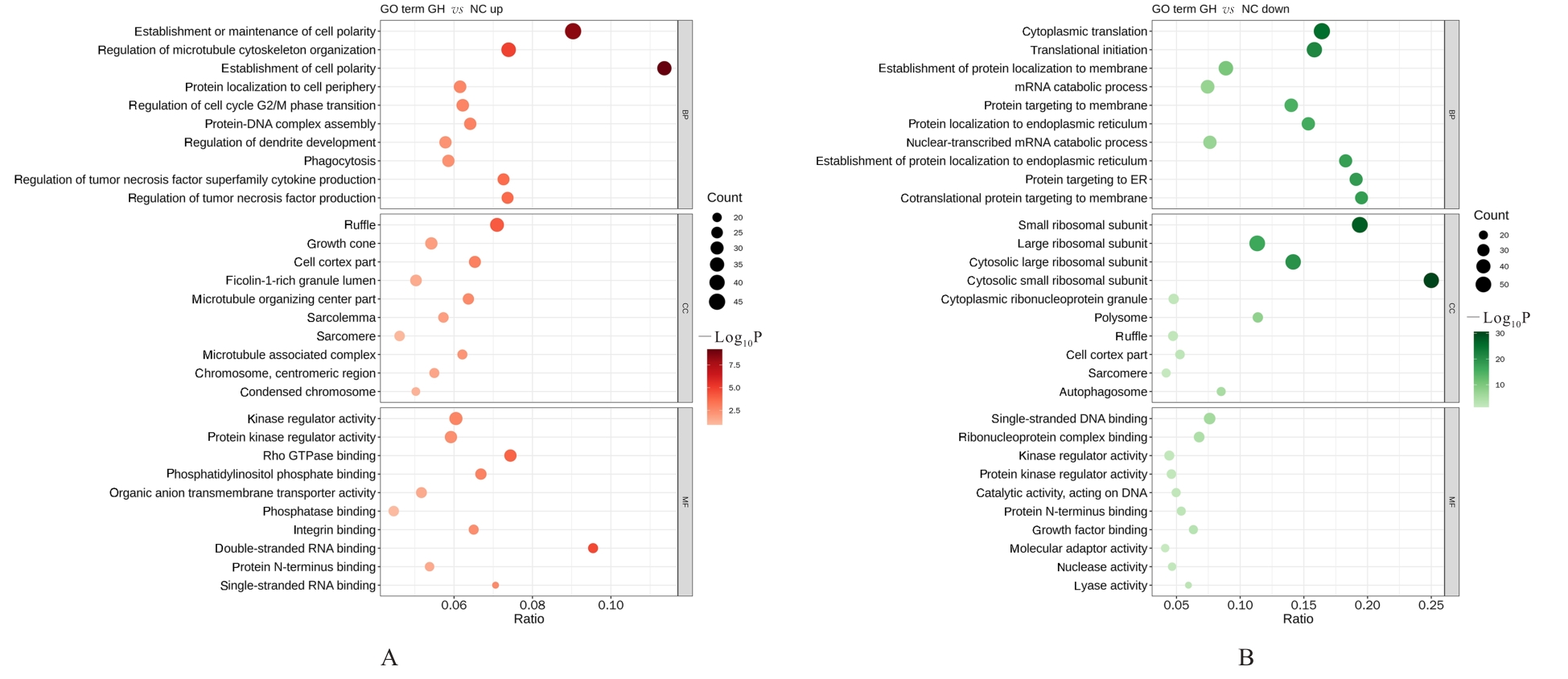
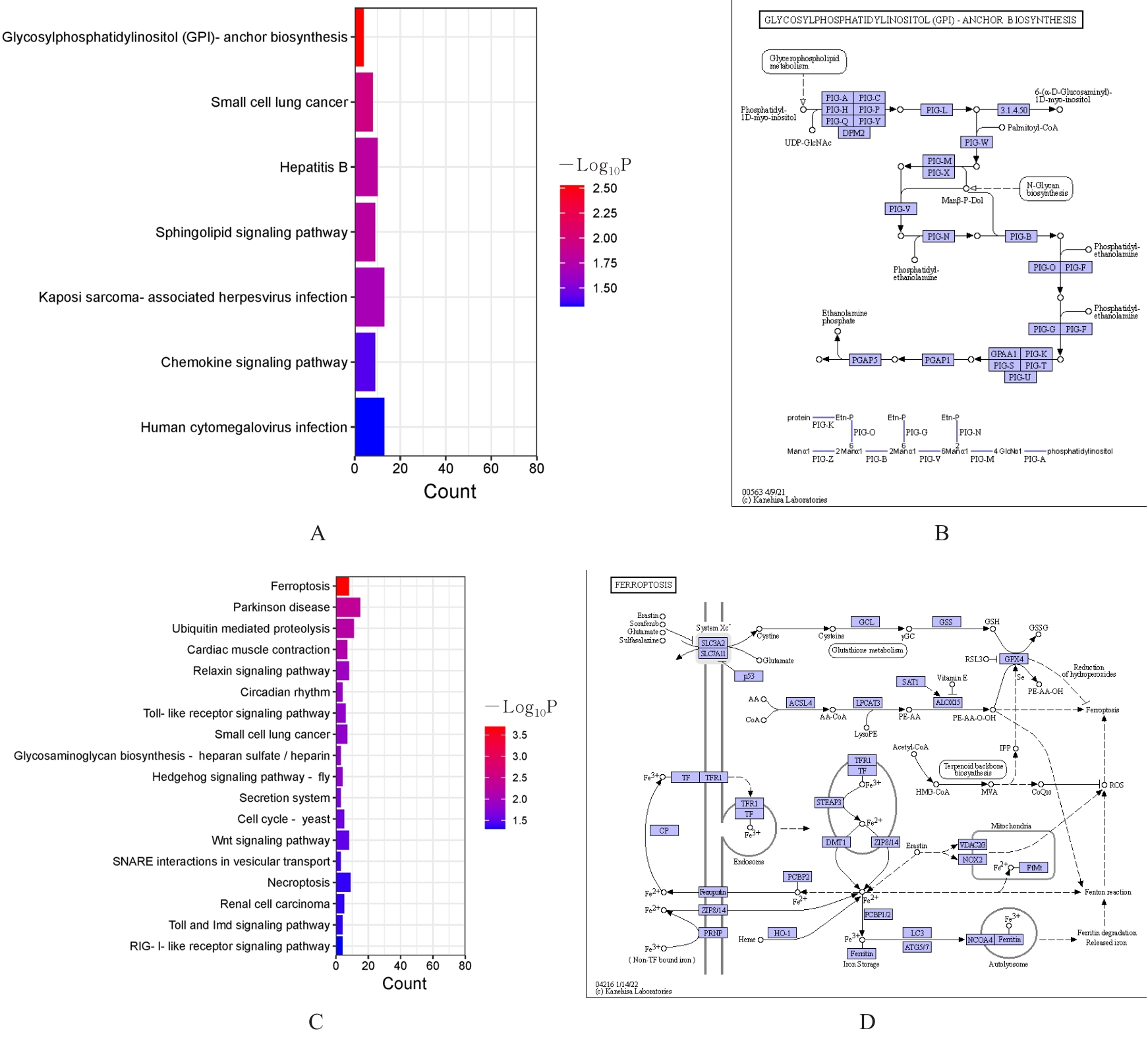
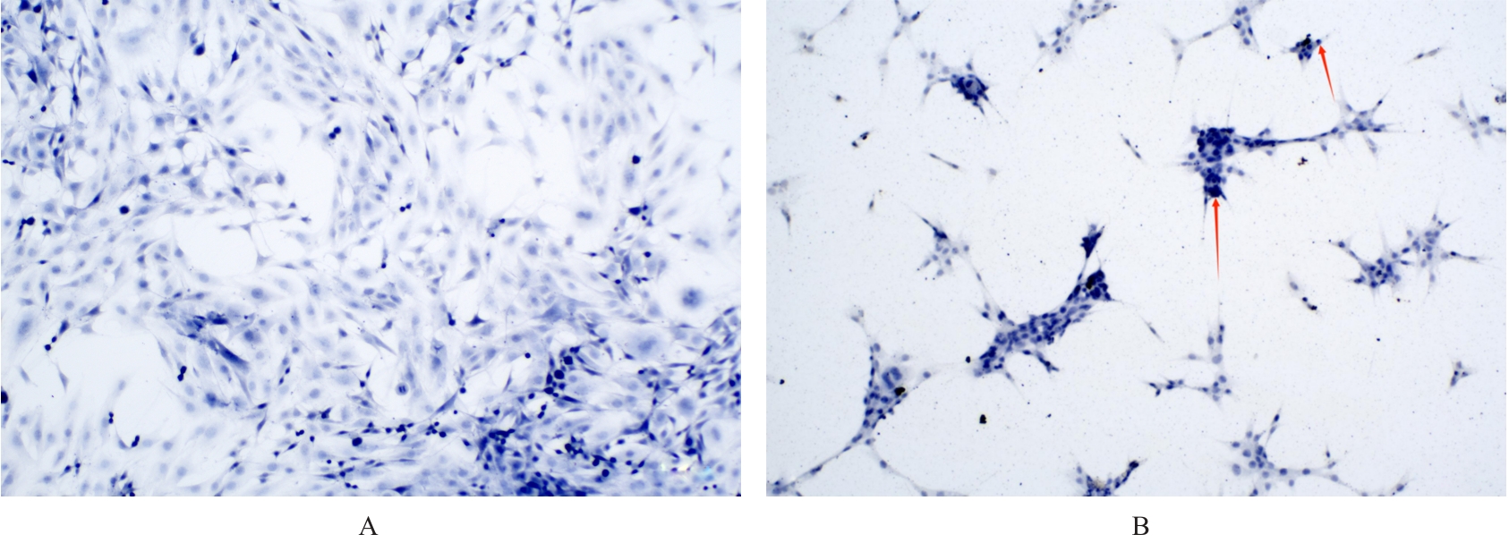
目的 采用mRNA高通量测序技术筛选高磷诱导下肢血管平滑肌细胞(VSMCs)钙化差异表达基因(DEGs),分析VSMCs钙化的关键基因及信号通路。 方法 将人VSMCs分为对照组和模型组,模型组细胞中加入高磷培养基,对照组细胞采用含10%胎牛血清的DMEM培养基于相同条件下培养。调整2组稳定转染VSMCs的状态,培养12 d,倒置显微镜下观察细胞形态表现并拍照。采用Hisat2软件筛选DEGs,采用Stringtie软件从生物过程(BP)、分子功能(MF)和细胞成分(CC)3个方面进行基因本体论(GO)功能和京都基因与基因组百科全书(KEGG)信号通路富集分析。Von Kossa染色法观察各组细胞钙化情况,实时荧光定量PCR(RT-qPCR)法检测2组细胞中碱性磷酸酶(ALP)、骨形态发生蛋白2(BMP2)、α-平滑肌肌动蛋白(α-SMA)、肿瘤蛋白53(Tp53)、谷胱甘肽过氧化物酶4(GPX4)、铁蛋白轻链1(Ftl1)和糖基磷脂酰肌醇特异性磷脂酶D1(GPLD1)mRNA表达水平。 结果 与对照组比较,模型组共2 524个DEGs,其中1 368个DEGs表达上调,1 156个DEGs表达下调;2组细胞DEGs聚类分离明显。GO功能和KEGG信号通路富集分析表达上调的DEGs主要参与微管细胞骨架组织的调节、细胞极性、蛋白质定位和细胞周期调控等BP,构建细胞膜部分、微管组织、染色体和着丝粒区等CC,发挥与磷脂酰肌醇磷酸盐、Rho鸟苷酸三磷酸酶(GTPase)蛋白结合、参与跨膜转运和调节蛋白激酶活性等MF;表达下调的DEGs主要参与细胞质翻译、蛋白质膜定位、mRNA代谢和蛋白质内质网定位等BP,构建核糖体亚单位、细胞膜和自噬体等CC,发挥与单链DNA、核糖核蛋白复合物、生长因子结合、调节蛋白激酶活性和催化作用等MF。差异表达上调的基因富集7条信号通路,其中最为显著的是糖基磷脂酰肌醇(GPI)锚定的生物合成;差异表达下调的基因富集18条信号通路,其中最为显著的是铁死亡。RT-qPCR法,与对照组比较,模型组细胞中GPX4、Ftl1和Tp53 mRNA表达水平明显降低(P<0.01),GPLD1 mRNA表达水平明显升高(P<0.01);与对照组比较,模型组细胞中α-SMA mRNA表达水平明显降低(P<0.01),ALP和BMP2 mRNA表达水平明显升高(P<0.01)。 结论 钙化的VSMCs与正常细胞存在DEGs,铁死亡和GPI锚定的生物合成信号途径是高磷诱导下肢VSMCs钙化的关键信号通路,主要由GPX4、Ftl1、Tp53和GPLD1共同介导完成。

关键词: 血管平滑肌细胞, 成骨分化, 细胞钙化, mRNA测序, 铁死亡

Abstract:

Objective To screen the differentially expressed genes (DEGs) under high phosphate-induced calcification in the vascular smooth muscle cells (VSMCs) by mRNA high-throughput sequencing technology, and to analyze the key genes and signaling pathways involved in the VSMCs calcification. Methods The human VSMCs were divided into control group and model group. The cells in model group was exposed to the high-phosphate medium, while the cells in control group were cultured in DMEM supplemented with 10% fetal bovine serum under the same conditions. The VSMCs in two groups, stably transfected, were cultured for 12 d. The morphology of the cells in two groups were observed and photographed under inverted microscope. The DEGs were selected by Hisat2 software, and Gene Ontology (GO) functional and Kyoto Encyclopedia of Genes and Genomes (KEGG) signaling pathway enrichment analysis were performed by Stringtie software from three aspects, such as biological processes (BP), molecular functions (MF), and cellular components (CC). The calcification of the cells in two groups was observed by Von Kossa staining method. Real-time fluorescence quantitative PCR (RT-qPCR) method was used to analyze the expression levels of alkaline phosphatase (ALP), bone morphogenetic protein 2 (BMP2), alpha-smooth muscle actin (α-SMA), tumor protein 53 (Tp53), glutathione peroxidase 4 (GPX4), ferritin light chain 1 (Ftl1), and glycosylphosphatidylinositol-specific phospholipase D1 (GPLD1) mRNA in the cells in two groups. Results Compared with control group, there were 2 524 DEGs in the cells in model group, and there were 1 368 upregulated DEGs and 1 156 downregulated DEGs. Clustering of DEGs between the cells in two groups was distinct. The GO functional and KEGG pathway enrichment analysis results showed that the upregulated DEGs were primarily involved in regulating the microtubule cytoskeleton, cell polarity, protein localization, and cell cycle regulation among BPs; in constructing cell membrane, microtubule organization, chromosomes, and kinetochore among CCs; and functioning in phosphatidylinositol phosphate, Rho GTPase protein binding, transmembrane transport, and protein kinase regulatory activity among MFs. Downregulated DEGs were mainly involved in cytoplasmic translation, protein membrane localization, mRNA metabolism, and protein endoplasmic reticulum localization among BPs; in forming ribosome subunits, cell membrane, and autophagy among CCs; and functioning in single-stranded DNA, ribonucleoprotein complex, growth factor binding, regulating protein kinase activity, and catalytic activity among MFs. Seven signaling pathways were significantly enriched in upregulated genes, most notably in the biosynthesis of glycosylphosphatidylinositol (GPI) anchors; whereas 18 signaling pathways were significantly enriched in the downregulated genes, most notably in ferroptosis.The RT-qPCR results showed that compared with control group, the expression levels of GPX4, Ftl1, and Tp53 mRNA in the cells in model group were significantly decreased (P<0.01), while the expression level of GPLD1 mRNA was significantly increased (P<0.01); compared with control group, the expression level of α-SMA mRNA in the cells in model group was significantly decreased (P<0.01), and the expression levels of ALP and BMP2 mRNA were significantly increased (P<0.01). Conclusion The VSMCs underwent calcification and normal cells exhibit the DEGs.The key signaling pathways in the calcification induced by high phosphate in the VSMCs include ferroptosis and GPI anchor biosynthesis, mediated primarily through GPX4, Ftl1, Tp53, and GPLD1.

Key words: Vascular smooth muscle cell, Osteogenic differentiation, Cell calcification, mRNA sequencing, Ferroptosis

中图分类号: 

  • Q254