吉林大学学报(医学版) ›› 2024, Vol. 50 ›› Issue (3): 666-675.doi: 10.13481/j.1671-587X.20240310

• 基础研究 • 上一篇    

单核细胞趋化蛋白1对肺癌A549细胞迁移和侵袭的影响及其机制

王远1,王志娟2,张明姝2,王艺慧2,张晴2,叶丽平2,3()   

  1. 1.锦州医科大学基础医学院病理学教研室,辽宁 锦州 121001
    2.锦州医科大学基础医学院病理 生理学教研室,辽宁 锦州 121001
    3.锦州医科大学生物人类学研究所,辽宁 锦州 121001
  • 收稿日期:2023-07-19 出版日期:2024-05-28 发布日期:2024-07-01
  • 通讯作者: 叶丽平 E-mail:yeliping@jzmu.edu.cn
  • 作者简介:王 远(1988-),女,山东省临沂市人,副教授,医学博士,主要从事肿瘤发生发展机制方面的研究。
  • 基金资助:
    国家自然科学基金青年科学基金项目(82103624);吴阶平医学基金会研究基金课题项目(320.6750.2020-06-68)

Effects of monocyte chemoattractant protein-1 on invasion and migration of lung cancer A549 and their mechanisms

Yuan WANG1,Zhijuan WANG2,Mingshu ZHANG2,Yihui WANG2,Qing ZHANG2,Liping YE2,3()   

  1. 1.Department of Pathology,School of Basic Medical Sciences,Jinzhou Medical University,Jinzhou 121001,China
    2.Department of Pathophysiology,School of Basic Medical Sciences,Jinzhou Medical University,Jinzhou 121001,China
    3.Institute of Biological Anthropology,Jinzhou Medical University,Jinzhou 121001,China
  • Received:2023-07-19 Online:2024-05-28 Published:2024-07-01
  • Contact: Liping YE E-mail:yeliping@jzmu.edu.cn

摘要:

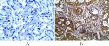
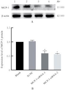
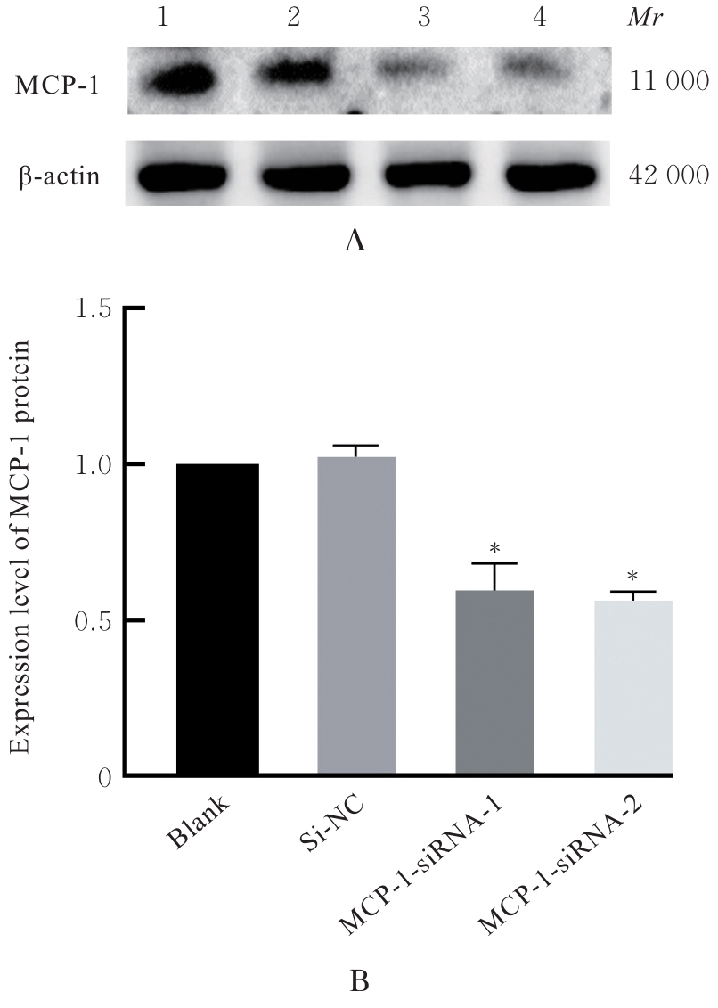
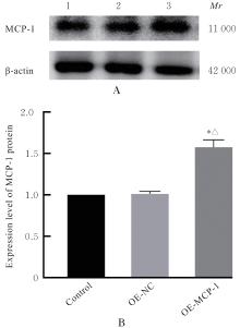
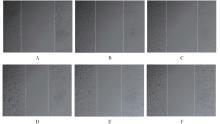
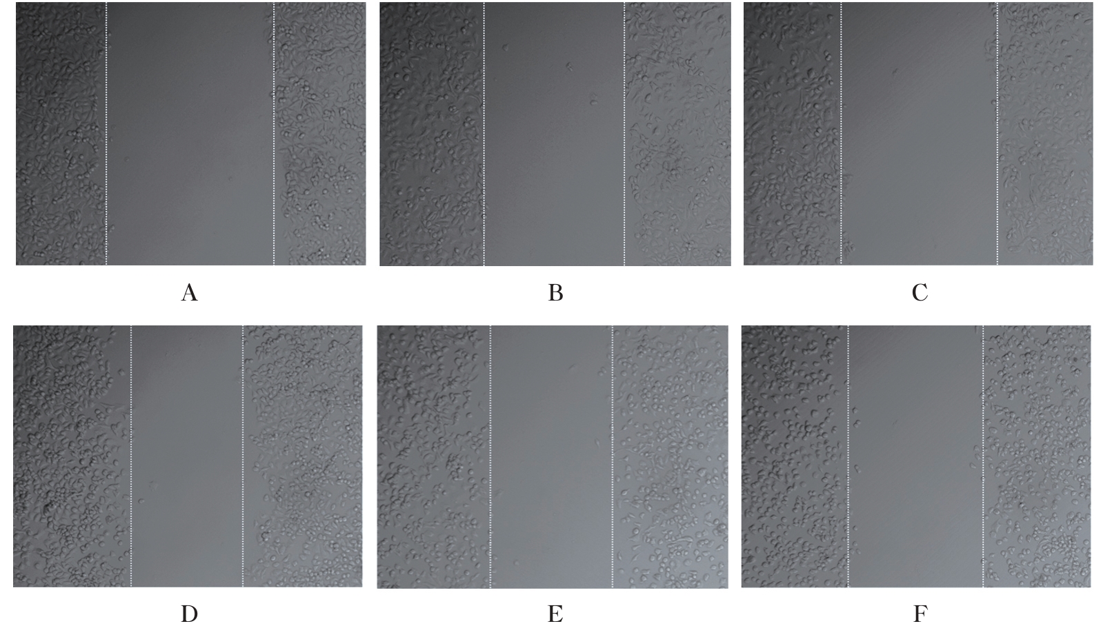
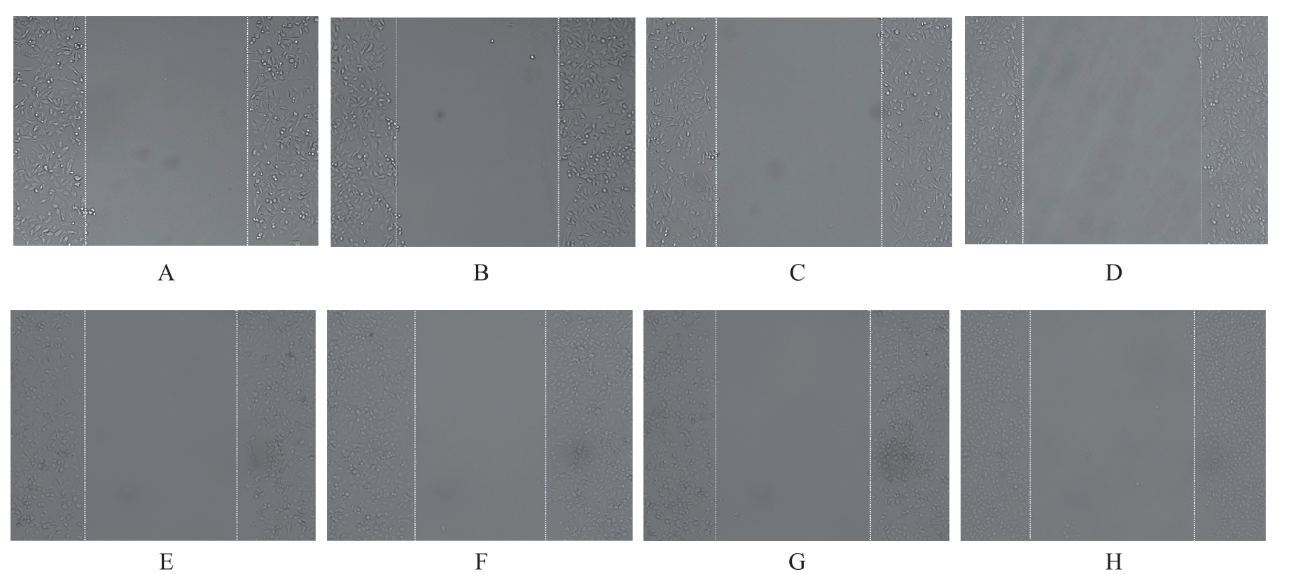
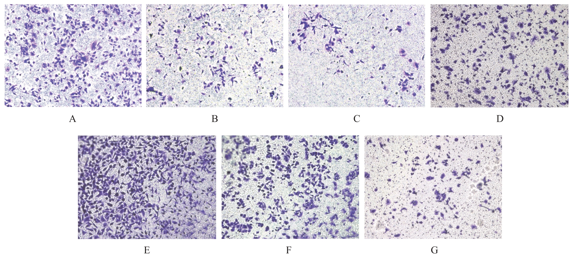
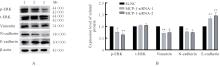
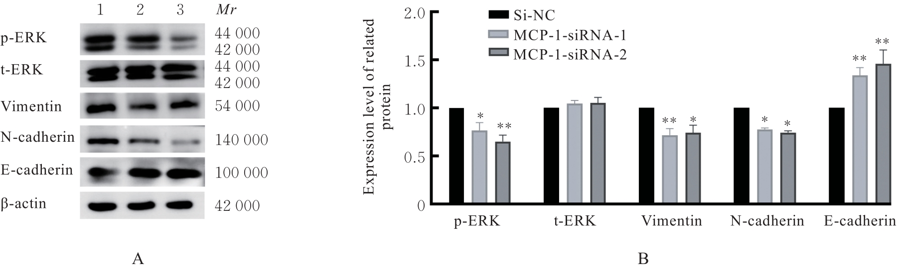
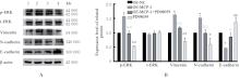
目的 探讨单核细胞趋化蛋白1(MCP-1)对肺癌A549细胞迁移和侵袭的影响,并阐明其作用机制。 方法 采用免疫组织化学法检测80例非小细胞肺癌(NSCLC)及癌旁正常肺组织中MCP-1蛋白表达情况。体外培养人肺癌A549细胞,MCP-1-小干扰RNA(siRNA)实验分为空白组、阴性对照组(si-NC组)、MCP-1-siRNA-1组和MCP-1-siRNA-2组;MCP-1过表达实验分为对照组、空载对照组(OE-NC组,转染MCP-1过表达空载质粒)、过表达MCP-1组(OE-MCP-1组,转染MCP-1过表达质粒)、过表达MCP-1+细胞外调节蛋白激酶(ERK)信号通路抑制剂PD98059组(OE-MCP-1+PD98059组,共转染MCP-1过表达质粒和PD98059)和PD98059组(转染PD98059)。分别将MCP-1-siRNA和质粒转染至肺癌A549细胞,Western blotting法验证各组A549细胞转染效率。细胞划痕实验和Transwell小室实验观察各组A549细胞迁移率和侵袭细胞数,Western blotting法检测各组A549细胞中磷酸化ERK(p-ERK)、 总ERK(t-ERK)和上皮-间质转化(EMT)相关蛋白表达水平。 结果 与癌旁组织比较,NSCLC组织中MCP-1蛋白阳性表达率明显升高(P<0.05);NSCLC组织中MCP-1蛋白表达水平与TNM分期和淋巴结转移有关联(P<0.05)。与si-NC组比较,MCP-1-siRNA-1组和MCP-1-siRNA-2组A549细胞中MCP-1蛋白表达水平均明显降低(P<0.01);与对照组和OE-NC组比较,OE-MCP-1组A549细胞中MCP-1蛋白表达水平明显升高(P<0.01)。细胞划痕实验,与si-NC组比较,MCP-1-siRNA-1组和MCP-1-siRNA-2组细胞迁移率均明显降低(P<0.01);与 OE-NC 组比较,OE-MCP-1 组细胞迁移率明显升高(P<0.01);与 OE-MCP-1 组比较,OE-MCP-1+PD98059 组细胞迁移率明显降低(P<0.01); 与 OE-MCP-1+PD98059 组比较,PD98059组细胞迁移率明显降低(P<0.01)。Transwell小室实验,与si-NC组比较,MCP-1-siRNA-1组和MCP-1-siRNA-2组侵袭细胞数明显减少(P<0.01);与OE-NC组比较,OE-MCP-1组侵袭细胞数明显增加(P<0.01);与OE-MCP-1组比较, OE-MCP-1+PD98059组侵袭细胞数明显减少(P<0.01); 与 OE-MCP-1+PD98059 组比较,PD98059 组侵袭细胞数明显减少(P<0.01)。 Western blotting 法,与si-NC组比较,MCP-1-siRNA-1组和MCP-1-siRNA-2组A549细胞中p-ERK、波形蛋白(Vimentin)和N-钙黏蛋白(N-cadherin)表达水平均明显降低(P<0.05或P<0.01),E-钙黏蛋白(E-cadherin)表达水平均明显升高(P<0.01); 与 OE-NC 组比较, OE-MCP-1 组 A549 细胞中p-ERK、Vimentin和N-cadherin蛋白表达水平均明显升高(P<0.01),E-cadherin蛋白表达水平均明显降低(P<0.01);与OE-MCP-1组比较,OE-MCP-1+PD98059组A549细胞中p-ERK、Vimentin和N-cadherin蛋白表达水平均明显降低(P<0.01),E-cadherin蛋白表达水平均明显升高(P<0.05);与OE-MCP-1+PD98059组比较,PD98059组A549细胞中p-ERK、Vimentin和N-cadherin蛋白表达水平均明显降低(P<0.05 或 P<0.01), E-cadherin蛋白表达水平明显升高(P<0.01)。 结论 MCP-1蛋白可上调肺癌A549细胞中EMT相关蛋白表达,促进肺癌A549细胞的迁移和侵袭,其作用机制与激活ERK信号通路有关。

关键词: 单核细胞趋化蛋白1, 细胞外信号调节激酶, 癌,非小细胞肺, 细胞侵袭, 细胞迁移

Abstract:

Objective To discuss the effects of monocyte chemoattractant protein-1 (MCP-1) on the migration and invasion of lung cancer A549 cells, and to clarify the mechanisms. Methods Immunohistochemistry method was used to detect the expression of MCP-1 protein in 80 cases of non-small cell lung cancer (NSCLC) and adjacent normal lung tissues. The human lung cancer A549 cells were cultured in vitro. The MCP-1-small interfering RNA (siRNA) experiment was divided into blank group, negative control group (si-NC group), MCP-1-siRNA-1 group, and MCP-1-siRNA-2 group. The MCP-1 over-expression experiment was divided into control group, empty vector control group (OE-NC, transfected with MCP-1 over-expression empty vector), over-expression MCP-1 group (OE-MCP-1 group, transfected with MCP-1 over-expression plasmid), over-expression MCP-1+extracellular regulated protein kinase (ERK) pathway inhibitor PD98059 group (OE-MCP-1+PD98059 group, co-transfected with MCP-1 over-expression plasmid and PD98059), and PD98059 group (transfected with PD98059).The MCP-1 siRNA and plasmids were transfected into the lung cancer A549 cells; Western blotting method was used to verify the transfection efficiencies of the cells in various groups; the migration rate and the number of invasion cells in various groups were observed by wound healing assay and Transwell chamber assay, respectively; Western blotting method was also used to detect the expression levels of phosphorylated ERK (p-ERK), total ERK (t-ERK), and epithelial-mesenchymal transition (EMT)-related proteins in the A549 cells in various groups. Results Compared with adjacent tissue, the positive expression rate of MCP-1 protein in NSCLC tissue was significantly increased (P<0.05), and the expression level of MCP-1 protein was related to TNM stage and lymph node metastasis (P<0.05). Compared with si-NC group, the expression level of MCP-1 protein in the cells in MCP-1-siRNA-1 and MCP-1-siRNA-2 groups was significantly decreased (P<0.01). Compared with control group and OE-NC group, the expression level of MCP-1 protein in the cells in OE-MCP-1 group was significantly increased (P<0.01). The wound healing assay results showed that compared with si-NC group, the migration rate of the cells in MCP-1-siRNA-1 and MCP-1-siRNA-2 groups were significantly decreased (P<0.01). Compared with OE-NC group, the migration rate of the cells in OE-MCP-1 group was significantly increased (P<0.01); compared with OE-MCP-1 group, the migration rate of the cells in OE-MCP-1+PD98059 group was significantly decreased (P<0.01). Compared with OE-MCP-1+PD98059 group, the migration rate of the cells in PD98059 group was significantly decreased (P<0.01). The Transwell chamber assay results showed that compared with si-NC group, the number of invasion cells in MCP-1-siRNA-1 and MCP-1-siRNA-2 groups was significantly decreased (P<0.01). Compared with OE-NC group, the number of invasion cells in OE-MCP-1 group was significantly increased (P<0.01); compared with OE-MCP-1 group, the number of invasion cells in OE-MCP-1+PD98059 group was significantly decreased (P<0.01); compared with OE-MCP-1+PD98059 group, the number of invasion cells in PD98059 group was significantly decreased (P<0.01).The Western blotting results showed that compared with si-NC group, the expression levels of p-ERK, Vimentin, and N-cadherin protein in the cells in MCP-1-siRNA-1 and MCP-1-siRNA-2 groups were significantly decreased (P<0.05 or P<0.01), and the expression level of E-cadherin proteins was significantly increased (P<0.01). Compared with OE-NC group, the expression levels of p-ERK, Vimentin, and N-cadherin proteins in the cells in OE-MCP-1 group were significantly increased (P<0.01), and the expression level of E-cadherin protein was significantly decreased (P<0.01). Compared with OE-MCP-1 group, the expression levels of p-ERK, Vimentin, and N-cadherins proteins in the OE-MCP-1+PD98059 group were significantly decreased (P<0.01), and the expression level of E-cadherin protein was significantly increased (P<0.05). Compared with OE-MCP-1+PD98059 group, the expression levels of p-ERK, Vimentin, and N-cadherin proteins in the cells in PD98059 group were significantly decreased (P<0.05 or P<0.01), and the expression level of E-cadherin protein was increased (P<0.01). Conclusion MCP-1 protein can upregulate the expression of EMT-related proteins in the lung cancer A549 cells, and promote the migration and invasion of the lung cancer A549 cells; its mechanism may be related to the activation of the ERK signaling pathway.

Key words: Monocyte chemoattractant protein-1, Extracellular-signal regulated protein kinase, Cancer,non-small cell lung, Cell invasion, Cell migration

中图分类号: 

  • R734.2