吉林大学学报(医学版) ›› 2024, Vol. 50 ›› Issue (5): 1227-1234.doi: 10.13481/j.1671-587X.20240505

• 基础研究 • 上一篇    

液体复苏抑制血管组织铁死亡对爆炸损伤并发失血性休克大鼠血管细胞结构损伤的减轻作用

彭小勇1,朱娱1,张双博2,朱英国2,李涛1,刘良明1,王建民2,杨光明2()   

  1. 1.陆军军医大学大坪医院野战外科研究部战伤休克与输血研究室,创伤、烧伤与复合伤国家重点实验室,重庆 400042
    2.陆军军医大学大坪医院野战外科研究部武器杀伤生物效应评估实验室,创伤、烧伤与复合伤国家重点实验室,重庆 400042
  • 收稿日期:2023-09-05 出版日期:2024-09-28 发布日期:2024-10-28
  • 通讯作者: 杨光明 E-mail:yanggm971@163.com
  • 作者简介:彭小勇(1986-),男,重庆市永川区人,助理研究员,医学硕士,主要从事战伤休克心血管功能变化方面的研究。
  • 基金资助:
    国家自然科学基金面上项目(82072164);军队后勤科研重大项目(ALJ18J00)

Alleviative effect of fluid resuscitation on damage of structure injury of vascular cells after blast injury complicated with hemorrhagic shock in rats by inhibiting ferroptosis of vascular tissue

Xiaoyong PENG1,Yu ZHU1,Shuangbo ZHANG2,Yingguo ZHU2,Tao LI1,Liangming LIU1,Jianmin WANG2,Guangming YANG2()   

  1. 1.State Key Laboratory of Trauma,Burns and Combined Injury,Department of Shock and Transfusion,Research Institute of Surgery,Daping Hospital,Army Medical University,Chongqing 400042,China
    2.State Key Laboratory of Trauma,Burns and Combined Injury,Department of Weapon Bioeffect Assessment,Research Institute of Surgery,Daping Hospital,Army Medical University,Chongqing 400042,China
  • Received:2023-09-05 Online:2024-09-28 Published:2024-10-28
  • Contact: Guangming YANG E-mail:yanggm971@163.com

摘要:

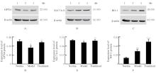
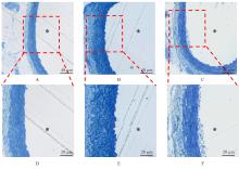
目的 探讨液体复苏对爆炸损伤并发失血性休克大鼠血管铁死亡发生情况和血管细胞结构的影响,并阐明其作用机制。 方法 取54只健康成年SD大鼠,随机分为正常组、爆炸损伤并发失血性休克(模型)组和液体复苏(治疗)组,每组18只,每组随机取10只大鼠观察存活情况,另8只大鼠用于检测其他指标。观察各组大鼠平均存活时间(ST)、24 h及72 h存活情况,观察各组大鼠血压(BP)、心率(HR)和呼吸频率(RR),检测各组大鼠血清中血肌酐(Scr)、血尿素氮(BUN)、乳酸(LAC)、血糖(GLU)、铁离子、谷胱甘肽(GSH)和丙二醛(MDA)水平及天冬氨酸氨基转移酶(AST)、丙氨酸氨基转移酶(ALT)和乳酸脱氢酶(LDH)活性,Western blotting法检测各组大鼠肠系膜上动脉组织中铁死亡标志蛋白谷胱甘肽过氧化物酶4(GPX4)、溶质载体家族7成员11(SLC7A11)和铁代谢调节蛋白血红素加氧酶1(HO-1)蛋白表达水平,观察各组大鼠肠系膜上动脉组织病理形态表现。 结果 正常组大鼠全部存活72 h,模型组大鼠最长ST不超过9 h。与模型组比较,治疗组大鼠的ST和24 h存活率(SR)明显升高(P<0.05)。与正常组比较,模型组大鼠BP、HR和RR均明显降低(P<0.01);与模型组比较,治疗组大鼠的BP、HR和RR明显升高(P<0.05)。与正常组比较,模型组大鼠血清中AST和ALT活性及Scr和BUN水平明显升高(P<0.01);与模型组比较,治疗组大鼠血清中LAC和GLU水平明显降低(P<0.01)。与正常组比较,模型组大鼠血清铁离子浓度、GSH水平、MDA水平和LDH活性明显升高(P<0.05);与模型组比较,治疗组大鼠血清铁离子浓度和LDH活性明显降低(P<0.01)。与正常组比较,模型组大鼠肠系膜上动脉组织中GPX4和SLC7A11蛋白表达水平明显降低(P<0.05);与模型组比较,治疗组大鼠肠系膜上动脉组织中GPX4和SLC7A11表达水平明显升高(P<0.05)。与正常组比较,模型组大鼠肠系膜上动脉组织中HO-1蛋白表达水平升高(P<0.01);与模型组比较,治疗组大鼠肠系膜上动脉组织中HO-1蛋白表达水平升高(P<0.01)。显微病理,模型组大鼠肠系膜上动脉各层细胞排列紊乱,明显肿胀,厚度明显增加;治疗组大鼠肠系膜上动脉组织病理变化减轻。超微病理,正常组大鼠血管内皮细胞结构完整,内皮细胞下基质无肿胀;模型组大鼠血管内皮细胞膜破坏,细胞质溶解破碎,且内皮细胞下基质明显肿胀;治疗组大鼠血管内皮细胞肿胀减轻。 结论 爆炸损伤并发失血性休克大鼠血管组织发生铁死亡,液体复苏能够通过抑制血管组织铁死亡减轻血管细胞结构损伤。

关键词: 爆炸损伤, 失血性休克, 液体复苏, 铁死亡, 血管

Abstract:

Objective To discuss the effect of fluid resuscitation on the occurrence of ferroptosis in vascular tissue and the structure of vascular cells in the rats with blast injury complicated with hemorrhagic shock, and to clarify its mechanism. Methods A total of 54 healthy adult SD rats were randomly divided into normal group, blast injury complicated with hemorrhagic shock (model) group, and the fluid resuscitation (treatment) group, and there were 18 rats in each group. Among them,10 rats were randomly selected to observe the surival status and another 8 rats were selected to detect the other indexes. The average survival time(ST), 24 h and 72 h survival rates of the rats in various groups were observed; the blood pressure (BP), heart rate (HR), and respiratory rate (RR) of the rats in various groups were observed; the levels of serum creatinine (Scr), blood urea nitrogen (BUN), lactate (LAC), glucose (GLU), iron ions, glutathione (GSH), and malondialdehyde (MDA) and the activities of aspartate aminotransferase (AST), alanine aminotransferase (ALT) and lactate dehydrogenase (LDH) in serum of the rats in various groups were detected;Western blotting method was used to detect the expression levels of ferroptosis marker proteins glutathione peroxidase 4 (GPX4), solute carrier family 7 member 11 (SLC7A11), and heme oxygenase 1 (HO-1) proteins in superior mesenteric artery tissue of the rats in various groups; the pathomorphology of the superior mesenteric artery of the rats in various groups was observed. Results All the rats in normal group survived for 72 h, while the longest ST of the rats in model group did not exceed 9 h. Compared with model group, the ST and 24 h survival rate (SR) of the rats in treatment group were significantly increased (P<0.05). Compared with normal group, the BP, HR, and RR of the rats in model group were significantly decreased (P<0.01). Compared with model group, the BP, HR, and RR of the rats in treatment group were significantly increased after fluid resuscitation (P<0.05). Compared with normal group, the activities of AST and ALT, and the levels of Scr and BUN in serum of the rats in model group were significantly increased (P<0.01). Compared with model group, the serum levels of LAC and GLU of the rats in treatment group were significantly decreased (P<0.01). Compared with normal group, the concentration of iron ion, GSH level, MDA level, LDH activity in serum of the rats in model group were significantly increased (P<0.05); compared with model group, the concentration of iron ion and LDH activity in serum of the rats in treatment group was significantly decreased (P<0.01). Compared with normal group, the expression levels of GPX4 and SLC7A11 in superior mesenteric artery tissue of the rats in model group were significantly decreased (P<0.05);compared with model group, the expression levels of GPX4 and SLC7A11 in superior mesenteric artery tissue of the rats in treatment group were significantly increased (P<0.05). Compared with normal group, the expression level of HO-1 protein in superior mesenteric artery tissue of the rats in model group was increased (P<0.01); compared with model group, the expression level of HO-1 protein in superior mesenteric artery tissue of the rats in treatment group was increased (P<0.01). The microscopic pathology results showed that the cell arrangement in the layers of the superior mesenteric artery tissue of the rats in model group was disordered, the swelling was significant and the thickness was increased; the pathological changes in superior mesenteric artery tissue of the rats in treatment group was alleviated. The ultramicroscopic pathology results showed that the endothelial cell structure of blood vessels of the rats in normal group was intact, and there was no swelling in the subendothelial matrix; the vascular endothelial cell membrane of the rats in model group was damaged, there were cytoplasmic dissolution and fragmentation, and the swelling of the subendothelial matrix was significant; the swelling of the vascular endothelial cells in treatment group was alleviated. Conclusion Ferroptosis occurs in vascular tissue of the rats with blast injury complicated with hemorrhagic shock, and fluid resuscitation can alleviate the structural damage of the vascular cells by inhibiting the vascular tissue ferroptosis.

Key words: Blast injury, Hemorrhagic shock, Fluid resuscitation, Ferroptosis, Vascular

中图分类号: 

  • R136.37