吉林大学学报(医学版) ›› 2024, Vol. 50 ›› Issue (5): 1438-1449.doi: 10.13481/j.1671-587X.20240531

• 方法学 • 上一篇    

新生大鼠原代软骨细胞分离和培养方法的改进

杨丹聃1,陈骄阳,王馨珩2,赵泽彤,潘莹3,薛百功3,高长曌4()   

  1. 1.吉林大学基础医学院病原免疫细胞遗传学实验中心,吉林 长春 130021
    2.吉林大学中日联谊医院皮肤科,吉林 长春 130033
    3.吉林大学基础医学院细胞学系,吉林 长春 130021
    4.吉林大学中日联谊医院放疗科,吉林 长春 130033
  • 收稿日期:2023-11-25 出版日期:2024-09-28 发布日期:2024-10-28
  • 通讯作者: 高长曌 E-mail:gaochangzhao@jlu.edu.cn
  • 作者简介:杨丹聃(1989-),女,吉林省长春市人,工程师,生物医学工程学硕士,主要从事细胞生物学和免疫学方面的教学及研究。
  • 基金资助:
    国家自然科学基金项目(8227091657);吉林省教育厅科研项目(JJKH20221050KJ)

Improvement of isolation and culture methods for primary chondrocytes of neonatal rats

Dandan YANG1,Jiaoyang CHEN,Xinheng WANG2,Zetong ZHAO,Ying PAN3,Baigong XUE3,Changzhao GAO4()   

  1. 1.Experimental Center of Pathogenobiology Immunology Cytobiology and Genetics,School of Basic Medical Sciences,Jilin University,Changchun 130021,China
    2.Department of Dermatology,China-Japan Union Hospital,Jilin University,Changchun 130033,China
    3.Department of Biology,School of Basic Medical Sciences,Jilin University,Changchun 130021,China
    4.Department of Radiotherapy,China-Japan Union Hospital,Jilin University,Changchun 130033,China
  • Received:2023-11-25 Online:2024-09-28 Published:2024-10-28
  • Contact: Changzhao GAO E-mail:gaochangzhao@jlu.edu.cn

摘要:

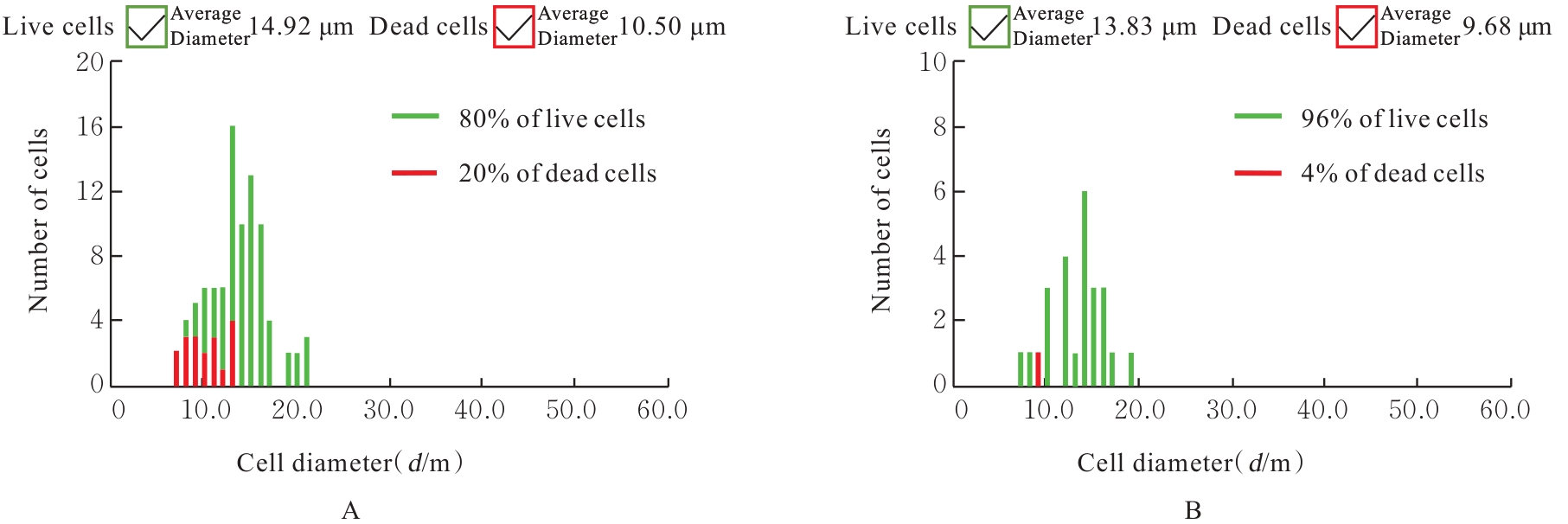
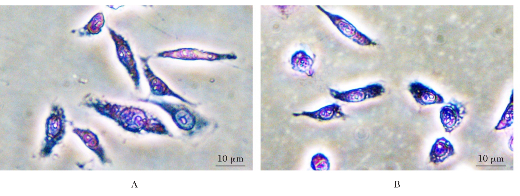
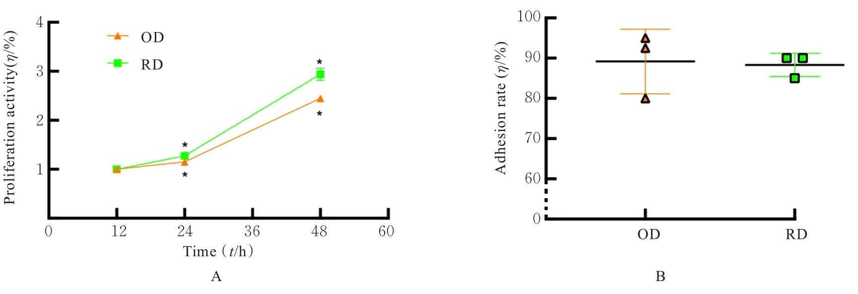
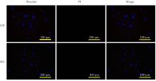
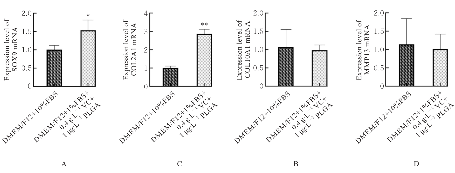
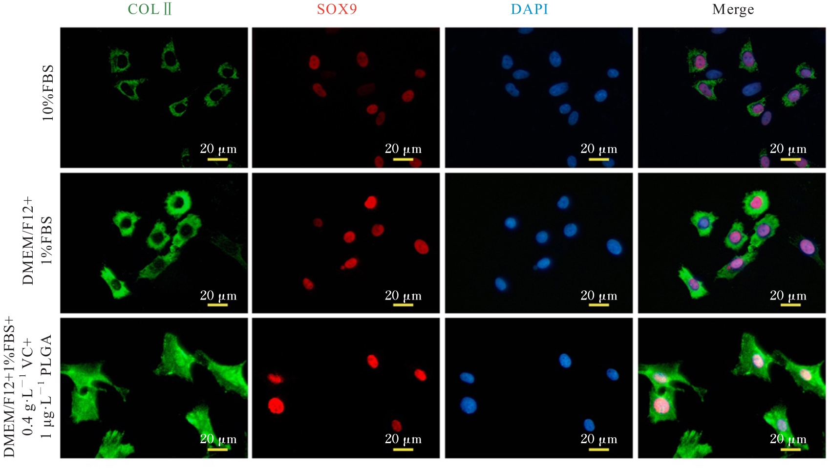
目的 探讨新生大鼠原代软骨细胞分离和培养的改进方法,以建立高效经济的体外软骨细胞培养体系。 方法 从新生大鼠关节中分离原代软骨细胞,分为过夜消化(OD)组和快速消化(RD)组进行分离,OD组软骨细胞采用Ⅱ 型胶原酶过夜消化,RD组软骨细胞采用预消化的物理化学消化相结合的手段分离细胞。采用含0%(空白组1)、1%、2%、4%和10%胎牛血清(FBS),0(空白组2)、0.1、0.2、0.4、0.8、1.0、2 .0 g·L-1维生素C(VC)和0(空白组3)、0.5、1.0、2.0、4.0、8.0、10.0 μg·L-1聚乳酸-羟基乙酸共聚体(PLGA)纳米粒子的改良培养液培养软骨细胞。将杜氏改良Eagle培养基F12营养混合液(DMEM/F12)与含不同浓度FBS、VC和PLGA的培养液分别混合,并按照各成分浓度进行相应分组。采用细胞计数仪计数各组细胞并检测各组细胞存活率和直径,采用甲苯胺蓝特异性染色法检测各组细胞形态表现,采用CCK-8法检测各组细胞增殖活性,采用细胞黏附实验检测各组细胞黏附率,采用Hoechst/碘化丙碇(PI)染色检测各组细胞凋亡情况,采用MTT法检测改良培养液培养后各组细胞增殖活性,将细胞分为DMEM/F12+10%FBS组(对照组)、DMEM/F12+1%FBS组、DMEM/F12+1%FBS+0.4 g·L-1 VC+1 μg·L-1 PLGA组,采用实时荧光定量PCR(RT-qPCR)法检测改良培养液培养后各组细胞中性别决定区域Y框转录因子9(SOX9)、Ⅱ型胶原α1链(Col2A1)、Ⅹ型胶原α1链(Col10A1)和基质金属蛋白酶13(MMP13)mRNA表达水平,采用免疫荧光染色检测改良培养液培养后各组细胞中Ⅱ型胶原(COLⅡ)和SOX9表达情况。 结果 OD组原代软骨细胞存活率小于RD组,细胞平均直径大于RD组。OD组原代软骨细胞形态较大,呈梭形,大多数细胞出现伪足;RD组原代软骨细胞形态较小,大多数细胞呈菱形,仅部分细胞出现伪足。2组原代软骨细胞经甲苯胺蓝特异性染色均显色明显,但RD组消化时间较短,软骨细胞实际培养时间较OD组缩短9~13 h,原代软骨细胞形态更为幼稚。OD组原代软骨细胞在培养24 h时增殖较为缓慢,培养48 h时增殖速度升高,较培养12 h时增殖活性明显升高(P<0.01)。RD组原代软骨细胞在培养24 h时增殖稍缓,培养48 h时增殖速度加快,较培养12 h时增殖活性明显升高(P<0.01);培养24和48 h时,与OD组比较,RD组原代细胞增殖速度升高(P<0.05)。RD组软骨凋亡细胞数少于OD组,2组均无坏死软骨细胞。大鼠软骨细胞增殖活性随着培养液中FBS浓度升高而升高,与空白组1比较,培养液中含1%、2%、4%和10%FBS时,软骨大鼠细胞增殖活性明显升高(P<0.05)。与空白组2比较,培养液中含0.2~1.0 g·L-1 VC时大鼠软骨细胞增殖活性明显升高(P<0.05),其中含0.4 g·L-1 VC时大鼠软骨细胞增殖活性最高(P<0.01)。与空白组3比较,培养液中含1~4 μg·L-1 PLGA时,大鼠软骨细胞增殖活性明显升高(P<0.05),其中含1 μg·L-1 PLGA时大鼠软骨细胞增殖活性最高(P<0.05)。与DMEM/F12+10%FBS组比较,DMEM/F12+1%FBS组大鼠软骨细胞中SOX9 mRNA和COL2A1 mRNA表达水平均明显升高(P<0.05或P<0.01)。与DMEM/F12+10%FBS组比较,DMEM/F12+1%FBS+0.4 g·L-1 VC+1 μg·L-1 PLGA组大鼠软骨细胞中SOX9 mRNA和COL2A1 mRNA表达水平明显升高(P<0.01)。免疫荧光染色,荧光显微镜下DMEM/F12+10%FBS组部分软骨细胞中出现COLⅡ绿色荧光信号和SOX9红色荧光信号,荧光强度弱;DMEM/F12+1%FBS组大多数软骨细胞中出现COLⅡ绿色荧光信号和SOX9红色荧光信号,荧光强度明显强于DMEM/F12+10%FBS组;DMEM/F12+1%FBS+0.4 g·L-1 VC+1 μg·L-1 PLGA组软骨细胞中均出现COLⅡ绿色荧光信号和SOX9红色荧光信号,荧光强度较DMEM/F12+10%FBS组和DMEM/F12+1%FBS组明显升高。DMEM/F12+1%FBS组软骨细胞中COLⅡ和SOX9蛋白表达量明显高于DMEM/F12+10% FBS组,DMEM/F12+1%FBS+0.4 g·L-1 VC+1 μg·L-1 PLGA组软骨细胞中COLⅡ和SOX9蛋白表达量明显高于DMEM/F12+10%FBS组。 结论 改良后的大鼠原代软骨细胞分离和培养方法可以弥补传统方法的缺陷,缩短原代软骨细胞的分离时间,提高原代软骨细胞的体外培养质量。

关键词: 软骨细胞, 原代细胞培养, 体外技术, 条件培养基, 血清

Abstract:

Objective To discuss the improved methods for the isolation and culture of primary chondrocytes from the neonatal rats,and to establish an efficient and economical in vitro chondrocyte culture system. Methods The primary chondrocytes were isolated from the joints of neonatal rats and divided into overnight digestion (OD) group and rapid digestion (RD) group for separation. The chondrocytes in OD group were digested overnight by typeⅡ collagenase, while the chondrocytes in RD group were separated by the combination of pre-digestion with physical and chemical digestion methods. The chondrocytes were cultured in modified media containing 0% (blank group 1), 1%, 2%, 4%, and 10% fetal bovine serum (FBS), 0 (blank group 2), 0.1, 0.2, 0.4, 0.8, 1.0, and 2.0 g·L-1 vitamin C(VC), and 0 (blank group 3), 0.5, 1.0, 2.0, 4.0, 8.0, 10.0 μg·L-1 poly(lactic-co-glycolic acid) (PLGA) nanoparticles. The media containing different concentrations of FBS, VC, and PLGA were mixed with Dulbecco’s modified Eagle’s medium/nutrient mixture F-12(DMEM/F12), and were divided into related groups based on the concentrations of ingredients. Cell counter was used to count the chondrocytes in various groups and the survival rates and diameters of the chondrocytes in various groups were detected; Toluidine blue staining was used to detect the morphology of the chondrocytes in various groups; CCK-8 method was used to detect the proliferative activities of the chondrocytes in various groups; cell adhesion assay was used to detect the adhesion rates of the chondrocytes in various groups; Hoechst/propidium iodide(PI) staining was used to detect the apoptosis of the chondrocytes in various groups; MTT assay was used to detect the proliferation activities of the chondrocytes in various groups after treated with modified media.The cells were divided into DMEM/F12+10%FBS group, DMEM/F12+1%FBS group, and DMEM/F12+1% FBS+0.4 g·L-1 VC+1 μg·L-1 PLGA group. Real-time fluorescence quantitative PCR(RT-qPCR) method was used to detect the expression levels of sex-determining region Y-box 9 (SOX9), collagen type Ⅱ alpha 1 chain (Col2A1), collagen type Ⅹ alpha 1 chain (Col10A1), and matrix metallopeptidase 13 (MMP13) mRNAs in the chondrocytes in various groups after treated with modified media;immunofluorescence staining was used to detect the expressions of type Ⅱ collagen (COLⅡ) and SOX9 in the chondrocytes in various groups after treated with modified media. Results The survival rate of primary chondrocytes in OD group was lower than that in RD group, and the average cell diameter was larger than that in RD group. The primary chondrocytes in OD group were larger and spindle-shaped, and most cells exhibited pseudopodia; in RD group, the primary chondrocytes were smaller, mostly rhomboid in shape, with only a portion of the cells showing pseudopodia. The Toluidine blue staining results showed significant coloration in both groups, but the digestion time of the chondrocytes in RD group was shorter, and compared with OD group, the actual culture time of the chondrocytes was reduced by 9-13 h, and more immature morphology of the primary chondrocytes were observed. The proliferation activity of the primary chondrocytes in OD group was slow at 24 h of culture but increased at 48 h of culture, and the proliferation activity of the primary chondrocytes was significantly higher at 48 h of culture compared with 12 h of culture (P<0.01). Compared with 12 h of culture,the proliferation rates of the primary chondrocytes in RD group were increased at 24 and 48 h of culture (P<0.01). At 24 and 48 h of culture, compared with OD group, the proliferation rates of the primary chondrocytes in RD group were increased (P<0.05). The number of apoptotic chondrocytes in RD group was lower than that in OD group, and no necrotic chondrocytes were observed in either group. The proliferation activities of chondrocytes of the rats were increased with the rising of FBS concentration in the culture medium. Compared with blank group 1, the proliferation activities of chondrocytes of the rats after treated with culture mediums containing 1%, 2%, 4%, and 10% FBS were significantly increased (P<0.05). Compared with blank group 2, the proliferative activities of chondrocytes of the rats after treated with culture mediums containing 0.2-1.0 g·L-1 VC were significantly increased (P<0.05), and the highest proliferation activity was found when the concentration of VC was 0.4 g·L-1 P<0.01). Compared with blank group 3, the proliferation activities of chondrocytes of the rats after treated with culture mediums containing 1-4 μg·L-1 PLGA were significantly increased (P<0.05), and the highest proliferation activity was found after treated with culture medium containing 1 μg·L-1 PLGA (P<0.05). Compared with DMEM/F12+10%FBS group, the expression levels of SOX9 mRNA and Col2A1 mRNA in the chondrocytes in DMEM/F12+1%FBS group were significantly increased (P<0.05 or P<0.01). Compared with DMEM/F12+10%FBS group, the expression levels of SOX9 mRNA and Col2A1 mRNA in the chondrocytes in DMEM/F12+1%FBS+0.4 g·L-1 VC+1 μg·L-1 PLGA group were significantly increased (P<0.01). The immunofluorescence staining results showed that the green fluorescence signal of COLⅡ and the red fluorescence signal of SOX9 were observed in some chondrocytes in DMEM/F12+10%FBS group under fluorescence microscope, and the fluorescence intensity was weak. In DMEM/F12+1%FBS group, most chondrocytes exhibited COLⅡ green fluorescence signal and SOX9 red fluorescence signal, and the fluorescence intensity was significantly stronger than that in DMEM/F12+10% FBS group. In DMEM/F12+1% FBS+0.4 g·L-1 VC+1 μg·L-1 PLGA group, the COLⅡ green fluorescence signal and SOX9 red fluorescence signal were found in all the chondrocytes, and the fluorescence intensity was significantly higher than those in DMEM/F12+10%FBS and DMEM/F12+1%FBS groups. The expression levels of COLⅡ and SOX9 proteins in the chondrocytes in DMEM/F12+1%FBS group were significantly higher than those in DMEM/F12+10%FBS group, and the expression levels of COLⅡ and SOX9 proteins in the chondrocytes in DMEM/F12+1%FBS+0.4 g·L-1 VC+ 1 μg·L-1 PLGA group were significantly higher than those in DMEM/F12+10%FBS group. Conclusion The improved methods for the isolation and culture of primary chondrocytes of the rats can overcome the shortcomings of traditional methods, shorten the isolation time of primary chondrocytes, and improve the quality of in vitro culture of primary chondrocytes.

Key words: Chondrocytes, Primary cell culture, In vitro techniques, Conditioned culture media, Serum

中图分类号: 

  • Q813.1