吉林大学学报(医学版) ›› 2024, Vol. 50 ›› Issue (3): 628-637.doi: 10.13481/j.1671-587X.20240306

• 基础研究 • 上一篇    

SGK1对Cyclin B/Cdc2通路介导小鼠G1期受精卵卵裂的调控作用及其机制

张慧灵,韩迪,郭文秀,庞海垚,孟峻()   

  1. 内蒙古医科大学附属医院检验科,内蒙古 呼和浩特 010050
  • 收稿日期:2023-12-26 出版日期:2024-05-28 发布日期:2024-07-01
  • 通讯作者: 孟峻 E-mail:nmfrank@163.com
  • 作者简介:张慧灵(1998-),女,内蒙古自治区呼和浩特市人,在读硕士研究生,主要从事临床生物化学及生殖分子生物学方面的研究。
  • 基金资助:
    国家自然科学基金项目(81360109);内蒙古自治区科技厅自然科学基金项目(2021MS08158)

Regulatory effect of SGK1 on oocyte cleavage in fertilized eggs in mice at G1 stage mediated by Cyclin B/Cdc2 pathway and its mechanism

Huiling ZHANG,Di HAN,Wengxiu GUO,Haiyao PANG,Jun MENG()   

  1. Department of Laboratory,Affiliated Hospital,Inner Mongolia Medical University,Hohhot 010050,China
  • Received:2023-12-26 Online:2024-05-28 Published:2024-07-01
  • Contact: Jun MENG E-mail:nmfrank@163.com

摘要:

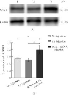
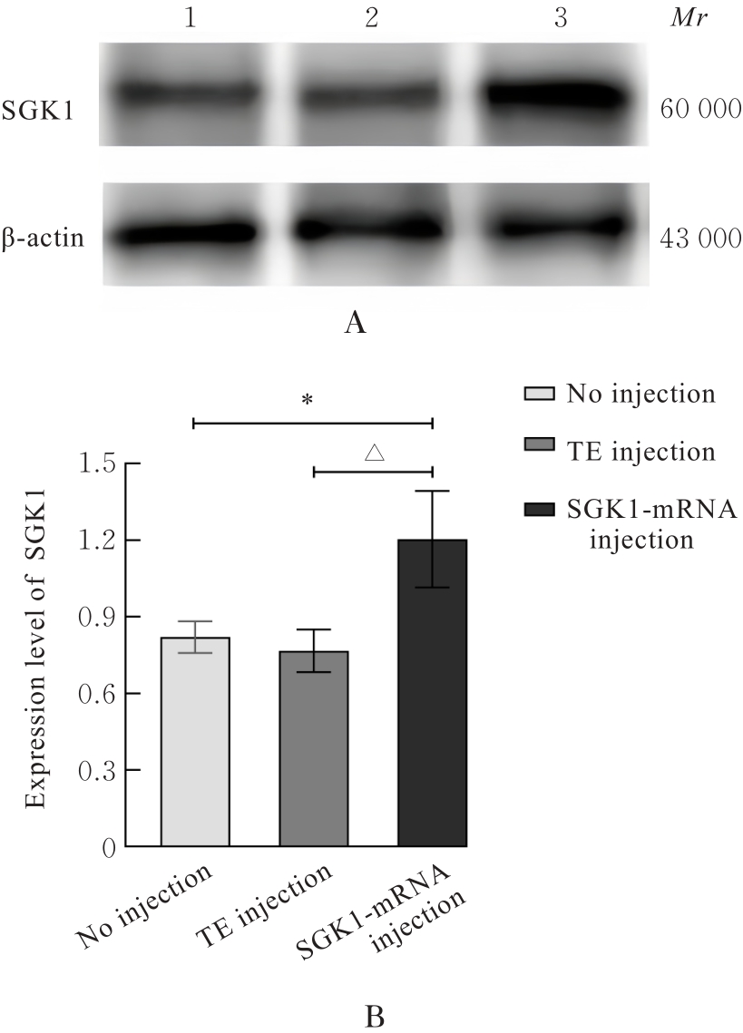
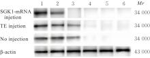
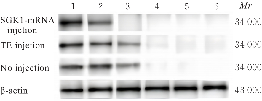
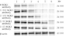
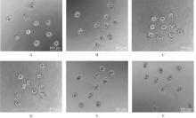
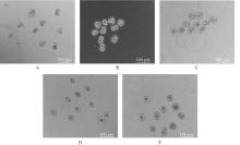
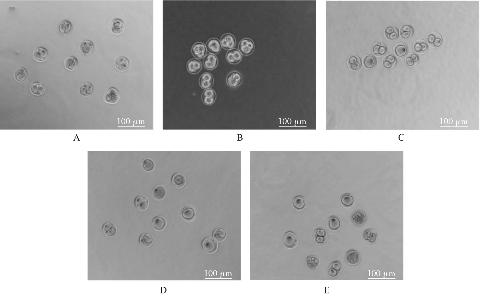
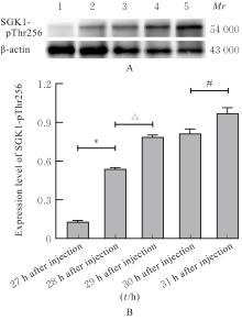
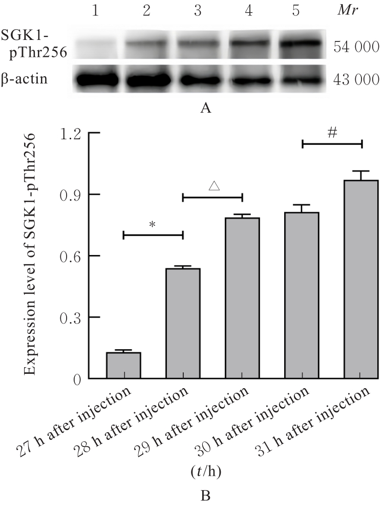
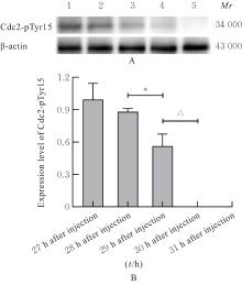
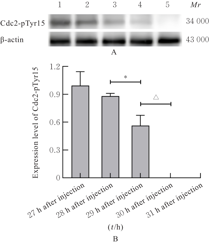
目的 探讨血清和糖皮质激素诱导蛋白激酶1(SGK1)在小鼠细胞周期G1期受精卵早期发育过程中的调控作用,并阐明相关机制。 方法 取若干只4~6周龄且体质量约为20 g的雌鼠和若干只8周龄以上且体质量约为30 g的雄鼠,雌鼠腹腔注射孕马血清促性腺激素(PMSG),每只10 IU,48 h后腹腔注射人绒毛膜促性腺激素(HCG),每只10 IU,并将注射HCG的雌鼠与雄鼠1∶1合笼过夜。取交配成功的雌鼠受精卵,注射HCG后分别于12~21 h、21~26 h、26~28 h和28~30 h收集细胞周期G1期、S期、G2期及M期的受精卵,并于光学显微镜下观察不同细胞周期的细胞形态表现。收集小鼠超排卵后 G1 期受精卵,体外转录生成 mRNA 后,分为未注射组、Tris-EDTA缓冲液注射组(TE注射组)和SGK1-mRNA注射组。采用SGK1抗体与KSOM培养液配置1∶25、1∶50、1∶100、1∶200和0共5种不同浓度SGK1抗体组的培养液,培养小鼠G1期受精卵。Western blotting法检测各组小鼠受精卵中SGK1蛋白表达水平和各组小鼠及不同浓度SGK1抗体组HCG注射不同时间受精卵中磷酸化细胞分裂周期因子2(Cdc2)酪氨酸15位点(Cdc2-pTyr15)去磷酸化情况,相差显微镜观察各组小鼠和不同浓度SGK1抗体组受精卵发育情况,Western blotting法检测HCG注射不同时间小鼠受精卵中磷酸化SGK1-苏氨酸256位点(SGK1-pThr256)和Cdc2-pTyr15蛋白表达水平。 结果 与未注射组和TE注射组比较,SGK1-mRNA注射组SGK1蛋白表达水平明显升高(P<0.01)。HCG注射后27~28 h,SGK1-mRNA注射组小鼠受精卵中Cdc2-pTyr15磷酸化信号逐渐消失,至HCG注射29 h,Cdc2-pTyr15磷酸化信号完全消失;HCG注射后28~29 h,未注射组和TE注射组小鼠受精卵中Cdc2-pTyr15磷酸化信号逐渐消失,至HCG注射后30 h,Cdc2-pTyr15磷酸化信号完全消失;随着SGK1抗体浓度升高,不同浓度SGK1抗体组受精卵中Cdc2-pTyr15磷酸化信号减弱和磷酸化信号消失的时间逐渐延长。HCG注射后27 h,SGK1-mRNA注射组小鼠受精卵开始卵裂;HCG注射后31 h,SGK1-mRNA注射组受精卵几乎全部分裂为G2期细胞受精卵;HCG注射后33 h,0 和 1∶200 SGK1 抗体组受精卵全部发生卵裂;随着 SGK1 抗体浓度升高, 1∶25、1∶50 和1∶100 SGK1抗体组受精卵卵裂逐渐减少,在1∶25 SGK1抗体组受精卵卵裂减少最明显。HCG注射后31 h,与未注射组和TE注射组比较,SGK1-mRNA注射组小鼠受精卵死亡率明显降低(P<0.05),卵裂率明显升高(P<0.05)。注射HCG后31和33 h,随着SGK1抗体浓度升高,与1∶200 SGK1抗体组比较,1∶100、1∶50和1∶25 SGK1抗体组受精卵死亡率逐渐升高(P<0.05),卵裂时间延长,受精卵卵裂率降低(P<0.05),并呈剂量依赖性,其中1∶25 SGK1抗体组受精卵卵裂率最低。HCG注射后27 h,小鼠受精卵中SGK1-pThr256蛋白表达水平逐渐升高(P<0.05或P<0.01),并呈时间依赖性;HCG注射后28~29 h,小鼠受精卵中Cdc2-pTyr15蛋白表达水平逐渐降低(P<0.01),并呈时间依赖性,并于HCG注射后30 h完全消失。 结论 过表达或抑制SGK1均会影响小鼠G1期受精卵进入M期的时间,SGK1蛋白可能是小鼠G1期受精卵早期发育的调控因子之一,其可能通过Cdc2调节G1期受精卵发育。

关键词: 血清和糖皮质激素诱导蛋白激酶1, 受精卵, 细胞分裂, 磷脂酰肌醇3-激酶, 卵裂

Abstract:

Objective To discuss the regulatory effect of serum and glucocorticoid-induced protein kinase 1 (SGK1) in the early development of fertilized eggs at G1 phase of the mice, and to clarify the related mechanism. Methods Some female mice aged 4-6 weeks and weighed about 20 g, and several male mice aged over 8 weeks and weighed about 30 g were selected. The female mice were intraperitoneally injected with 10 IU of pregnant mare serum gonadotropin (PMSG), followed by 10 IU of human chorionic gonadotropin (HCG) after 48 h. After HCG injection, the female mice were caged overnight with the male mice at a ratio of 1∶1.The fertilized eggs at G1, S, G2, and M phases were collected at 12-21 h, 21-26 h, 26-28 h, and 28-30 h after injected with HCG, and their cellular morphology at different cell cycles were observed under light microscope. The mouse fertilized eggs at G1 phase after superovulation were collected, the mRNA was synthesized in vitro, and divided into no injection group, Tris-EDTA buffer injection group (TE injection group), and SGK1-mRNA injection group.The SGK1 antibodies were mixed with KSOM culture medium with the concentrations of 1∶25, 1∶50, 1∶100, 1∶200, and 0 to culture the mouse fertilized eggs at G1 phase. Western blotting method was used to detect the expression levels of SGK1 protein in fertilized eggs of the mice in various groups and the dephosphorylation for phosphorylated SGK1-Threonine 256 site tyrosine15 site of cell diusion cyclin 2(Cdc2)(Cdc2-pTyr15) in the fertilized eggs of the mice in various groups and different concentrations of SGK1 antibody groups and the developmental states of the fertilized eggs in the fertilized eggs of the mice in various groups and different concentrations of SGK1 antibody groups were observed under phase contrast microscope; the expression levels of phosphorylated SGK1-Thr256 (SGK1-pThr256) and Cdc2-pTyr15 proteins in fertilized eggs at different post-HCG injection times were detected by Western blotting method. Results Compared with no injection and TE injection groups, the expression level of SGK1 protein in the cells in SGK1-mRNA injection group was significantly increased (P<0.01). 27-28 h after injected with HCG, the phosphorylation signaling of Cdc2-pTyr15 in fertilized eggs of the mice in SGK1-mRNA injection group was gradually disappeared, and there was no phosphorylation signaling 29 h after injected with HCG. At 28-29 h after injected with HCG,the phosphorylation signaling of Cdc2-pTyr15 in fertilized eggs of the mice in no injection and TE injection groups gradually disappeared, completely disappeared at 30 h after injected with HCG. With the increasing of the concentration of SGK1 antibody, the disappearing time of the Cdc2-pTyr15 phosphorylation signaling was increased. At 27 h after injected with HCG,the fertilized eggs of the mice in SGK1-mRNA injection group was initiated cleavage; at 31 h after injected with HCG, nearly all the fertilized eggs turned into G2 phase; at 33 h after injected with HCG, all the fertilized eggs in 0 and 1∶200 SGK1 antibody groups underwent cleavage. However, with the increasing of SGK1 antibody concentration, the cleavage of the fertilized eggs in 1∶25, 1∶50, and 1∶100 SGK1 antibody groups was gradually decreased, particularly at 1∶25 SGK1 antibody group. Compared with no injection and TE injection groups, the death rate of the fertilized eggs of the mice in SGK1-mRNA injection group was significantly decreased at 31 h after injected with HCG (P<0.05), and the cleavage rate was increased(P<0.05). With the increasing of the SGK1 antibody concentration, the death rates of the fertilized eggs in different concentrations of SGK1 antibody group were increased (P<0.05), with the extending of cleavage time was increased, and the cleavage rate of the fertilized eggs was decreased in a dose-dependent manner, and the cleavage rate of fertilized eggs in 1∶25 SGK1 antibody group was the lowest.The expression level of SGK1-pThr256 protein in fertilized eggs of the mice was gradually increased from 27 h after injected with HCG (P<0.05 or P<0.01) in a time-dependent manner; at 28 to 29 h after injected with HCG, the expression levels of Cdc2-Tyr15 protein were gradually decreased (P<0.05) in a time-dependent manner, and had completely disappeared at 30 h after injected with HCG. Conclusion Both the over-expression and inhibition of SGK1 can affect the time for the fertilized eggs at G1 phase to entry into M phase, suggesting that SGK1 protein may be one of the regulatory factors in the early development of fertilized eggs at G1 phase of the mice, and it may regulate the development of the fertilized eggs at G1 phase through regulation of Cdc2.

Key words: Serum and glucocorticoid-regulated protein kinases 1, Fertilized egg, Cell division, Phosphatidylinositol 3-kinase, Cleavage

中图分类号: 

  • Q132