吉林大学学报(医学版) ›› 2025, Vol. 51 ›› Issue (1): 85-95.doi: 10.13481/j.1671-587X.20250111

• 基础研究 • 上一篇    下一篇

积雪草酸通过Nrf2/HO-1信号通路对脂多糖诱导大鼠海马神经元损伤的改善作用

白燕燕,周禹彤,隋海娟,刘卓()   

  1. 锦州医科大学基础医学院药理学教研室,辽宁 锦州 121000
  • 收稿日期:2024-03-25 接受日期:2024-05-14 出版日期:2025-01-28 发布日期:2025-03-06
  • 通讯作者: 刘卓 E-mail:liuz@jzmu.edu.cn
  • 作者简介:白燕燕(1996-),女,甘肃省平凉市人,医学硕士,主要从事神经药理学方面的研究。
  • 基金资助:
    辽宁省科技厅自然科学基金指导项目(2019-ZD-0818);锦州市科技局指导性科技计划项目(JZ2023B055);辽宁省教育厅大学生创新创业训练计划项目(20201016029)

Improvement effect of asiatic acid on damage of lipopolysaccharide-induced hippocampum neuron in rats through Nrf2/HO-1 signaling pathway

Yanyan BAI,Yutong ZHOU,Haijuan SUI,Zhuo LIU()   

  1. Department of Pharmacology,School of Basic Medical Sciences,Jinzhou Medical University,Jinzhou 121000,China
  • Received:2024-03-25 Accepted:2024-05-14 Online:2025-01-28 Published:2025-03-06
  • Contact: Zhuo LIU E-mail:liuz@jzmu.edu.cn

摘要:

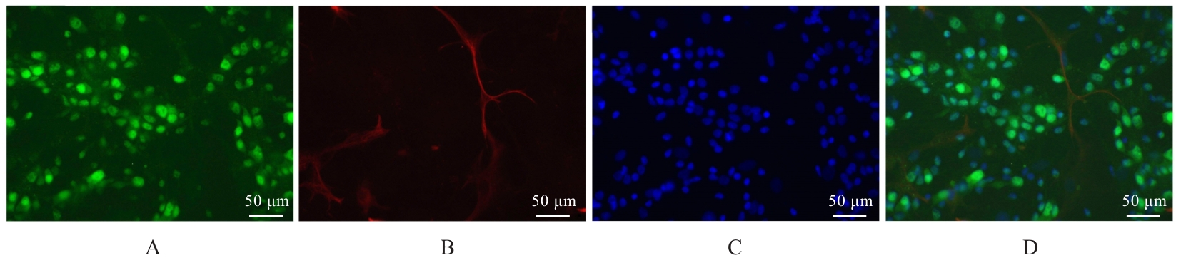
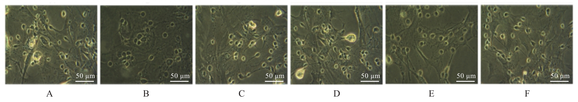
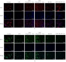
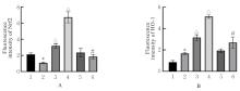
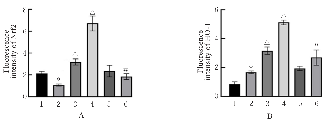
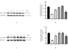
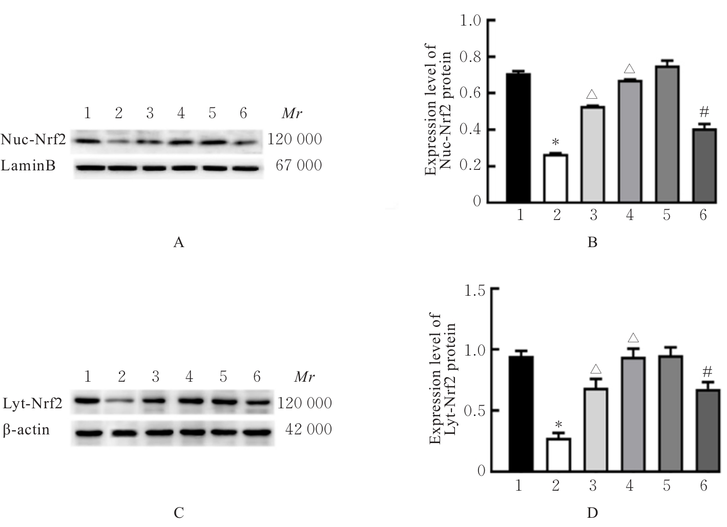
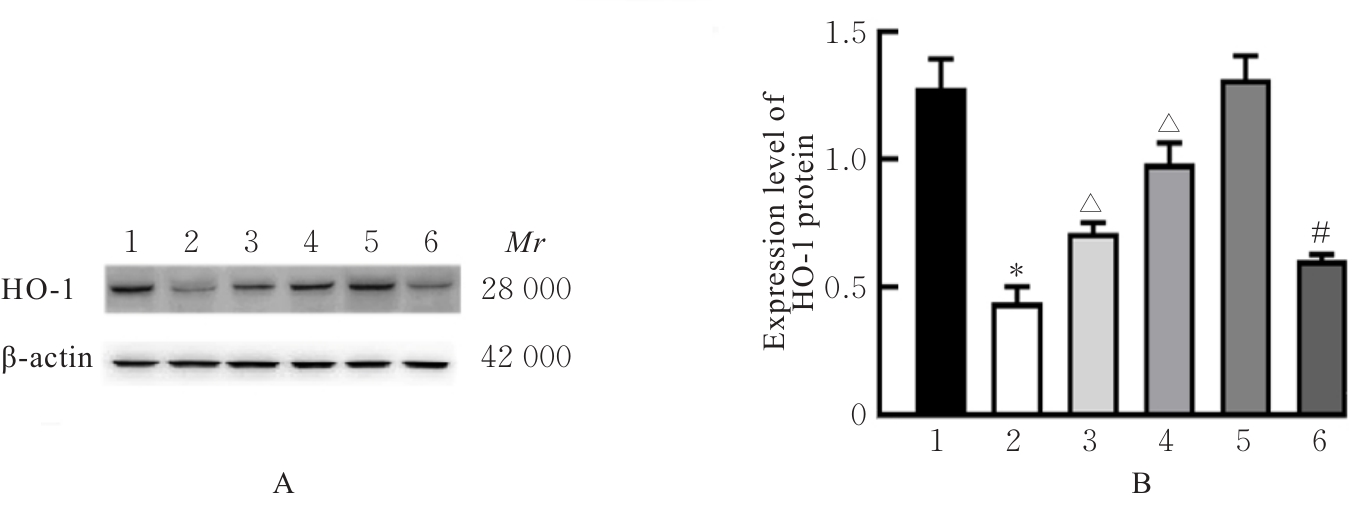
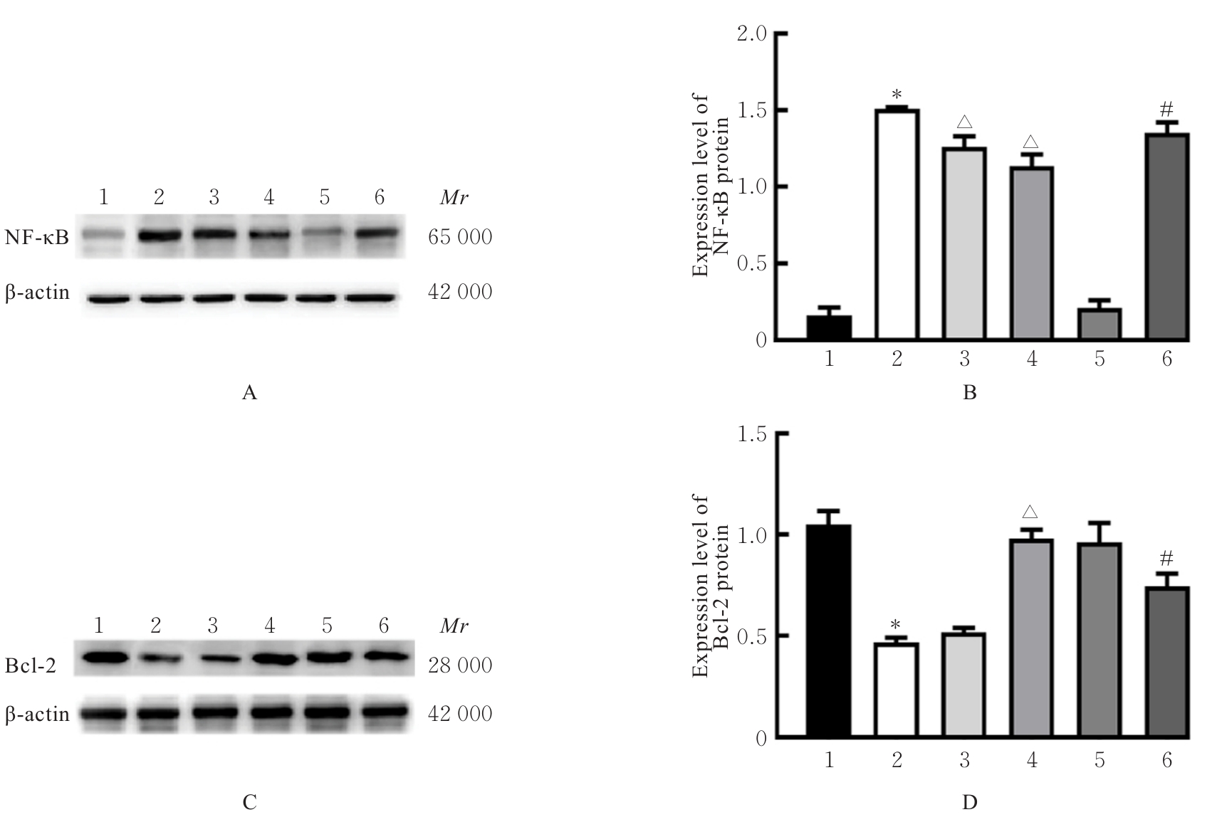
目的 探讨积雪草酸(AA)对脂多糖(LPS)诱导大鼠原代海马神经元炎症和氧化应激损伤的作用,并阐明其作用机制。 方法 原代培养大鼠海马神经元(免疫荧光染色法鉴定细胞纯度)分为对照组、LPS组(10 mg·L-1 LPS)、LPS+AA组(10 mg·L-1 LPS+10、20和40 μmol·L-1 AA)、AA组(20 μmol·L-1 AA)、ML385组[10 μmol·L-1核因子E2相关因子2(Nrf2)抑制剂]和LPS+ML385+AA组(10 mg·L-1 LPS+10 μmol·L-1 ML385+20 μmol·L-1 AA);给药处理后,噻唑蓝(MTT)法检测各组大鼠海马神经元的存活率,乳酸脱氢酶(LDH)试剂盒检测各组大鼠海马神经元LDH漏出率,酶联免疫吸附试验(ELISA)试剂盒检测各组大鼠海马神经元上清液中炎症因子[白细胞介素(IL)-1β和肿瘤坏死因子(TNF)-α]水平以及各组大鼠海马神经元中超氧化物歧化酶(SOD)活性和丙二醛(MDA)水平,Griess法测定各组大鼠海马神经元上清液中一氧化氮(NO)水平,免疫荧光法检测各组大鼠海马神经元中Nrf2和血红素氧合酶1(HO-1)蛋白表达情况;Western blotting法检测各组大鼠海马神经元中Nrf2、HO-1、核因子κB(NF-κB)和B细胞淋巴瘤2(Bcl-2)蛋白表达水平。 结果 与对照组比较,LPS组大鼠海马神经元中的海马神经元存活率、SOD活性和Bcl-2表达水平明显降低(P<0.01),LDH漏出率、IL-1β和TNF-α水平、MDA水平、NO水平以及NF-κB蛋白表达水平均明显升高(P<0.01),Nrf2和HO-1的荧光强度明显减弱,Nrf2和HO-1蛋白表达水平明显降低(P<0.01);与LPS组比较,LPS+10 μmol?L-1 AA组和LPS+20 μmol?L-1 AA组大鼠海马神经元存活率、SOD活性和Bcl-2表达水平明显升高(P<0.01),LDH漏出率、IL-1β和TNF-α水平、MDA水平、NO水平以及NF-κB蛋白表达水平明显降低(P<0.05或P<0.01),Nrf2和HO-1的荧光强度明显升高(P<0.01),Nrf2和HO-1蛋白表达水平明显升高(P<0.01);与LPS+20 μmol?L-1 AA组比较,LPS+ML385+AA组海马神经元中Nrf2和HO-1荧光强度明显减弱(P<0.01),细胞核和细胞质中Nrf2、细胞中HO-1和Bcl-2蛋白表达水平明显降低(P<0.01),NF-κB蛋白表达水平明显升高(P<0.01)。 结论 AA可改善LPS诱导的原代培养大鼠海马神经元炎症和氧化应激损伤,其机制可能与激活Nrf2/HO-1信号通路有关。

关键词: 阿尔茨海默病, 积雪草酸, 海马神经元, 炎症, 氧化应激, 核因子E2相关因子2, 血红素氧合酶1

Abstract:

Objective To discuss the effect of asiatic acid (AA) on the inflammation and oxidative stress damage induced by lipopolysaccharide (LPS) in the primary cultured hippocampus neurons, and to clarify its mechanism. Methods The primary cultured rat hippocampus neurons (cell purity identified by immunofluorescence staining) were divided into control group, LPS (10 mg·L-1) group, and LPS+AA group (10 mg·L-1 LPS+10, 20, and 40 μmol·L-1 AA), AA group (20 μmol·L-1 AA), ML385 group [10 μmol·L-1 nuclear factor erythroid 2-related factor (Nrf2) inhibitor], and LPS+ML385+AA group (10 mg·L-1 LPS+10 μmol·L-1 ML385+20 μmol·L-1 AA). After drug treatment, methylthiazolyldiphenyl-tetrazolium bromide (MTT) method was used to detect the survival rates of the hippocampus neurons in various groups; lactate dehydrogenase (LDH) kit was used to detect the LDH leakage rates of the hippocampus neurons in various groups; enzyme linked immunosorbent assay (ELISA) kit was used to detect the expression levels of inflammatory factors [interleukin (IL)-1β and tumor necrosis factor (TNF)-α] and the activities of superoxide dismutase (SOD) and malondialdehyde (MDA) levels in the hippocampus neurons in various groups; Griess method was used to detect the nitric oxide (NO) levels in supernatant of the hippocampus neurons in various groups; immunofluorescence staining was used to detect the expressions of Nrf2 and heme oxygenase-1 (HO-1) proteins in the hippocampus neurons in various groups; Western blotting method was used to detect the expression levels of Nrf2, HO-1, nuclear factor-kappa B(NF-κB), and B-cell lymphoma 2 (Bcl-2) proteins in the hippocampus neurons in various groups. Results Compared with control group, the survival rate of the hippocampus neurons, SOD activity, and Bcl-2 expression level in the cells in LPS group were significantly decreased (P<0.01), while the LDH leakage rate, expression levels of IL-1β and TNF-α, MDA level, and NO level, as well as the expression level of NF-κB protein, were significantly increased (P<0.01); the fluorescence intensities and expression levels of Nrf2 and HO-1 proteins in hippocampus neurons were significantly decreased (P<0.01). Compared with LPS group, the survival rates of hippocampus neurons, SOD activities, and expression levels of Bcl-2 in the cells in LPS+10 μmol·L-1 AA group and 20 μmol·L-1 AA group were significantly increased (P<0.01), while the LDH leakage rates, expression levels of IL-1β and TNF-α, MDA levels, and NO levels, as well as expression levels of NF-κB protein, were significantly decreased (P<0.05 or P<0.01), and the fluorescence intensities and protein expression levels of Nrf2 and HO-1 in the cells were significantly increased (P<0.01). Compared with LPS+20 μmol·L-1 AA group, the fluorescence intensities of Nrf2 and HO-1 in the cells in LPS+ML385+AA group were significantly decreased (P<0.01), and the expression levels of Nrf2 protein in the nucleus and cytoplasm, the expression levels of HO-1 and Bcl-2 proteins in the cells were significantly decreased (P<0.01), while the expression level of NF-κB protein was significantly increased (P<0.01). Conclusion AA can improve LPS-induced inflammation and oxidative stress damage in the primary cultured rat hippocampus neurons, and its mechanism may be related to the activation of the Nrf2/HO-1 signaling pathway.

Key words: Alzheimer’s disease, Asiatic acid, Hippocampus neuron, Inflammation, Oxidative stress, Nuclear factor erythroid 2-related factor, Heme oxygenase-1

中图分类号: 

  • R742