吉林大学学报(医学版) ›› 2024, Vol. 50 ›› Issue (3): 689-696.doi: 10.13481/j.1671-587X.20240313

• 基础研究 • 上一篇    

五加生脉饮对小鼠的抗疲劳作用及其机制

韩迦南1,刘倬睿1,曾沛涌1,姜爽2,李洪宇1()   

  1. 1.北华大学药学院药物分析教研室,吉林 吉林 132013
    2.长春中医药大学健康管理学院 预防医学教研室,吉林 长春 130117
  • 收稿日期:2023-07-11 出版日期:2024-05-28 发布日期:2024-07-01
  • 通讯作者: 李洪宇 E-mail:26149236@qq.com
  • 作者简介:韩迦南(2000-),女,吉林省长春市人,在读硕士研究生,主要从事天然药物抗疲劳作用方面的研究。
  • 基金资助:
    吉林省教育厅“十三五”科学技术项目(JJKH20200078KJ)

Anti-fatigue effect of Wujia Shengmai Yin in mice and its mechanism

Jianan HAN1,Zhuorui LIU1,Peiyong ZENG1,Shuang JIANG2,Hongyu LI1()   

  1. 1.Department of Pharmaceutical Analysis,School of Pharmacy,Beihua University,Jilin 132013,China
    2.Department of Preventive Medicine,School of Health Management,Changchun University of Chinese Medicine,Changchun 130117,China
  • Received:2023-07-11 Online:2024-05-28 Published:2024-07-01
  • Contact: Hongyu LI E-mail:26149236@qq.com

摘要:

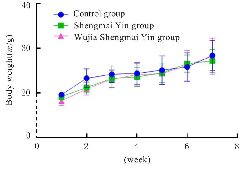
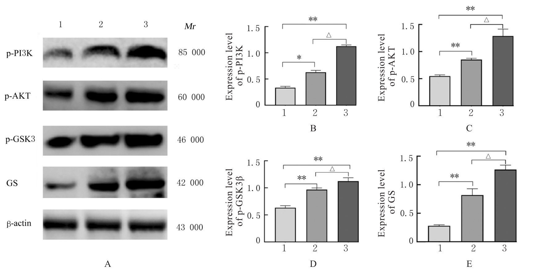
目的 探讨五加生脉饮的抗疲劳作用,并阐明其作用机制。 方法 36只雄性ICR小鼠随机分为对照组(等体积蒸馏水)、生脉饮组(500 mg·kg-1生脉饮)和五加生脉饮组(600 mg·kg-1五加生脉饮)。每隔7 d测定各组小鼠体质量,观察其精神状态。采用疲劳转棒实验和力竭负重游泳实验检测各组小鼠转棒停留时间和力竭游泳时间;试剂盒检测各组小鼠血清中血尿素氮(BUN)和乳酸(LA)水平及乳酸脱氢酶(LDH)活性、肝脏组织中肝糖原(LG)水平、肌肉组织中肌糖原(MG)和丙二醛(MDA)水平及谷胱甘肽过氧化物酶(GSH-Px)和超氧化物歧化酶(SOD)活性;Western blotting法检测各组小鼠肝脏组织中糖代谢相关蛋白表达水平。 结果 与实验前比较,实验后各组小鼠体质量均呈现增长趋势,但差异均无统计学意义(P>0.05)。疲劳转棒实验,与对照组比较,五加生脉饮组小鼠转棒停留时间明显增加(P<0.01);力竭负重游泳实验,与对照组比较,生脉饮组和五加生脉饮组小鼠力竭游泳时间均明显增加(P<0.01)。与对照组比较,生脉饮组和五加生脉饮组小鼠血清中BUN水平均明显降低(P<0.01),LDH活性均明显升高(P<0.01);五加生脉饮组LA水平明显降低(P<0.01)。与生脉饮组比较,五加生脉饮组小鼠血清中BUN和LA水平均明显降低(P<0.01),LDH活性明显升高(P<0.01)。与对照组比较,生脉饮组和五加生脉饮组小鼠肝脏组织中LG水平和肌肉组织中MG水平均明显升高(P<0.01);与生脉饮组比较,五加生脉饮组小鼠肝脏组织中LG水平和肌肉组织中MG水平均明显升高(P<0.01)。与对照组比较,生脉饮组和五加生脉饮组小鼠肌肉组织中GSH-Px和SOD活性均明显升高(P<0.01),MDA水平明显降低(P<0.01);与生脉饮组比较,五加生脉饮组小鼠肌肉组织中GSH-Px和SOD活性均明显升高(P<0.01),MDA水平明显降低(P<0.01)。Western blotting法,与对照组比较,生脉饮组和五加生脉饮组小鼠肝脏组织中磷酸化磷脂酰肌醇3-激酶(p-PI3K)、磷酸化蛋白激酶B(p-AKT)、磷酸化糖原合成酶激酶3β(p-GSK3β)及糖原合成酶(GS)蛋白表达水平均明显升高(P<0.05或P<0.01);与生脉饮组比较,五加生脉饮组小鼠肝脏组织中p-PI3K、p-AKT、p-GSK3β和GS蛋白表达水平均明显升高(P<0.01)。 结论 五加生脉饮可通过激活磷脂酰肌醇3激酶(PI3K)/蛋白激酶B(AKT)/糖原合成酶激酶3β(GSK3β)信号通路,提高机体抗氧化能力并增加糖原合成,发挥抗疲劳作用。

关键词: 五加生脉饮, 氧化应激, 抗疲劳, 糖代谢, 尿素氮, 乳酸

Abstract:

Objective To discuss the anti-fatigue effect of Wujia Shengmai Yin,and to clarify its mechanism. Methods Thirty-six male ICR mice were randomly devided into control group (equivalent volume of distilled water), Shengmai Yin group (500 mg·kg?1 of Shengmai Yin), and Wujia Shengmai Yin group (600 mg·kg?1 of Wujia Shengmai Yin). The body weights of the mice in various groups were detected every 7 d, and the mental states were observed. The rotating rod test and exhaustive swimming test were used to detect the duration on the rod and the swimming time to exhaustion of the mice in various groups, respectively;the levels of urea nitrogen (BUN) and lactate (LA), and the activities of lactate dehydrogenase (LDH) in serum, the levels of liver glycogen (LG), muscle glycogen (MG), and malondialdehyde (MDA), and activities of glutathione peroxidase (GSH-Px) and superoxide dismutase (SOD) in muscle tissue of the mice in various groups were detected by kits; the expression levels of glucose metabolism-related proteins in liver tissue of the mice in various groups were detected by Western blotting method. Results Compared with before experiment, the body weights after experiment of the mice in various groups showed a increasing trend but the differences were not statistically significant (P>0.05). The rotating rod test results showed that compared with control group, the duration on the rod of the mice in Wujia Shengmai Yin group was significantly increased (P<0.01). The exhaustive swimming test results showed that compared with control group, the swimming time to exhaustion of the mice in Shengmai Yin group and Wujia Shengmai Yin group was significantly increased (P<0.01). Compared with control group, the levels of BUN in serum of the mice in Shengmai Yin group and Wujia Shengmai Yin group were significantly decreased (P<0.01), and the activities of LDH were significantly increased (P<0.01); the level of LA of the mice in Wujia Shengmai Yin group was significantly decreased(P<0.01). Compared with Shengmai Yin group,the levels of BUN and LA in the serum and LDH activity of the mice in Wujia Shengmai Yin group were significantly decreased (P<0.01). Compared with control group, the levels of LG in liver tissue and the levels of MG in muscle tissue of the mice in Shengmai Yin group and Wujia Shengmai Yin group were significantly increased (P<0.01); compared with Shengmai Yin group, the level of LG in liver tissue and the level of MG in muscle tissue of the mice in Wujia Shengmai Yin group were increased (P<0.01). Compared with control group, the activities of GSH-Px and SOD in muscle tissue of the mice in Shengmai Yin group and Wujia Shengmai Yin group were significantly increased, and the levels of MDA in Shengmai Yin group and Wujia Shengmai Yin group was significantly decreased (P<0.01); compared with Shengmai Yin group, the activities of GSH-Px and SOD in muscle tissue of the mice in Wujia Shengmai Yin group were significantly increased and the level of MDA was decreased (P<0.01). The Western blotting results showed that compared with control group, the expression levels of phosphorylated phosphoinositide 3-kinase (p-PI3K), phosphorylated protein kinase B (p-AKT), phosphorylated glycogen synthase kinase 3 beta (p-GSK3β), and glycogen synthase (GS) proteins in liver tissue of the mice in Shengmai Yin group and Wujia Shengmai Yin group were significantly increased (P<0.05 or P<0.01); compared with Shengmai Yin group, the expression levels of p-PI3K, p-AKT, p-GSK3β, and GS proteins in liver tissue of the mice in Wujia Shengmai Yin group were significantly increased(P<0.01). Conclusion Wujia Shengmai Yin enhances the anti-fatigue effect by activating the phosphoinositide 3-kinase (PI3K)/protein kinase B (AKT)/glycogen synthase kinase 3 beta (GSK3β) signaling pathways, and improves the body’s antioxidant capacity, and increases the glycogen synthesis.

Key words: Wujia Shengmai Yin, Oxidative stress, Anti-fatigue, Glycometabolism, Urea nitrogen, Lactate

中图分类号: 

  • R289.1