吉林大学学报(医学版) ›› 2025, Vol. 51 ›› Issue (3): 672-679.doi: 10.13481/j.1671-587X.20250312

• 基础研究 • 上一篇    

水蛭素对小鼠脑卒中后抑郁的改善作用及其机制

赵丹(),史博,魏志玄,崔群建   

  1. 南阳医学高等专科学校第一附属医院神经外科,河南 南阳 473058
  • 收稿日期:2024-06-14 接受日期:2024-08-09 出版日期:2025-05-28 发布日期:2025-07-18
  • 通讯作者: 赵丹 E-mail:zhaod820518@163.com
  • 作者简介:赵 丹(1982-),男,河南省南阳市人,主治医师,医学硕士,主要从事脑血管相关疾病方面的研究。
  • 基金资助:
    河南省卫健委医科科技攻关计划联合共建项目(LHGJ20210983)

Improvement effect of hirudin on post-stroke depression in mice and its mechanism

Dan ZHAO(),Bo SHI,Zhixuan WEI,Qunjian CUI   

  1. Department of Neurosurgery,First Affiliated Hospital,Nanyang Medical College,Nanyang 473058,China
  • Received:2024-06-14 Accepted:2024-08-09 Online:2025-05-28 Published:2025-07-18
  • Contact: Dan ZHAO E-mail:zhaod820518@163.com

摘要:

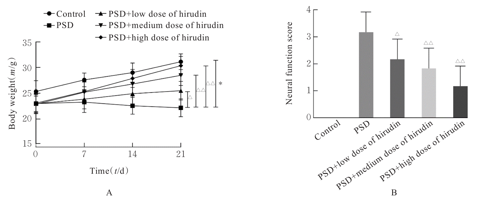
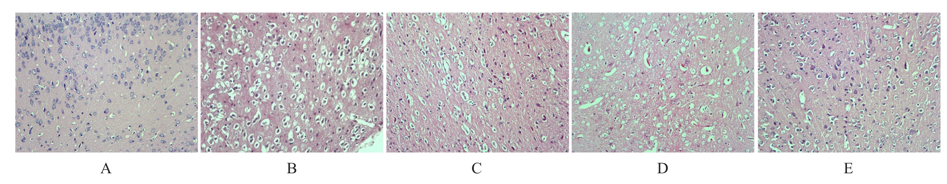
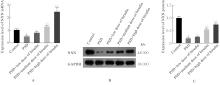
目的 探讨水蛭素对小鼠脑卒中后抑郁(PSD)的影响,并阐明其可能的作用机制。 方法 60只雄性C57BL/6小鼠随机分为对照组、PSD组、PSD+低剂量水蛭素组、PSD+中剂量水蛭素组和PSD+高剂量水蛭素组,每组12只。采用大脑中动脉栓塞(MCAO)法建立小鼠脑卒中模型,采用慢性不可预见的中等应激刺激(CUMS)结合孤养法建立小鼠抑郁模型,PSD+低剂量水蛭素组、PSD+中剂量水蛭素组和PSD+高剂量水蛭素组小鼠在建立抑郁模型的同时分别经尾静脉注射10、20及40 U·kg-1水蛭素干预,对照组和PSD组小鼠经尾静脉注射等量生理盐水。记录CUMS刺激第0、7、14和21天时各组小鼠体质量,计算各组小鼠LONGA神经功能评分,糖水偏好实验、悬尾实验和强迫游泳实验检测各组小鼠糖水偏好率、悬尾及强迫游泳不动时间,HE染色观察各组小鼠内侧前额叶皮质(mPFC)脑区组织病理形态表现,生化试剂盒检测各组小鼠内侧前额叶皮层(mPFC)脑区组织中丙二醛(MDA)和还原性谷胱甘肽(GSH)水平及超氧化物歧化酶(SOD)活性,2',7'-二氯二氢荧光素二乙酸酯(DCFH-DA)荧光探针法检测各组小鼠mPFC脑区组织中活性氧(ROS)阳性率,实时荧光定量PCR(RT-qPCR)和Western blotting法检测各组小鼠mPFC脑区组织中核氧化还原蛋白(NXN)mRNA及蛋白表达水平。 结果 与对照组比较,CUMS刺激第0、7、14和21天PSD组小鼠体质量均明显降低(P<0.05或P<0.01);与PSD组比较,CUMS刺激第14和21天PSD+低剂量水蛭素组、PSD+中剂量水蛭素组及PSD+高剂量水蛭素组小鼠体质量均明显升高(P<0.05或P<0.01),神经功能评分均明显降低(P<0.05或P<0.01)。糖水偏好实验、悬尾实验和强迫游泳实验,与对照组比较,PSD组小鼠糖水偏好率明显降低(P<0.01),悬尾和强迫游泳不动时间均明显增加(P<0.01);与PSD组比较,PSD+低剂量水蛭素组、PSD+中剂量水蛭素组和PSD+高剂量水蛭素组小鼠糖水偏好率均明显升高(P<0.05或P<0.01),悬尾和强迫游泳不动时间均明显减少(P<0.05或P<0.01)。HE染色,对照组小鼠mPFC脑区组织细胞形态正常,结构清晰,大小分布均匀;PSD组和PSD+低剂量水蛭素组小鼠mPFC脑区组织细胞明显减少,并出现严重空泡样变性,核仁固缩;PSD+中剂量水蛭素组和PSD+高剂量水蛭素组小鼠mPFC脑区组织细胞较PSD组明显增多,空泡样变性和核仁固缩明显改善。生化试剂盒和DCFH-DA荧光探针法,与对照组比较,PSD组小鼠mPFC脑区组织中GSH水平和SOD活性均明显降低(P<0.01),MDA水平和ROS阳性率均明显升高(P<0.01);与PSD组比较,PSD+低剂量水蛭素组、PSD+中剂量水蛭素组和PSD+高剂量水蛭素组小鼠mPFC脑区组织中GSH水平及SOD活性均明显升高(P<0.05或P<0.01),MDA水平和ROS阳性率均明显降低(P<0.05或P<0.01)。RT-qPCR和Western blotting法,与对照组比较,PSD组小鼠mPFC脑区组织中NXN mRNA和蛋白表达水平均明显降低(P<0.01);与PSD组比较,PSD+低剂量水蛭素组、PSD+中剂量水蛭素组和PSD+高剂量水蛭素组小鼠mPFC脑区组织中NXN mRNA及蛋白表达水平均明显升高(P<0.05或P<0.01)。 结论 水蛭素可促进PSD小鼠mPFC脑区组织氧化还原平衡,修复其神经功能损伤,改善PSD。

关键词: 水蛭素, 脑卒中, 抑郁, 内侧前额叶皮质, 核氧化还原蛋白

Abstract:

Objective To discuss the effect of hirudin on post-stroke depression (PSD) in the mice, and to clarify its potential mechanism. Methods Sixty male C57BL/6 mice were randomly divided into control group, PSD group, PSD+low dose of hirudin group, PSD+medium dose of hirudin group, and PSD+high dose of hirudin group, and there were 12 mice in each group. The stroke model was established by middle cerebral artery occlusion (MCAO), and the depression model was induced by chronic unpredictable mild stress (CUMS) combined with solitary housing. The mice in PSD+low dose of hirudin, PSD+medium dose of hirudin, and PSD+high dose of hirudin groups were intravenously injected with 10, 20, and 40 U·kg-1 hirudin, respectively, while the mice in control and PSD groups received equal volumes of saline. The body weights of the mice were recorded on days 0, 7, 14, and 21 of CUMS. The LONGA neurological function score was calculated. Sucrose preference test, tail suspension test, and forced swimming test were used to detect the sucrose preference rate, immobility time in tail suspension, and forced swimming in various groups, respectively; HE staining was used to observe the histopathological changes in the medial prefrontal cortex (mPFC); biochemical kits were used to detect the levels of malondialdehyde (MDA) and reduced glutathione (GSH) as well as superoxide dismutase (SOD) activity in mPFC tissue of the mice in various groups; 2',7'-dichlorodihydrofluorescein diacetate (DCFH-DA) fluorescence probe method was used to detect the reactive oxygen species (ROS) positive rate in mPFC tissue of the mice in various groups; real-time fluorescence quantitative PCR (RT-qPCR) and Western blotting method were used to detect the expression levels of nucleoredoxin (NXN) mRNA and protein in mPFC tissue of the mice in various groups. Results Compared with control group, the body weight of the mice in PSD group was significantly decreased on days 0, 7, 14, and 21 of CUMS (P<0.05 or P<0.01). Compared with PSD group, the body weights of the mice in PSD+low dose of hirudin, PSD+medium dose of hirudin, and PSD+high dose of hirudin groups were significantly increased on days 14 and 21 of CUMS (P<0.05 or P<0.01), and the neurological function scores were significantly decreased (P<0.05 or P<0.01). The sucrose preference test, tail suspension test, and forced swimming test results showed that compared with control group, the sucrose preference rate of the mice in PSD group was significantly decreased (P<0.01), while the immobility times in tail suspension and forced swimming were significantly increased (P<0.01). Compared with PSD group, the sucrose preference rates of the mice in PSD+low dose of hirudin, PSD+medium dose of hirudin, and PSD+high dose of hirudin groups were significantly increased (P<0.05 or P<0.01), and the immobility times were significantly decreased (P<0.05 or P<0.01). The HE staining showed normal cell morphology, clear structure, and uniform size distribution in mPFC tissue in control group. In PSD and PSD+low dose of hirudin groups, the number of the cells in mPFC tissue was significantly reduced, with severe vacuolar degeneration and pyknotic nuclei. Compared with PSD group, the numbers of the cells in PSD+medium dose of hirudin and PSD+high dose of hirudin groups were significantly increased, and the vacuolar degeneration and nuclear pyknosis were alleviated. The Biochemical and DCFH-DA fluorescence probe assays results showed that compared with control group, the GSH level and SOD activity in mPFC tissue of the mice in PSD group were significantly decreased (P<0.01), while the MDA level and ROS positive rate were significantly increased (P<0.01). Compared with PSD group, the GSH levels and SOD activities of the mice in PSD+low dose of hirudin, PSD+medium dose of hirudin, and PSD+high dose of hirudin groups were significantly increased (P<0.05 or P<0.01), while the MDA levels and ROS positive rates were significantly decreased (P<0.05 or P<0.01). The RT-qPCR and Western blotting results showed that compared with control group, the expression levels of NXN mRNA and protein in mPFC tissue of the mice in PSD group were significantly decreased (P<0.01). Compared with PSD group, the expression levels of NXN mRNA and protein in mPFC tissue of the mice in PSD+low dose of hirudin, PSD+medium dose of hirudin, and PSD+high dose of hirudin groups were significantly increased (P<0.05 or P<0.01). Conclusion Hirudin promotes redox balance in mPFC of the PSD mice, repairs neurological damage, and improves PSD.

Key words: Hirudin, Stroke, Depression, Medial prefrontal cortex, Nucleoredoxin

中图分类号: 

  • R749.13