| [1] |
Ruiyun LU,Jingfeng GU,Jian ZHANG,Xin ZHANG,Fei XU.
Reverse effect of miR-30a-5p by targeting TRIM31 expression on 5-fluorouracil resistance in colorectal cancer cells and its mechanism
[J]. Journal of Jilin University(Medicine Edition), 2021, 47(3): 714-723.
|
| [2] |
Dong CHEN,Zhiyao LI,Haitao CHEN,Zhirui CHUAN,Yingxian ZHANG,Xin JIN,Shicong TANG,Xiaomao LUO.
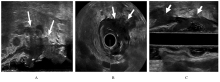
Diagnostic value of three-dimensional transrectal ultrasound in preoperative N-staging of middle and lower rectal cancer
[J]. Journal of Jilin University(Medicine Edition), 2021, 47(2): 497-504.
|
| [3] |
Dong CHEN,Haitao CHEN,Zhiyao LI,Fengming RANG,Xi ZHANG,Zhirui CHUAN,Shicong TANG,Xiaomao LUO.
Effects of three-dimensional transrectal ultrasound and MRI in evaluation of extramural vascular invasion degrees of middle and lower rectal cancer
[J]. Journal of Jilin University(Medicine Edition), 2021, 47(1): 203-209.
|
| [4] |
Xia LI,Yi YU,Haiwei ZUO,Fengjuan ZHOU,Yong XIN.
Inhibitory effect of circRNA on colorectal cancer and its bioinformatics analysis
[J]. Journal of Jilin University(Medicine Edition), 2020, 46(6): 1283-1287.
|
| [5] |
Lingxue SHI,Shuo LIU,Xuewei ZHENG,Xiaoliang CHENG,Zhaohui CHEN,Baolin MA,Hongguo ZHANG,Jun DING.
Application of magnetic resonance diffusion weighted imaging ADC and rADC in differential diagnosis of benign and malignant parotid gland tumors
[J]. Journal of Jilin University(Medicine Edition), 2020, 46(6): 1309-1314.
|
| [6] |
WANG Dan, LIAO Dan, LI Hong, XIONG Liqiu, WU Ying, DONG Ying, GAI Xiaodong.
Expressions of plasmacytoid dendritic cells and Foxp3+ regulatory T cells in colorectal cancer tissue and their significances
[J]. Journal of Jilin University(Medicine Edition), 2020, 46(04): 834-838.
|
| [7] |
ZHOU Dongkui, LU Mingqian, FENG Xuesong, LIU Yufei, SONG Hao, XU Liang.
Follicular dendritic cell sarcoma of small intestine and ileocecal region: A case report and literature review
[J]. Journal of Jilin University(Medicine Edition), 2020, 46(04): 858-862.
|
| [8] |
MENG Shuang, LI Yingjie, ZANG Xiaozhen, ZHAO Qianfang, ZHANG Jin, LI Jing.
Inhibitory effect of knockdown of TLR2 gene on proliferation of colorectal cancer cells and its mechanism
[J]. Journal of Jilin University(Medicine Edition), 2020, 46(02): 316-322.
|
| [9] |
WANG Dan, SONG Ziqi, LI Yifei, LI Chun, DONG Zhiheng, DONG Ying, GAI Xiaodong.
Expressions of Foxp3+ regulatory T cells and myeloid dentritic cells in human colorectal cancer and tumor draining lymph node tissues and their significances
[J]. Journal of Jilin University(Medicine Edition), 2019, 45(03): 621-626.
|
| [10] |
DUAN Jiqing, SUO Jian, SUN Donghui.
Application of two-step procedure to turn rectum out from abdominal cavity in radical resection of low rectal carcinoma by natural orifice specimen extraction surgery: A report of 39 cases
[J]. Journal of Jilin University(Medicine Edition), 2019, 45(03): 683-687.
|
| [11] |
ZHANG Ping, ZHOU Jia, TANG Yan, LIU Hongyu.
Preparation of PR-SPIO-PLGA molecular probe and its property
[J]. Journal of Jilin University(Medicine Edition), 2018, 44(06): 1307-1311.
|
| [12] |
ZHU Guangwei, ZHENG Wei, HUANG Yongjian, HUA Jin, YANG Shugang, YE Jianxin.
Expression of MyD88 in cancer tissue of patients with colorectal cancer and clinical significance
[J]. Journal of Jilin University(Medicine Edition), 2018, 44(05): 1047-1051.
|
| [13] |
WANG Dan, LI Yifei, LI Chun, DONG Zhiheng, DONG Ying, GAI Xiaodong.
Relationships between expressions of B7-H1 and B7-H4 and Foxp3+ regulated T-cell infiltration in colorectal cancer tissue
[J]. Journal of Jilin University Medicine Edition, 2018, 44(03): 543-547.
|
| [14] |
YANG Xueliang, SUN Xuemei, ZHAO Xiaohui, YANG Lijuan, SHEN Weigao, XIAO Zishen, GUO Chong, LIU Yanbo.
Expressions of IL-17E, IL-17F and their receptors in colorectal carcinoma tissue and their significances
[J]. Journal of Jilin University Medicine Edition, 2018, 44(03): 574-578.
|
| [15] |
CAO Yansha, LI Hua, CHEN Minghong, LIU Baoqin, WANG Huaqin, LI Ning.
Expression of BAG3 protein in colon cancer tissue detected by tissue microarray method and its clinical significance
[J]. Journal of Jilin University Medicine Edition, 2017, 43(06): 1177-1181.
|
 ),Xiaomao LUO1(