Journal of Jilin University(Medicine Edition) ›› 2021, Vol. 47 ›› Issue (4): 874-881.doi: 10.13481/j.1671-587X.20210408
• Research in basic medicine • Previous Articles Next Articles
Induction of 3C protein of Coxsackievirus A16 in apoptosis of human rhabdomyosarcoma cells and its mechanism
Yingying SHI1,Shitong CHEN2,Bo HU2,Duo SHEN2,Kuang ZHU2,Siwei YE2,Jinmei FENG3( )
)
- 1.Department of Immunology,School of Medical Sciences,Jianghan University,Wuhan 430056,China
2.School of Medical Sciences,Jianghan University,Wuhan 430056,China
3.Department of Pathogen Biology,School of Medicsl Sciences,Jianghan University,Wuhan 430056,China
-
Received:2020-12-12Online:2021-07-28Published:2021-07-22 -
Contact:Jinmei FENG E-mail:fengjm@jhun.edu.cn
CLC Number:
- R373.23
Cite this article
Yingying SHI,Shitong CHEN,Bo HU,Duo SHEN,Kuang ZHU,Siwei YE,Jinmei FENG. Induction of 3C protein of Coxsackievirus A16 in apoptosis of human rhabdomyosarcoma cells and its mechanism[J].Journal of Jilin University(Medicine Edition), 2021, 47(4): 874-881.
share this article
Tab. 1
Primer sequences of PCR"
| Gene | Primer sequence(5'-3') |
|---|---|
| Bak | Forward AGGGCTTAGGACTTGGTTTG |
| Reverse GGGATTCCTAGTGGTGTTGATAG | |
| Bad | Forward GGAGGATGAGTGACGAGTTTG |
| Reverse GGAGCTTTGCCGCATCT | |
| PUMA | Forward GTGACCACTGGCATTCATTTG |
| Reverse TCCTCCCTCTTCCGAGATTT | |
| β-actin | Forward CACGATGGAGGGGCCGGACTCATC |
| Reverse TAAAGACCTCTATGCCAACACAGT |
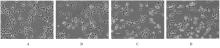
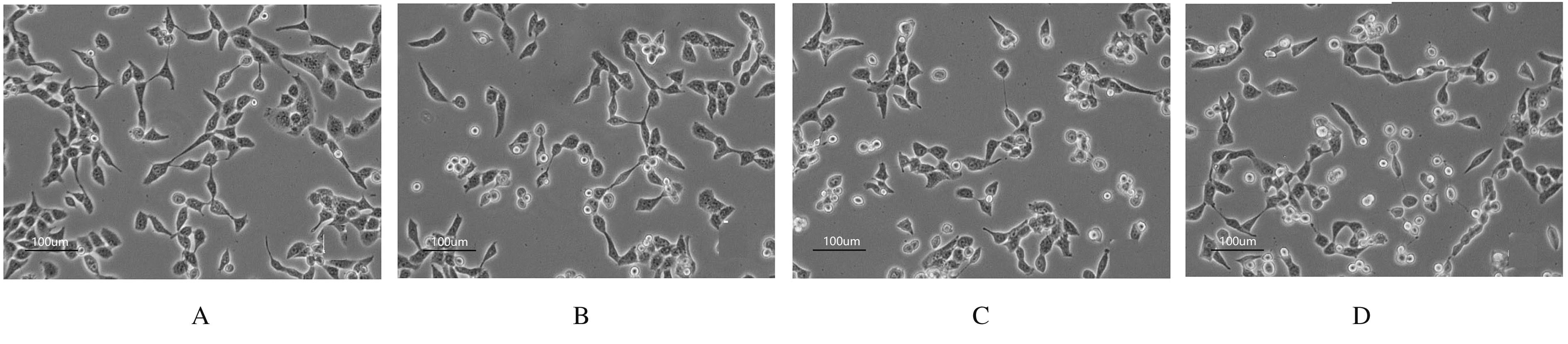
Fig. 1
Morphology of RD cells in various groups(×100)"
Tab. 2
Survival rates of RD cells in various groups"
| Group | Survival rate |
|---|---|
| Control | 90.00±5.98 |
pCMV-HA-3C 0.1 μg | 85.83±5.71 |
| 0.2 μg | 82.68±6.02 |
| 0.4 μg | 65.09±3.78* |
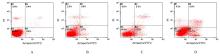
Fig. 2
Apoptotic rates of RD cells in various groups detected by flow cytometry"
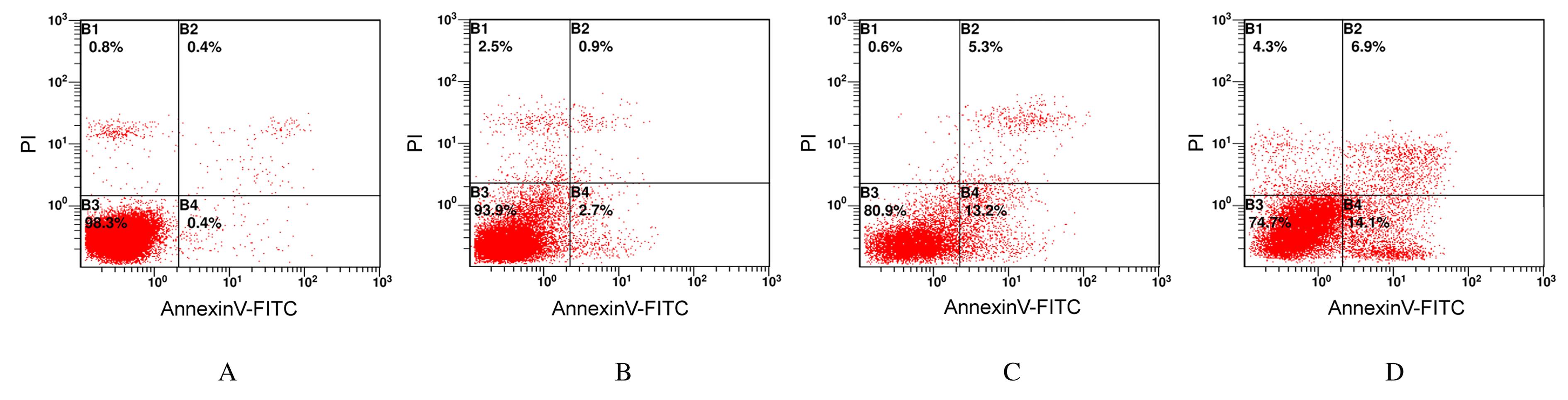

Fig. 3
Electrophoregram of expressions of Bak,Bad and PUMA proteins in RD cells in various groups"

Tab. 3
Expression levels of Bak, Bad and PUMA mRNA and proteins in RD cells in various groups"
| Group | Bak | Bad | PUMA | |||
|---|---|---|---|---|---|---|
| mRNA | Protein | mRNA | Protein | mRNA | Protein | |
| Control | 1.02±0.14 | 0.69±0.01 | 1.02±0.12 | 0.77±0.01 | 1.01±0.10 | 0.80±0.01 |
| 0.2 μg pCMV-HA-3C | 1.54±0.11* | 0.78±0.01** | 1.72±0.05** | 0.83±0.01* | 1.73±0.19* | 0.85±0.01* |

Fig.4
Electrophoregram of expressions of PARP, cleaved-PARP and cleaved-caspase-3 proteins in RD cells in various groups"
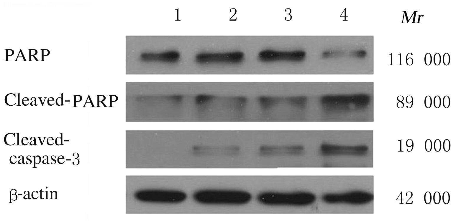
Tab.4
Expression levels of PARP,cleaved-PARP and cleaved-caspase-3 proteins in RD cells in various groups"
| Group | PARP | Cleaved- PARP | Cleaved- caspase-3 |
|---|---|---|---|
Control pCMV-HA-3C | 0.88±0.01 | 0.71±0.01 | 0.55±0.01 |
| 0.1 μg | 0.86±0.01 | 0.75±0.01* | 0.60±0.01* |
| 0.2 μg | 0.82±0.07 | 0.77±0.01* | 0.68±0.01* |
| 0.4 μg | 0.70±0.02** | 0.99±0.03** | 0.88±0.03** |
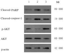
Fig. 5
Electrophoregram of expressions of AKT signaling pathway proteins in RD cells in AKT activator SC79 treatment experiment in various groups"

Tab. 5
Expression levels of AKT signaling pathway proteins in RD cells in various groups in AKT activator SC79 treatment experiment (n=3,x±s)"
| Group | Cleaved-PARP | Cleaved-caspase-3 | AKT | p-AKT |
|---|---|---|---|---|
| Control | 0.82±0.01 | 0.81±0.01 | 1.02±0.02 | 0.98±0.01 |
| 0.2 μg pCMV-HA-3C | 1.07±0.01* | 1.16±0.02* | 1.10±0.01* | 0.82±0.01** |
| SC79 treatment | 0.84±0.01ΔΔ | 0.97±0.04Δ | 0.94±0.01ΔΔ | 1.10±0.01ΔΔ |
| 1 | MAO Q Y, WANG Y P, YAO X, et al. Coxsackievirus A16: epidemiology, diagnosis, and vaccine[J]. Hum Vaccines Immunother, 2014, 10(2): 360-367. |
| 2 | ZHANG M, ZHAO Y L, ZHANG H H, et al. Molecular characterization of Coxsackievirus A16 strains isolated from children with severe hand, foot, and mouth disease in Yunnan, Southwest China, during 2009-2015[J]. J Med Virol, 2019, 91(1): 155-160. |
| 3 | ASTRUP B S, JOHNSEN I B, ENGSBRO A L. The role of Coxsackievirus A16 in a case of sudden unexplained death in an infant-A SUDI case[J]. Forensic Sci Int, 2016,259: e9-e13. |
| 4 | KVANSAKUL M. Viral infection and apoptosis[J]. Viruses, 2017, 9(12): 356. |
| 5 | SHIM J M, KIM J, TENSON T, et al. Influenza virus infection, interferon response, viral counter-response, and apoptosis[J]. Viruses, 2017, 9(8): E223. |
| 6 | ZHU G, ZHENG Y, ZHANG L, et al. Coxsackievirus A16 infection triggers apoptosis in RD cells by inducing ER stress[J]. Biochem Biophys Res Commun, 2013,441(4):856-861. |
| 7 | SHI Y Y, HE X H, ZHU G G, et al. Coxsackievirus A16 elicits incomplete autophagy involving the mTOR and ERK pathways[J]. PLoS One, 2015, 10(4): e0122109. |
| 8 | LU G W, QI J X, CHEN Z J, et al. Enterovirus 71 and coxsackievirus A16 3C proteases: binding to rupintrivir and their substrates and anti-hand, foot, and mouth disease virus drug design[J]. J Virol, 2011, 85(19): 10319-10331. |
| 9 | RAMAJAYAM R, TAN K P, LIANG P H. Recent development of 3C and 3CL protease inhibitors for anti-coronavirus and anti-picornavirus drug discovery[J]. Biochem Soc Trans, 2011,39(5):1371-1375. |
| 10 | RUI Y J, SU J M, WANG H, et al. Disruption of MDA5-mediated innate immune responses by the 3C proteins of coxsackievirus A16, coxsackievirus A6, and enterovirus D68[J]. J Virol, 2017, 91(13): e00546. |
| 11 | LI J, YAO Y F, CHEN Y, et al. Enterovirus 71 3C promotes apoptosis through cleavage of PinX1, a telomere binding protein[J]. J Virol, 2017, 91(2): e02016. |
| 12 | REN Y J, SHU T, WU D, et al. The ORF3a protein of SARS-CoV-2 induces apoptosis in cells[J]. Cell Mol Immunol, 2020, 17(8): 881-883. |
| 13 | XU Y S, VICTORIO C B L, MENG T, et al. The saffold virus-Penang 2B and 3C proteins, but not the L protein, induce apoptosis in HEp-2 and vero cells[J]. Virol Sin, 2019, 34(3): 262-269. |
| 14 | ADAMS J M, CORY S. The Bcl-2 protein family: arbiters of cell survival[J]. Science, 1998,281(5381):1322-1326. |
| 15 | KAZI A, SUN J, DOI K, et al. The BH3 alpha-helical mimic BH3-M6 disrupts Bcl-X(L), Bcl-2, and MCL-1 protein-protein interactions with Bax, Bak, Bad, or Bim and induces apoptosis in a Bax- and Bim-dependent manner[J]. J Biol Chem, 2011,286(11):9382-9392. |
| 16 | DANG Y N, ZHANG Y F, XU L Y, et al. PUMA-mediated epithelial cell apoptosis promotes Helicobacter pylori infection-mediated gastritis[J]. Cell Death Dis, 2020, 11(2): 139. |
| 17 | BOULARES A H, YAKOVLEV A G, IVANOVA V,et al. Role of poly(ADP-ribose) polymerase (PARP) cleavage in apoptosis. Caspase 3-resistant PARP mutant increases rates of apoptosis in transfected cells[J]. J Biol Chem, 1999,274(33):22932-22940. |
| 18 | CHOUDHARY G S, AL-HARBI S, ALMASAN A. Caspase-3 activation is a critical determinant of genotoxic stress-induced apoptosis[J]. Methods Mol Biol, 2015,1219: 1-9. |
| 19 | FRANKE T F, HORNIK C P, SEGEV L, et al. PI3K/Akt and apoptosis: size matters[J]. Oncogene, 2003,22(56):8983-8998. |
| 20 | BROTELLE T, BAY J O. PI3K-AKT-mTOR pathway: Description, therapeutic development, resistance, predictive/prognostic biomarkers and therapeutic applications for cancer[J]. Bull Cancer, 2016,103(1):18-29. |
| 21 | PONNUSAMY L, KOTHANDAN G, MANOHARAN R. Berberine and Emodin abrogates breast cancer growth and facilitates apoptosis through inactivation of SIK3-induced mTOR and Akt signaling pathway[J]. Biochim Biophys Acta Mol Basis Dis, 2020,1866(11):165897. |
| 22 | ZHAI H, KANG Z H, ZHANG H B, et al. Baicalin attenuated substantia nigra neuronal apoptosis in Parkinson’s disease rats via the mTOR/AKT/GSK-3β pathway[J]. J Integr Neurosci, 2019, 18(4): 423-429. |
| [1] | Qingxu LANG,Xueshuang NIU,Kaiwen YANG,Ren ZHANG,Siteng WANG, ZUMIRETIGULI·Wumaier,Zhenqi WANG. Effects of sodium butyrate combined with ionizing radiation on apoptosis of lung cancer A549 cells and its mechanism [J]. Journal of Jilin University(Medicine Edition), 2022, 48(4): 915-921. |
| [2] | Guanhu LI,Qingxu LANG,Chunyan LIU,Qin LIU,Mengrou GENG,Xiaoqian LI,Zhenqi WANG. Inhibitory effect of valproic acid combined with X-ray irradiation on proliferation of breast cancer MDA-MB-231 cells and its mechanism [J]. Journal of Jilin University(Medicine Edition), 2022, 48(3): 622-629. |
| [3] | Qiuting CAO,Jingchun HAN,Xiaofei ZHANG. Effect of silencing helicase BLM gene on chemotherapy sensitivity of irinotecan in colorectal cancer cells and its mechanism [J]. Journal of Jilin University(Medicine Edition), 2022, 48(3): 657-667. |
| [4] | Cuilan LIU,Fengai HU,Jing LIU,Dan WANG,Changyun QIU,Dunjiang LIU,Di ZHAO. Effect of adiponectin receptor agonist AdiopRon on biological behaviors of glioma cells and its mechanism [J]. Journal of Jilin University(Medicine Edition), 2022, 48(3): 702-710. |
| [5] | Ming xing YANG,Wen DONG,Ji LI. Inductive effect of peiminine on apoptosis of lung cancer A549 cells and its mechanism [J]. Journal of Jilin University(Medicine Edition), 2022, 48(3): 711-717. |
| [6] | Ming LI,Qiuting WANG,Shan CHEN,Huifang SHI. Improvement effect of p38 MAPK inhibitor on chronic obstructive pulmonary disease injury in mice through inhibiting cell pyrotosis mediated by NLRP3 pathway [J]. Journal of Jilin University(Medicine Edition), 2022, 48(3): 744-754. |
| [7] | Suxian CHEN,Zehui GU,Yangfei MA,Qi TAN,Qi LI,Yadi WANG. Promotion effect of rutin on apoptosis of human colon cancer SW480 cells and its mechanism [J]. Journal of Jilin University(Medicine Edition), 2022, 48(2): 356-363. |
| [8] | Zhihui ZHAO,Xianghua BAI,Jinling HE,Weiqin DUAN,Min LIU,Shengmao ZHANG. Inhibitory effect of sufentanil on apoptosis of myocardial cells in myocardial ischemia-reperfusion injury rats and its mechanism [J]. Journal of Jilin University(Medicine Edition), 2022, 48(2): 364-373. |
| [9] | Guangsong XU,Haibing JIANG,Jing PAN,Guoqing LI. Inhibitory effects of betulinic acid on migration and invasion of gastric cancer MGC-803 cells and their mechanisms [J]. Journal of Jilin University(Medicine Edition), 2022, 48(1): 122-128. |
| [10] | Wenxiong SUN,Pu LI. Expression of SOCS3 in peripheral blood mononuclear cells of patients with diffuse large B-cell lymphoma and its effect on autophagy and apoptosis of OCI-LY7 cells [J]. Journal of Jilin University(Medicine Edition), 2022, 48(1): 172-179. |
| [11] | Leihua CUI,Yubo HOU,Chang SU,Minghe LI,Xin NIE. Effect of N-acetylcysteine on apoptosis of MC3T3-E1 cells induced by nicotine and its mechanism [J]. Journal of Jilin University(Medicine Edition), 2022, 48(1): 26-32. |
| [12] | Yu ZHU,Jingjing WANG,Fang WU. Expression of miR-150-5p in kidney tissue of diabetic nephropathy model mice and its effect on MPC5 mouse podocyte injury and mechanism [J]. Journal of Jilin University(Medicine Edition), 2022, 48(1): 44-51. |
| [13] | Zhaohui WAN,Liang ZENG,Hui ZHOU. Effect of overexpression of Bax inhibitor 1 on cardiomyocyte apoptosis in rats with acute myocardial infarction and its mechanism [J]. Journal of Jilin University(Medicine Edition), 2022, 48(1): 74-81. |
| [14] | Daiqiang HUANG,Pengfei LIU,Jianbin HE,Jingxue SUN,Lin YUAN,Jing YUAN,Lei ZHAO. Effect of sevoflurane exposure during pregnancy period on maternal behavior of offspring of mice and protection mechanism of H2 [J]. Journal of Jilin University(Medicine Edition), 2021, 47(6): 1347-1352. |
| [15] | Jingjing MA,Yu LIU,Yan LIANG,Ying LI. Promotion effect of total alkaloids of leonurus on decidual villus excretion in drug abortion rats and its mechanism [J]. Journal of Jilin University(Medicine Edition), 2021, 47(6): 1476-1484. |