Journal of Jilin University(Medicine Edition) ›› 2021, Vol. 47 ›› Issue (5): 1187-1193.doi: 10.13481/j.1671-587X.20210515
• Research in basic medicine • Previous Articles Next Articles
Effect of human umbilical cord mesenchymal stem cells on proliferation and apoptosis of cervical cancer HeLa cells and its mechanism
Chen WANG( ),Jun TIAN,Hailing CHENG
- Department of Gynecology,Huaihe Hospital,Henan University,Kaifeng 475000,China
-
Received:2021-01-26Online:2021-09-28Published:2021-10-26 -
Contact:Chen WANG E-mail:qingtianxiayu_88@126.com
CLC Number:
- R285.5
Cite this article
Chen WANG,Jun TIAN,Hailing CHENG. Effect of human umbilical cord mesenchymal stem cells on proliferation and apoptosis of cervical cancer HeLa cells and its mechanism[J].Journal of Jilin University(Medicine Edition), 2021, 47(5): 1187-1193.
share this article

Fig. 1
Morphology of MSCs isolated from umbilical cord (×40)"
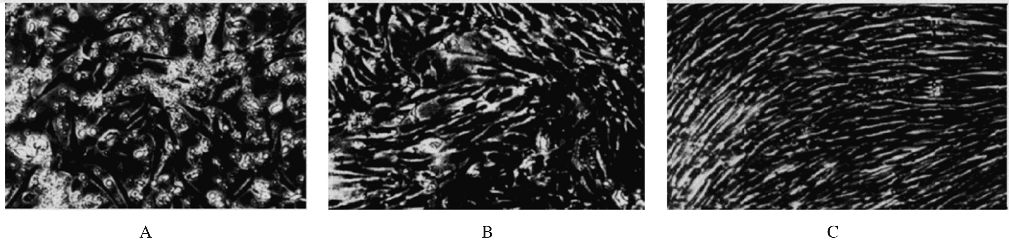

Fig. 2
Expressions of surface antigen markers of MSCs"


Fig. 3
Colony forming abilities of HeLa cells in various groups after treated with MSCs conditioned medium"


Fig. 4
Inhibitory rates of proliferation of HeLa cells in various groups"


Fig. 5
Apoptotic rates of HeLa cells in two groups"

Tab. 1
Expression levels of caspase-3, P53 and Bcl-2 in HeLa cells in various groups"
| Time(t/h) | Caspase-3 | P53 | Bcl-2 |
|---|---|---|---|
| 0 | 0.09±0.01 | 0.08±0.01 | 0.59±0.08 |
| 24 | 0.33±0.05* | 0.52±0.09* | 0.42±0.07* |
| 48 | 0.66±0.09*△ | 0.71±0.06*△ | 0.31±0.06*△ |
| F | 68.888 | 79.653 | 20.034 |
| P | <0.01 | <0.01 | <0.01 |

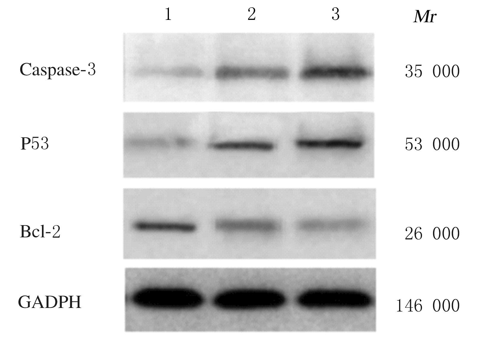
Fig. 6
Electrophoregram of expressions of caspase-3, P53 and Bcl-2 proteins in HeLa cells after treated with MSCs for 0, 24,and 48 h"
| 1 | MUNIR S, BASU A, MAITY P, et al. TLR4-dependent shaping of the wound site by MSCs accelerates wound healing[J].EMBO Rep,2020,21(5):e48777. |
| 2 | JIANG D, SINGH K, MUSCHHAMMER J, et al. MSCs rescue impaired wound healing in a murine LAD1 model by adaptive responses to low TGF-β1 levels[J]. EMBO Rep,2020, 21(4):e49115. |
| 3 | CAO X, DUAN L, HOU H, et al. IGF-1C hydrogel improves the therapeutic effects of MSCs on colitis in mice through PGE2-mediated M2 macrophage polarization[J]. Theranostics,2020, 10(17):7697-7709. |
| 4 | LIU S, LIU F, ZHOU Y, et al. Immunosuppressive property of MSCs mediated by cell surface receptors[J]. Front Immunol, 2020, 11:1076. |
| 5 | ZHAO Q, ZHANG L, WEI Y, et al. Systematic comparison of hUC-MSCs at various passages reveals the variations of signatures and therapeutic effect on acute graft-versus-host disease[J]. Stem Cell Res Ther,2019, 10(1):354. |
| 6 | BRISSON M, KIM JJ, CANFELL K, et al. Impact of HPV vaccination and cervical screening on cervical cancer elimination: a comparative modelling analysis in 78 low-income and lower-middle-income countries[J]. Lancet, 2020, 395(10224):575-590. |
| 7 | ANNAN FM, OPPONG ASANTE K, KUGBEY N. Perceived seriousness mediates the influence of cervical cancer knowledge on screening practices among female university students in Ghana[J]. BMC Womens Health, 2019, 19(1):140. |
| 8 | NIGUSSIE T, ADMASSU B, NIGUSSIE A. Cervical cancer screening service utilization and associated factors among age-eligible women in Jimma town using health belief model, South West Ethiopia[J]. BMC Womens Health, 2019, 19(1):127. |
| 9 | ASSEFA A A, ASTAWESEGN F H, ESHETU B. Cervical cancer screening service utilization and associated factors among HIV positive women attending adult ART clinic in public health facilities, Hawassa town, Ethiopia: a cross-sectional study[J]. BMC Health Serv Res, 2019, 19(1):847. |
| 10 | 郭娜娜, 高晨, 陈蕾. Polo样激酶1(PLK1)的敲低及其对宫颈癌HeLa细胞生长的抑制作用[J]. 细胞与分子免疫学杂志, 2018, 34(4):334-340. |
| 11 | HOMBACH AA, GEUMANN U, GÜNTHER C,et al. IL7-IL12 engineered mesenchymal stem cells (MSCs) improve A CAR T cell attack against colorectal cancer cells[J]. Cells, 2020, 9(4):873. |
| 12 | HULL R, MBELE M, MAKHAFOLA T, et al. Cervical cancer in low and middle-income countries[J]. Oncol Lett,2020, 20(3):2058-2074. |
| 13 | MCKENZIE BA, FERNANDES JP, DOAN MAL,et al.Activation of the executioner caspases-3 and -7 promotes microglial pyroptosis in models of multiple sclerosis[J]. J Neuroinflammation, 2020, 17(1):253. |
| 14 | TUMMERS B, MARI L, GUY CS, et al. Caspase-8-dependent inflammatory responses are controlled by its adaptor, FADD, and necroptosis[J]. Immunity, 2020, 52(6):994-1006. |
| 15 | LI Z, GUO D, YIN X, et al. Zinc oxide nanoparticles induce human multiple myeloma cell death via reactive oxygen species and Cyt-C/Apaf-1/Caspase-9/Caspase-3 signaling pathway in vitro [J]. Biomed Pharmacother, 2020, 122:109712. |
| 16 | JUNG JH, LEE H, Cao B, et al. RNA-binding motif protein 10 induces apoptosis and suppresses proliferation by activating P53[J]. Oncogene, 2020,39(5):1031-1040. |
| 17 | ZHANG Y, XU L, CHANG Y, et al. Therapeutic potential of ReACP53 targeting mutant P53 protein in CRPC[J]. Prostate Cancer Prostatic Dis, 2020, 23(1):160-171. |
| 18 | WEI W, LIU C. Prognostic and predictive roles of microRNA‑411 and its target STK17A in evaluating radiotherapy efficacy and their effects on cell migration and invasion via the P53 signaling pathway in cervical cancer[J]. Mol Med Rep, 2020, 21(1):267-281. |
| 19 | 金红, 杨岚, 张黎, 等. 双氢青蒿素和吉非替尼联用对肺癌NCI-H1975细胞凋亡相关蛋白Bax与Bcl-2表达的影响[J]. 中国老年学杂志, 2019, 39(23):5806-5810. |
| 20 | 陈浩然.血小板凋亡机制对抗肿瘤药物引起血小板减少症的作用研究进展[J].解放军医学杂志,2019,44(1):80-85. |
| 21 | 赖银璇, 王明蕊, 杨海丽, 等. 柚皮素通过ROS/JNK/Bcl2通路抑制宫颈癌HeLa细胞增殖和迁移[J]. 中药药理与临床, 2018, 34(1): 40-43. |
| [1] | Zhijuan WANG,Mingshu ZHANG,Liping YE. Effects of platelet-derived growth factor D on proliferation, migration and invasion of lung cancer H1299 cells through ERK signaling pathway and their mechanisms [J]. Journal of Jilin University(Medicine Edition), 2022, 48(4): 898-904. |
| [2] | Yandi MA,Xiangyun LU,Shangfeng HE,Xueyan YU,Yunhua HU,Haixia GAO,Yunzhao CHEN,Jie YU,Wenjie WANG,Feng LI,Xiaobin CUI. Expression of m6A methylation binding protein YTHDF2 in esophageal carcinoma tissue and its effect on proliferation and migration of esophageal carcinoma cells [J]. Journal of Jilin University(Medicine Edition), 2022, 48(4): 962-970. |
| [3] | Guowu WANG,Yuan YAO,Yu ZHANG,Na XU,Fang LIU. Inhibitory effect of miR-152 on proliferation and invasion of endometrial carcinoma cells by reducing low-density lipoprotein receptor expression [J]. Journal of Jilin University(Medicine Edition), 2022, 48(3): 591-599. |
| [4] | Guanhu LI,Qingxu LANG,Chunyan LIU,Qin LIU,Mengrou GENG,Xiaoqian LI,Zhenqi WANG. Inhibitory effect of valproic acid combined with X-ray irradiation on proliferation of breast cancer MDA-MB-231 cells and its mechanism [J]. Journal of Jilin University(Medicine Edition), 2022, 48(3): 622-629. |
| [5] | Peiyu YAN,Aichen ZHANG,Hong ZHANG,Yang LI,Mengmeng ZHANG,Mengze LUO,Ying PAN. Therapeutic effect of adipose-derived mesenchymal stem cells on premature ovarian failure model rats and its mechanism [J]. Journal of Jilin University(Medicine Edition), 2022, 48(3): 648-656. |
| [6] | Cuilan LIU,Fengai HU,Jing LIU,Dan WANG,Changyun QIU,Dunjiang LIU,Di ZHAO. Effect of adiponectin receptor agonist AdiopRon on biological behaviors of glioma cells and its mechanism [J]. Journal of Jilin University(Medicine Edition), 2022, 48(3): 702-710. |
| [7] | Zhuangzhi WU,Xiaoning HE,Siqi CHEN. Inhibitory effect of miR-124-3p on proliferation and invasion of oral squamous cell carcinoma cells and its mechanism [J]. Journal of Jilin University(Medicine Edition), 2022, 48(3): 718-727. |
| [8] | Xingyu ZHAO,Xin YANG,Zhihua ZHU,Han HE,Zitong SONG,Wei ZHANG. Inhibitory effect of juglone on proliferation of cervical cancer cells and its mechanism [J]. Journal of Jilin University(Medicine Edition), 2022, 48(2): 348-355. |
| [9] | Xiaoyan LI,Wei ZHANG,Jie HE. Promotion effect of REG1A on proliferation and migration of lung adenocarcinoma cells by regulating Wnt/β-catenin signaling pathway [J]. Journal of Jilin University(Medicine Edition), 2022, 48(2): 444-453. |
| [10] | Guangsong XU,Haibing JIANG,Jing PAN,Guoqing LI. Inhibitory effects of betulinic acid on migration and invasion of gastric cancer MGC-803 cells and their mechanisms [J]. Journal of Jilin University(Medicine Edition), 2022, 48(1): 122-128. |
| [11] | Chaofeng ZHOU,Shifan ZHOU,Qing TIAN,Sai WANG,Honglin LI,Chunzheng MA. Effect of lncRNA-NORAD overexpression on biological behaviors of esophageal cancer Eca-109 cells and its mechanism [J]. Journal of Jilin University(Medicine Edition), 2022, 48(1): 33-43. |
| [12] | Naigao TANG,Genjian ZHENG. Effects of miR-762 on proliferation and apoptosis of human tongue squamous cell carcinoma cells by targeting NCOR1 expression [J]. Journal of Jilin University(Medicine Edition), 2021, 47(5): 1099-1107. |
| [13] | Yajie CAO,Xiaohong BAO,Shuzhen LI,Haiying GENG,Zengxiaorui CAI,Chunmei DAI,Ning LI. Effects of insulin-like growth factor 1 receptor inhibitor NVP-AEW541 on proliferation, migration and invasion of ESCC cells [J]. Journal of Jilin University(Medicine Edition), 2021, 47(5): 1131-1138. |
| [14] | Meng QU,Shiya WENG,Hong ZHENG,Yan LI,Runze GAO,Shenggao WANG,Chunyan YU,Boxue CHEN,Zhiheng DONG. Protective effect of fermented red ginseng total saponins on rat myocardial interstitial fibroblasts cultured with high glucose and its mechanism [J]. Journal of Jilin University(Medicine Edition), 2021, 47(5): 1201-1208. |
| [15] | Siyu LI,Min ZHAO,Maoling YANG,Nong XIAO,Wei JIANG. Regulatory effect of retinoic acid on proliferation of hippocampal neural stem cells after hypoxic-ischemic brain damage in rats via GSK-3β [J]. Journal of Jilin University(Medicine Edition), 2021, 47(5): 1077-1085. |