Journal of Jilin University(Medicine Edition) ›› 2022, Vol. 48 ›› Issue (1): 216-221.doi: 10.13481/j.1671-587X.20220127
• Clinical medicine • Previous Articles Next Articles
Analysis on characteristics of 18F-FDG PET/CT imaging of patient with primary small cell osteosarcoma of coccyx:A case report and literature review
Ping HU,Nan JIANG,Yinghua LI,Hongguang ZHAO( )
)
- Department of Nuclear Medicine,First Hospital,Jilin University,Changchun 130021,China
-
Received:2021-06-11Online:2022-01-28Published:2022-01-17 -
Contact:Hongguang ZHAO E-mail:zhaohg@jlu.edu.cn
CLC Number:
- R734.2
Cite this article
Ping HU,Nan JIANG,Yinghua LI,Hongguang ZHAO. Analysis on characteristics of 18F-FDG PET/CT imaging of patient with primary small cell osteosarcoma of coccyx:A case report and literature review[J].Journal of Jilin University(Medicine Edition), 2022, 48(1): 216-221.
share this article
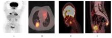
Fig. 1
Images of 18F-FDG PET/CT of a patient with small cell osteosarcoma of coccyx"


Fig. 2
Images of plain and enhanced MRI of cervical spine in a patient with small cell osteosarcoma of coccyx"
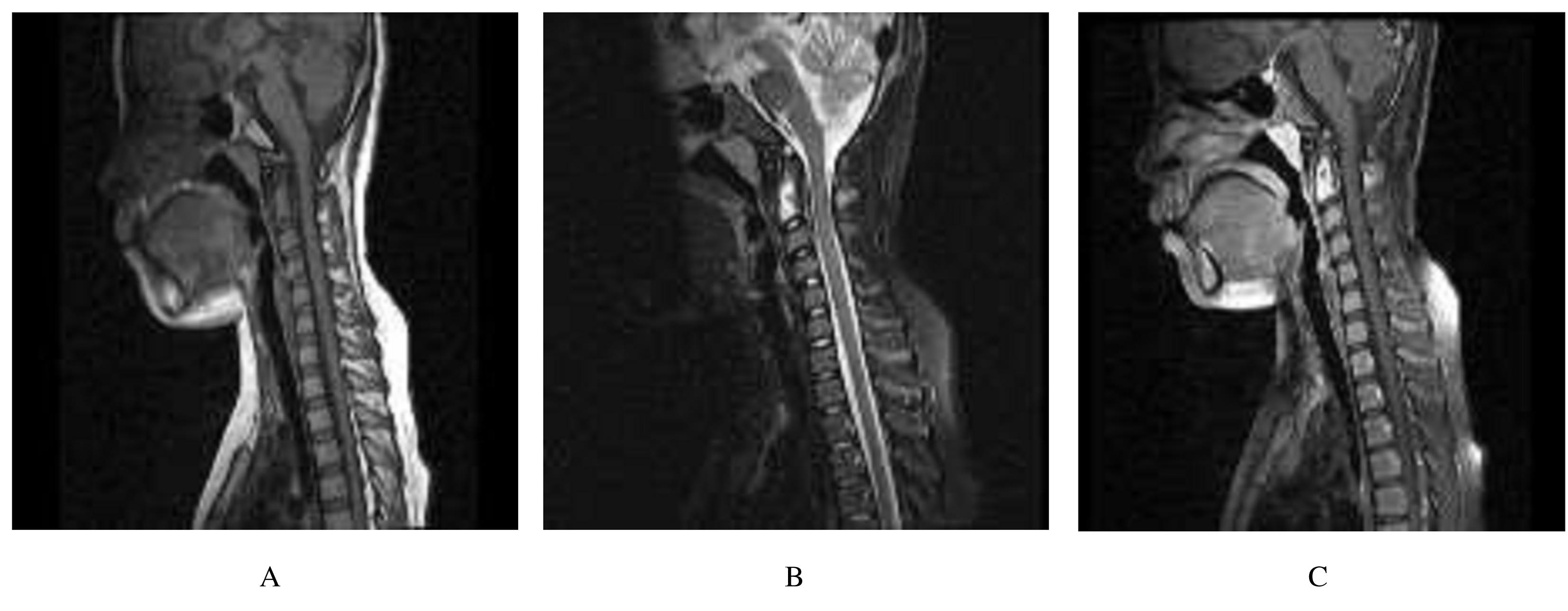

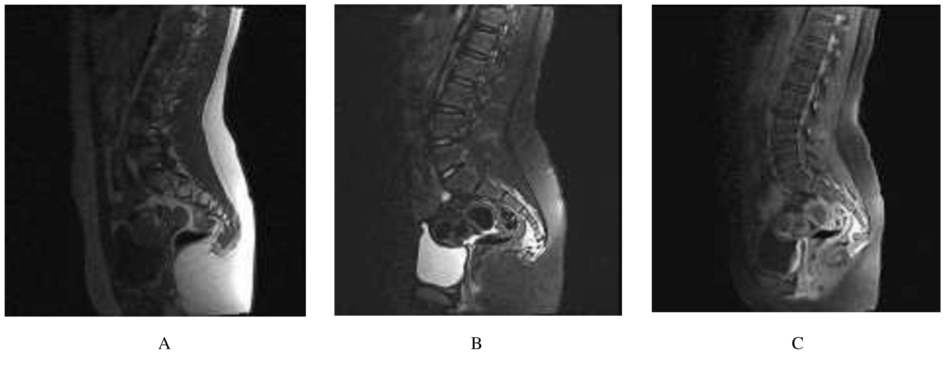
Fig. 3
Images of plain and enhanced MRI of sacrococcyx in a patient with small cell osteosarcoma of coccyx"
| 1 | PALMINI G, ZONEFRATI R, ROMAGNOLI C,et al. Establishment and characterization of a human small cell osteosarcoma cancer stem cell line: a new possible in vitro model for discovering small cell osteosarcoma biology[J]. Stem Cells Int, 2016, 2016: 3042198. |
| 2 | ZHANG M, ZHANG W, LI Q, et al. Small-cell extraskeletal osteosarcoma: case report and literature review[J]. Int J Clin Exp Pathol, 2014, 7(2): 797-800. |
| 3 | IARŬMOV N, VASILEV N, PETKOV K. Osteosarcoma of the coccyx[J]. Khirurgiia (Sofiia), 1997, 50(4): 55-56. |
| 4 | XIE L, LIAO Y, SHEN L, et al. Identification of the miRNA-mRNA regulatory network of small cell osteosarcoma based on RNA-seq[J]. Oncotarget, 2017, 8(26): 42525-42536. |
| 5 | MÜLLER D A, SILVAN U. On the biomechanical properties of osteosarcoma cells and their environment[J]. Int J Dev Biol, 2019, 63(1/2): 1-8. |
| 6 | KADAMKULAM S A, BHASKARLA A V, ELRIFAI M, et al. Endobronchial metastasis as an uncommon pattern of metastatic dissemination from small cell osteosarcoma [J]. BMJ Case Rep, 2019, 12(7):e229779. |
| 7 | LIU F, ZHANG Q, ZHU D, et al. Performance of positron emission tomography and positron emission tomography/computed tomography using fluorine-18-fluorodeoxyglucose for the diagnosis, staging, and recurrence assessment of bone sarcoma: a systematic review and meta-analysis[J]. Medicine (Baltimore), 2015, 94(36): e1462. |
| 8 | BISHOP J A, SHUM C H, SHETH S, et al. Small cell osteosarcoma: cytopathologic characteristics and differential diagnosis[J]. Am J Clin Pathol, 2010, 133(5): 756-761. |
| 9 | TSIAMBAS E, FOTIADES P P, SIOKA C, et al. Novel molecular and metabolic aspects in osteosarcoma [J]. J BUON, 2017, 22(6):1595-1598. |
| 10 | KABNURKAR R, AGRAWAL A, REKHI B, et al. Unusual sites of metastatic recurrence of osteosarcoma detected on fluorine-18 fluorodeoxyglucose positron emission tomography/computed tomography[J]. Indian J Nucl Med, 2015, 30(2): 171-173. |
| 11 | 杜晓光,谢新立,王 旭,等. 18F-FDG及18F-FLT PET/CT对多发骨结核的诊断价值[J].郑州大学学报(医学版),2020,55(4): 567-572. |
| 12 | PATIL K O. Pleuropulmonary blastoma in pediatric age group: A very rare case report and review of literature [J]. Open J Pediatr, 2020, 10(1):109-115. |
| 13 | BENBRAHIM Z, ARIFI S, DAOUDI K,et al. Askin’s tumor: a case report and literature review[J]. World J Surg Oncol, 2013, 11: 10. |
| 14 | XIA T T, GUAN Y B, CHEN Y X, et al. Askin tumor[J]. Medicine, 2014, 93(6): e42. |
| 15 | WU X, ZHOU C, JIN L, et al. Primary pulmonary lymphoma in children[J]. Orphanet J Rare Dis, 2019, 14(1): 35. |
| 16 |
PENNINGTON Z, EHRESMAN J, PITTMAN P D, et al. Chondrosarcoma of the spine: a narrative review[J].Spine J,2021.DOI:10.1016/j.spinee. 2021.04.021 .
doi: 10.1016/j.spinee. 2021.04.021 |
| 17 | HIROZANE T, MASUDA M, SUGANO T, et al. Direct conversion of osteosarcoma to adipocytes by targeting TNIK [J]. JCI Insight, 2021, 6(3): e137245. |
| 18 | ZHONG J, HU Y, SI L, et al. Clarifying prognostic factors of small cell osteosarcoma: a pooled analysis of 20 cases and the literature[J]. J Bone Oncol, 2020, 24: 100305. |
| [1] | Chengyuan HE,Hongyu YANG,Yujing TAN,Hang SU,Hongshu LI,Chun LI. Expression of IL-17A in non-small cell lung cancer tissue and its regulation on VEGF expression via NF-κB signaling pathway [J]. Journal of Jilin University(Medicine Edition), 2022, 48(4): 1003-1009. |
| [2] | Qingxu LANG,Xueshuang NIU,Kaiwen YANG,Ren ZHANG,Siteng WANG, ZUMIRETIGULI·Wumaier,Zhenqi WANG. Effects of sodium butyrate combined with ionizing radiation on apoptosis of lung cancer A549 cells and its mechanism [J]. Journal of Jilin University(Medicine Edition), 2022, 48(4): 915-921. |
| [3] | Junjie HOU,Xuguang MI,Xiaonan LI,Xiaonan LI,Ying YANG,Xianzhuo JIANG,Ying ZHOU,Zhiqiang NI,Ningyi JIN,Yanqiu FANG. Bronchopleural fistula complicated in treatment process of non-small cell lung cancer by bevacizumab combined with paclitaxel: A case report and literature review [J]. Journal of Jilin University(Medicine Edition), 2022, 48(2): 513-517. |
| [4] | Weihang LI,Ziyi DING,Dong WANG,Yikai PAN,Yuhui LIU,Shilei ZHANG,Jing LI,Ming YAN. Bioinformatics analysis based on screening of core driving genes in osteosarcoma and construction of gene model for prediction of survival time of patients [J]. Journal of Jilin University(Medicine Edition), 2021, 47(6): 1570-1580. |
| [5] | Dongkui ZHOU, Mingqian LU, Yaxin LIN, Xuesong FENG, Yan GAO, Hao SONG. Small cell carcinoma of gallbladder with liver and retroperitoneal lymph node metastasis: A case report and literature review [J]. Journal of Jilin University(Medicine Edition), 2021, 47(3): 747-752. |
| [6] | Dong CHEN, Zhiyao LI, Haitao CHEN, Zhirui CHUAN, Yingxian ZHANG, Xin JIN, Shicong TANG, Xiaomao LUO. Diagnostic values of three-dimensional transrectal ultrasound and MRI in diagnosis of T substages and circumferential resection margin of patients with middle and lower rectal cancer [J]. Journal of Jilin University(Medicine Edition), 2021, 47(3): 753-760. |
| [7] | Ye YUAN,Jinbao ZHUANG,Xu SHI,Changyuan LI. Influence of PGE2 on PD-1 expression in infiltrating T lymphocytes in non-small cell lung cancer tissue and its mechanism [J]. Journal of Jilin University(Medicine Edition), 2021, 47(2): 249-256. |
| [8] | Dong CHEN,Zhiyao LI,Haitao CHEN,Zhirui CHUAN,Yingxian ZHANG,Xin JIN,Shicong TANG,Xiaomao LUO. Diagnostic value of three-dimensional transrectal ultrasound in preoperative N-staging of middle and lower rectal cancer [J]. Journal of Jilin University(Medicine Edition), 2021, 47(2): 497-504. |
| [9] | Guanhu LI,Qingxu LANG,Chunyan LIU,Qin LIU,Mengrou GENG,Xiaoqian LI,Zhenqi WANG. Inhibitory effect of sodium butyrate combined with X- irradiation on proliferation of lung cancer A549 cells and its influence in cell cycle [J]. Journal of Jilin University(Medicine Edition), 2021, 47(1): 152-157. |
| [10] | Dong CHEN,Haitao CHEN,Zhiyao LI,Fengming RANG,Xi ZHANG,Zhirui CHUAN,Shicong TANG,Xiaomao LUO. Effects of three-dimensional transrectal ultrasound and MRI in evaluation of extramural vascular invasion degrees of middle and lower rectal cancer [J]. Journal of Jilin University(Medicine Edition), 2021, 47(1): 203-209. |
| [11] | Lingxue SHI,Shuo LIU,Xuewei ZHENG,Xiaoliang CHENG,Zhaohui CHEN,Baolin MA,Hongguo ZHANG,Jun DING. Application of magnetic resonance diffusion weighted imaging ADC and rADC in differential diagnosis of benign and malignant parotid gland tumors [J]. Journal of Jilin University(Medicine Edition), 2020, 46(6): 1309-1314. |
| [12] | CHEN Yan, PAN Dianzhu, LIU Zhong, MA Yanmei, ZHANG Lan. Inhibitory effect of sileneing Frat2 on NCI-H1688 cells and its Wnt/β catenin signaling pathway mechanism [J]. Journal of Jilin University(Medicine Edition), 2020, 46(04): 751-758. |
| [13] | ZHOU Dongkui, LU Mingqian, FENG Xuesong, LIU Yufei, SONG Hao, XU Liang. Follicular dendritic cell sarcoma of small intestine and ileocecal region: A case report and literature review [J]. Journal of Jilin University(Medicine Edition), 2020, 46(04): 858-862. |
| [14] | LIU Hui, MA Yunfei, LIU Bailong, LIU Min. Anlotinib combined with thoracic radiotherapy in treatment of recurrent and refractory small cell lung cancer: A case report and literature review [J]. Journal of Jilin University(Medicine Edition), 2020, 46(02): 394-398. |
| [15] | HE Yingying, XUE Jinhui, ZHAO Na. Effects of rhein on proliferation, migration and invasion abilities of non-small cell lung cancer A549 cells and their mechanisms [J]. Journal of Jilin University(Medicine Edition), 2020, 46(02): 302-308. |