Journal of Jilin University(Medicine Edition) ›› 2022, Vol. 48 ›› Issue (6): 1382-1388.doi: 10.13481/j.1671-587X.20220602
• Research in basic medicine • Previous Articles Next Articles
Effect of mediodorsal thalamic nucleus lesions on electrical activity in medial prefrontal cortex of rats with Parkinson’s disease
Lingling FAN1,2,Shuping DING3,Guomin SHEN1,2,Zhihong HU1,Aihong REN1,Bo DENG1
- 1.Department of Physiology,School of Basic Medical Sciences,Henan University of Science and Technology,Luoyang 471000,China
2.School of Basic Medical Sciences,Henan University of Science and Technology,Henan International Joint Laboratory of Thrombosis and Hemostasis,Luoyang 471000,China
3.Department of Basic Courses,Luoyang Polytechnic,Luoyang 471023,China
-
Received:2022-01-30Online:2022-11-28Published:2022-12-07 -
Contact:Guomin SHEN
CLC Number:
- R742.5
Cite this article
Lingling FAN,Shuping DING,Guomin SHEN,Zhihong HU,Aihong REN,Bo DENG. Effect of mediodorsal thalamic nucleus lesions on electrical activity in medial prefrontal cortex of rats with Parkinson’s disease[J].Journal of Jilin University(Medicine Edition), 2022, 48(6): 1382-1388.
share this article
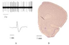
Fig. 1
Discharge characteristics of pyramidal neuron and recording sites of neurons in mPFC of rats"


Fig. 2
Diagrams of discharge patterns and ISI of pyramidal neurons in mPFC"
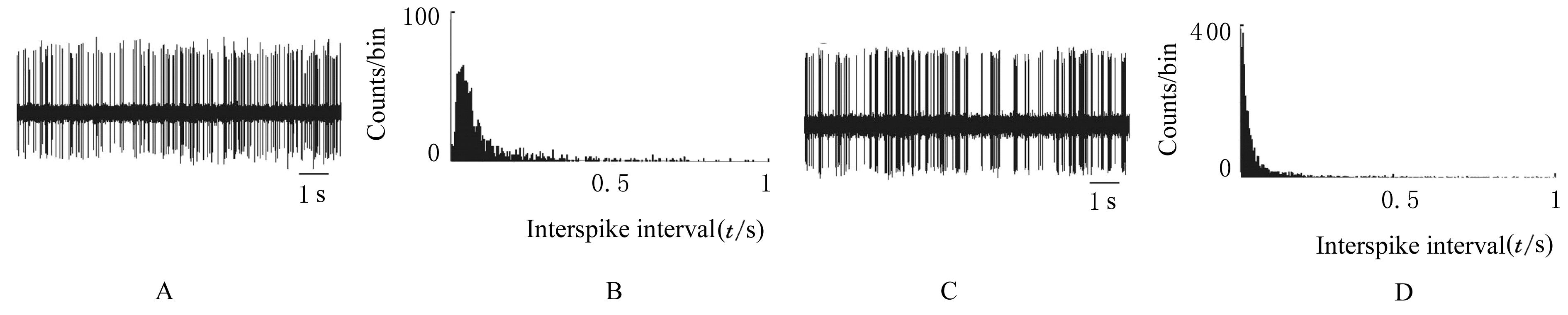

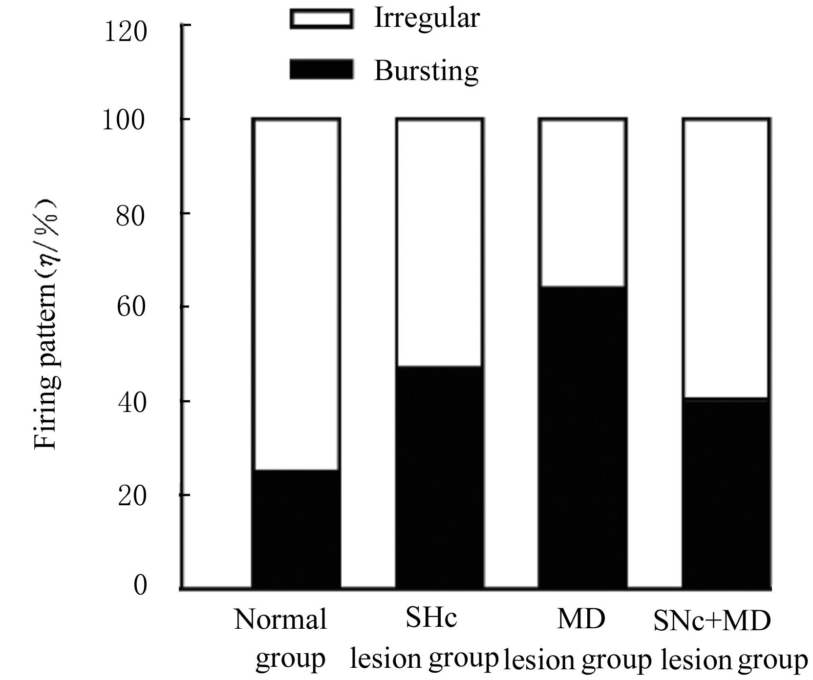
Fig. 3
Distributions of firing patterns of neurons in various groups"
Tab. 1
Firing frequencies and coefficients of variation of ISI of pyramidal neurons in mPFC of rats in various groups"
| Group | Number of neurons | Firing frequency (Hz) | Coefficient of variation of ISI |
|---|---|---|---|
| Normal | 59 | 1.26±0.12 | 1.20±0.07 |
| SNc lesion | 47 | 2.12±0.25* | 1.59±0.07** |
| MD lesion | 56 | 2.36±0.27** | 1.74±0.11** |
| SNc+MD lesion | 57 | 1.58±0.18 | 1.59±0.06** |
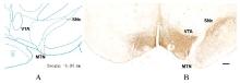
Fig. 4
Location map of SNc and VTA and morphology of tyrosine hydroxylase immunopositive neurons in SNc and VTA"


Fig. 5
Morphology of brain tissue of rats with MD lesion(Nissl staining,Bar=200 μm)"

| 1 | NIELSEN N S, SKOVBOLLING S L. Non-motor symptoms in Parkinson’s disease[J]. Ugeskr Laeger, 2021, 183(27): V01210089. |
| 2 | MARTINEZ B, PEPLOW P V. Neuroprotection by immunomodulatory agents in animal models of Parkinson’s disease[J].Neural Regen Res,2018,13(9): 1493-1506. |
| 3 | SQUIRE L R, MOORE R Y. Dorsal thalamic lesion in a noted case of human memory dysfunction[J]. Ann Neurol, 1979, 6(6): 503-506. |
| 4 | VICTOR M, ADAMS R D, COLLINS G H. The Wernicke-Korsakoff syndrome. A clinical and pathological study of 245 patients, 82 with post-mortem examinations[J]. Contemp Neurol Ser, 1971, 7: 1-206. |
| 5 | YOUNG K A, HOLCOMB L A, YAZDANI U, et al. Elevated neuron number in the limbic thalamus in major depression[J]. Am J Psychiatry, 2004, 161(7): 1270-1277. |
| 6 | LI W, LIU J, SKIDMORE F, et al. White matter microstructure changes in the thalamus in Parkinson disease with depression: a diffusion tensor MR imaging study[J]. AJNR Am J Neuroradiol, 2010, 31(10): 1861-1866. |
| 7 | CARDOSO E F, MAIA F M, FREGNI F, et al. Depression in Parkinson’s disease: convergence from voxel-based morphometry and functional magnetic resonance imaging in the limbic thalamus[J]. NeuroImage, 2009, 47(2): 467-472. |
| 8 | LI W J, XI Y, ZHANG Q J, et al. Neuronal firing activity changes in mediodorsal thalamus nucleus in rats with Parkinson’s disease [J]. J Xi’an Jiaotong Univ Med Sci, 2013,34(5): 614-618 |
| 9 | 范玲玲, 邓 博, 闫君宝, 等. 5-羟色胺-7受体激动剂对帕金森病模型大鼠内侧前额叶皮层锥体神经元兴奋性的影响[J]. 南方医科大学学报, 2016, 36(6): 756-762. |
| 10 | RIDDERINKHOF K R, ULLSPERGER M, CRONE E A, et al. The role of the medial frontal cortex in cognitive control[J].Science,2004,306(5695): 443-447. |
| 11 | GIL SALADIÉ D, DELGADO GONZÁLEZ M, OLIVERAS LEY C, et al. Depression in Parkinson’s disease and its relation to the cognitive and motor manifestations[J]. Neurologia, 1992, 7(7): 176-180. |
| 12 | CROSS L, BROWN M W, AGGLETON J P, et al. The medial dorsal thalamic nucleus and the medial prefrontal cortex of the rat function together to support associative recognition and recency but not item recognition[J]. Learn Mem, 2012, 20(1): 41-50. |
| 13 | CONSTANTINIDIS C, GOLDMAN-RAKIC P S. Correlated discharges among putative pyramidal neurons and interneurons in the primate prefrontal cortex[J]. J Neurophysiol, 2002, 88(6): 3487-3497. |
| 14 | WALSH J J, HAN M H. The heterogeneity of ventral tegmental area neurons: projection functions in a mood-related context[J]. Neuroscience, 2014, 282: 101-108. |
| 15 | LEWIS B L, O’DONNELL P. Ventral tegmental area afferents to the prefrontal cortex maintain membrane potential ‘up’ states in pyramidal neurons via D(1) dopamine receptors[J]. Cereb Cortex, 2000, 10(12): 1168-1175. |
| 16 | WANG J, O’DONNELL P. D(1) dopamine receptors potentiate nmda-mediated excitability increase in layer Ⅴ prefrontal cortical pyramidal neurons[J]. Cereb Cortex, 2001, 11(5): 452-462. |
| 17 | TSENG K Y, O'DONNELL P. Dopamine-glutamate interactions controlling prefrontal cortical pyramidal cell excitability involve multiple signaling mechanisms[J]. J Neurosci, 2004, 24(22): 5131-5139. |
| 18 | WANG Q, WANG P H, MCLACHLAN C, et al. Simvastatin reverses the downregulation of dopamine D1 and D2 receptor expression in the prefrontal cortex of 6-hydroxydopamine-induced Parkinsonian rats[J]. Brain Res, 2005, 1045(1/2): 229-233. |
| 19 | FILION M. Effects of interruption of the nigrostriatal pathway and of dopaminergic agents on the spontaneous activity of globus pallidus neurons in the awake monkey[J]. Brain Res, 1979, 178(2/3): 425-441. |
| 20 | CRUIKSHANK S J, URABE H, NURMIKKO A V, et al. Pathway-specific feedforward circuits between thalamus and neocortex revealed by selective optical stimulation of axons[J]. Neuron, 2010,65(2): 230-245. |
| 21 | FERRON A, THIERRY A M, LE DOUARIN C,et al. Inhibitory influence of the mesocortical dopaminergic system on spontaneous activity or excitatory response induced from the thalamic mediodorsal nucleus in the rat medial prefrontal cortex[J]. Brain Res, 1984, 302(2): 257-265. |
| 22 | GIGG J, TAN A M, FINCH D M. Glutamatergic excitatory responses of anterior cingulate neurons to stimulation of the mediodorsal thalamus and their regulation by GABA: an in vivo iontophoretic study[J]. Cereb Cortex, 1992, 2(6): 477-484. |
| 23 | KURODA M, YOKOFUJITA J, MURAKAMI K. An ultrastructural study of the neural circuit between the prefrontal cortex and the mediodorsal nucleus of the thalamus[J]. Prog Neurobiol, 1998, 54(4): 417-458. |
| 24 | DELEVICH K, TUCCIARONE J, HUANG Z J,et al. The mediodorsal thalamus drives feedforward inhibition in the anterior cingulate cortex via parvalbumin interneurons[J]. J Neurosci, 2015, 35(14): 5743-5753. |
| 25 | TIMBIE C, BARBAS H. Pathways for emotions: specializations in the amygdalar, mediodorsal thalamic, and posterior orbitofrontal network[J]. J Neurosci, 2015, 35(34): 11976-11987. |
| 26 | MANTZ J, GODBOUT R, TASSIN J P, et al. Inhibition of spontaneous and evoked unit activity in the rat medial prefrontal cortex by mesencephalic raphe nuclei[J]. Brain Res, 1990, 524(1): 22-30. |
| 27 | HASSLER R, HAUG P, NITSCH C, et al. Effect of motor and premotor cortex ablation on concentrations of amino acids, monoamines, and acetylcholine and on the ultrastructure in rat striatum. A confirmation of glutamate as the specific cortico-striatal transmitter[J]. J Neurochem, 1982, 38(4): 1087-1098. |
| [1] | Qi PAN,Zizhen WANG,Fuyue YE,Wei XING,Jiahan LIN,Jianyue LU,Kun YANG. Effect of proteasome inhibitor lactacystin on oxidative damage of dopaminergic neurons in substantia nigra of rats [J]. Journal of Jilin University(Medicine Edition), 2022, 48(3): 728-733. |
| [2] | Xiaochen HUANG,Hao LI,Baohua WANG,Kai LI. Protective effect of lidocaine on PC12 cells in Parkinson’s disease model and its mechanism [J]. Journal of Jilin University(Medicine Edition), 2022, 48(3): 638-647. |
| [3] | Lingna HAN,Chunlei WANG,Yongli CHANG,Li YUAN,Xiaojing LIU. Effect of electrical lesions of lateral habenular nucleus on spatial learning and memory functions in rats with Parkinson’s disease and its mechanism [J]. Journal of Jilin University(Medicine Edition), 2021, 47(5): 1108-1115. |
| [4] | Xiaofeng LUO,Yao LI,Jiang HU,Chenhao ZHANG. Regulatory effect of gardenoside on sleep disorder in rats with Parkinson’s disease and its mechanism [J]. Journal of Jilin University(Medicine Edition), 2020, 46(6): 1177-1181. |
| [5] | WANG Chunlei, CHANG Yongli, HAN Lingna. Improvement effects of repeated transcranial direct current stimulation on depression behaviors in rat models of Parkinson's disease and their mechanisms [J]. Journal of Jilin University Medicine Edition, 2018, 44(04): 693-697. |
| [6] | DU Na, MAO Xijing, YU Tingmin, YAO Gang. Clinical manifestations and early combined diagnosis of subacute combined degeneration of spinal cord:A report of 18 cases [J]. Journal of Jilin University Medicine Edition, 2018, 44(04): 806-809. |
| [7] | WANG Shuang, GAO Jie, GUO Yufang, WANG Xiang. Regulation effect of 5-HT7 receptor on electrical activity of 5-HT neurons in nucleus raphes dorsalis of rats with Parkinson's disease [J]. Journal of Jilin University Medicine Edition, 2015, 41(03): 573-577. |
| [8] | LIU Yan-tong,GAO Jie,WANG Shuang. Influence of intraventricular injection of 5,7-drhydroxytryptamine in 5-HT1A receptor sensitivity of pyramidal neurons in medial prefrontal cortex [J]. Journal of Jilin University Medicine Edition, 2014, 40(05): 958-961. |
| [9] | JIA Xiao-jing, ZHANG Zhi-liang, QU Ya-qin, ZHAI Cheng-wei, YANG Tai-quan, WOLF-DIETER Rausch, GONG Shou-liang. Establishment of cell models of dopaminergic neurons for Parkinson’s disease [J]. Journal of Jilin University(Medicine Edition), 2008, 34(5): 778-781. |
| [10] | ZHANG Hai-na,HU Guo-hua,CHEN Qiu-hui,SUN Dong,LIU Cai-xia,SUN Ya-juan. Establishment of cell model of Parkinson’〖KG-3〗s disease and toxic effect of rotenone on dopaminergic neurons [J]. J4, 2007, 33(5): 811-814. |
| [11] | Zhang Yu,XU Hui,ZHANG Ying,HAN Wei,HU Yi-hong,HU Lin-sen. Expression of ERp29 in model of Parkinson’s disease of PC12 cells induced by proteasome inhibitor [J]. J4, 2007, 33(2): 249-252. |
| [12] | ZHANG Zhi-xin, LU Lai-jin,LIU Zhi-gang,CHEN Lei. Long-term curative effects of one end-to-side and double end-to-sideneurorrhaphy in treatment of peripheral nerve defects [J]. J4, 2006, 32(4): 701-704. |
| [13] | JIANG Xin-mei, LIU Ke-hui,CUI Li,ZHOU Yan, XING Ying-qi, HU Yi-hong. Pathological characterization and neuroelectrophysiological changeson delayed neuropathy induced by methamidophos in hens [J]. J4, 2006, 32(1): 4-4. |
| [14] | XU Hai-yan, YU Lei, LI Hua-li, ZHAO Hua,LIULIU Yong-feng,ZHANG Cheng-wei. Electrophysiology of functional relationship between dorsal raphe and habenular nucleus [J]. J4, 2006, 32(1): 35-4. |
| [15] | CHEN Xia,LI Ying-ji,GE Jing-yan,ZHANG Wen-jie,ZHONG Guo-gan. Effects of oxymatrine on action potential in normal ratsand arrhythmic rats induced by aconitine [J]. J4, 2004, 30(5): 704-706. |
|