Journal of Jilin University(Medicine Edition) ›› 2023, Vol. 49 ›› Issue (3): 682-690.doi: 10.13481/j.1671-587X.20230317
• Research in basic medicine • Previous Articles Next Articles
Effects of HIF-1α/ROS on apoptosis and invasion of lung cancer A549 cells under hypoxia and its mechanism
Bo HUANG1( ),Jie DING2,Hongrong GUO1,Hongjuan WANG1,Jianqun XU1,Quan ZHENG1
- 1.Department of Respiratory and Critical Care Medicine,Wuhan Third Hospital,Hubei Province,Affiliated Tongren Hospital,Wuhan University,Wuhan 430070,China
2.Hemodialysis Room,Wuhan Third Hospital,Hubei Province,Affiliated Tongren Hospital,Wuhan University,Wuhan 430070,China
-
Received:2022-07-15Online:2023-05-28Published:2023-06-20 -
Contact:Bo HUANG E-mail:doctorhuang82@126.com
CLC Number:
- R734.2
Cite this article
Bo HUANG,Jie DING,Hongrong GUO,Hongjuan WANG,Jianqun XU,Quan ZHENG. Effects of HIF-1α/ROS on apoptosis and invasion of lung cancer A549 cells under hypoxia and its mechanism[J].Journal of Jilin University(Medicine Edition), 2023, 49(3): 682-690.
share this article
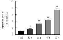
Fig. 1
Expression levels of HIF-1α mRNA in A549 cells under hypoxia condition"

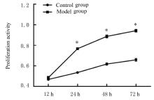
Fig. 2
Proliferation activities of A549 cells under hypoxia condition"
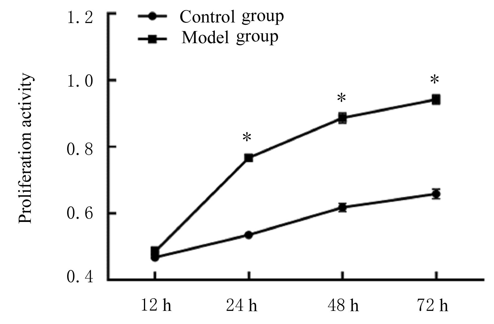
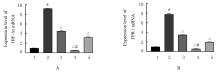
Fig. 3
Expression levels of HIF-1α(A) and FPR1(B) mRNA in cells in various groups"
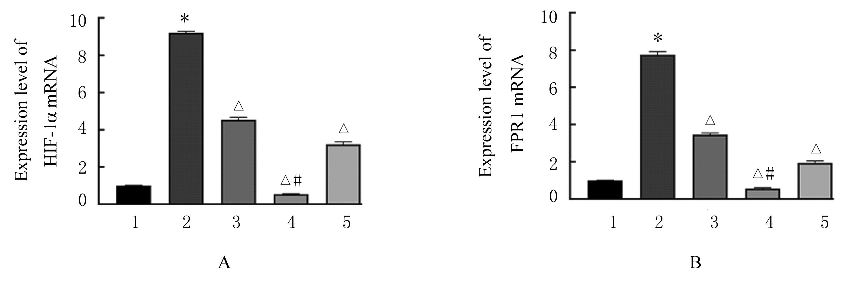
Fig. 4
Electrophoregram(A) and histograms(B,C) of expressions of HIF-1α and FPR1 proteins in cells in various groups"
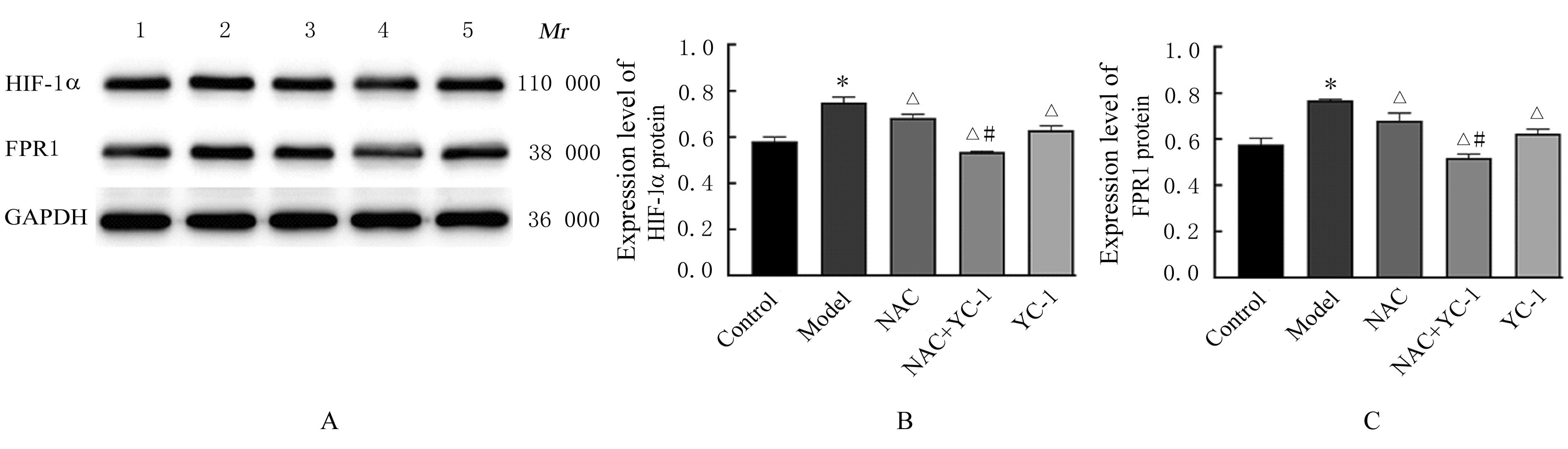
Fig. 5
ROS levels in cells in various groups detected by flow cytometry"
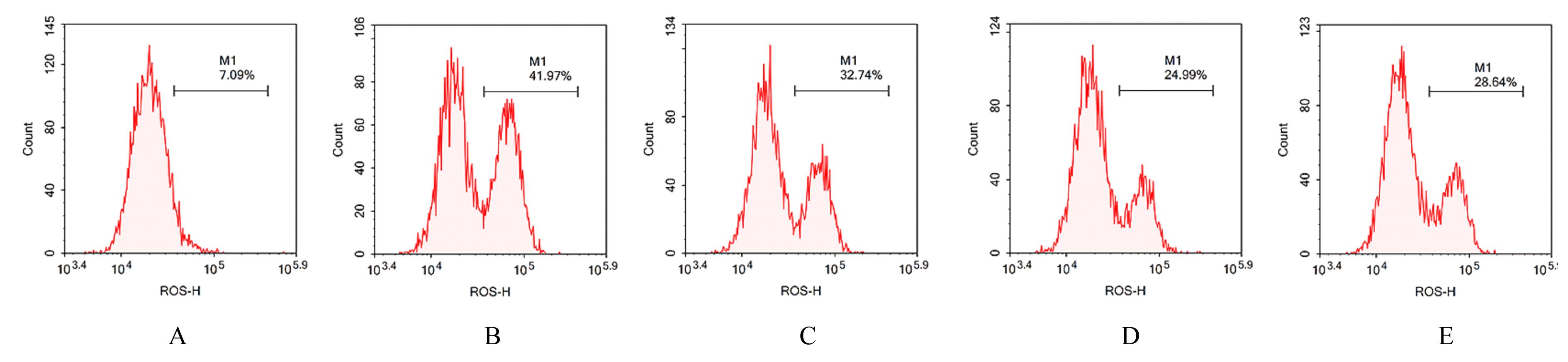

Fig. 6
ROS levels in cells in various groups"
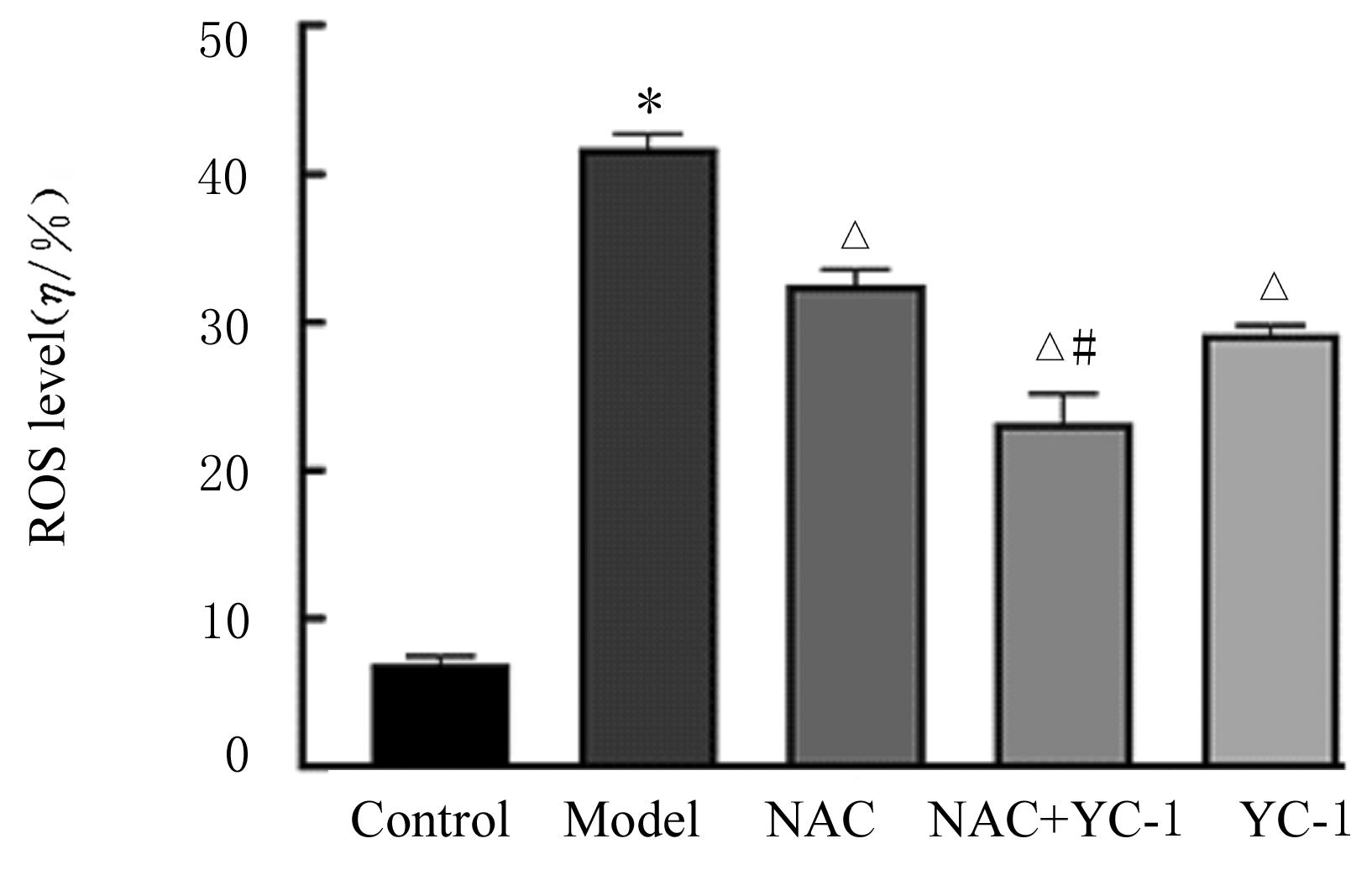
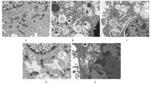
Fig. 7
Ultrastructures of autophagosomes in cells in various groups observed by transmission electron microscope (×12 000)"

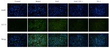
Fig. 8
Expression intensities of LC3-Ⅱ in cells in various groups detected by immunofluorescence method(×200)"
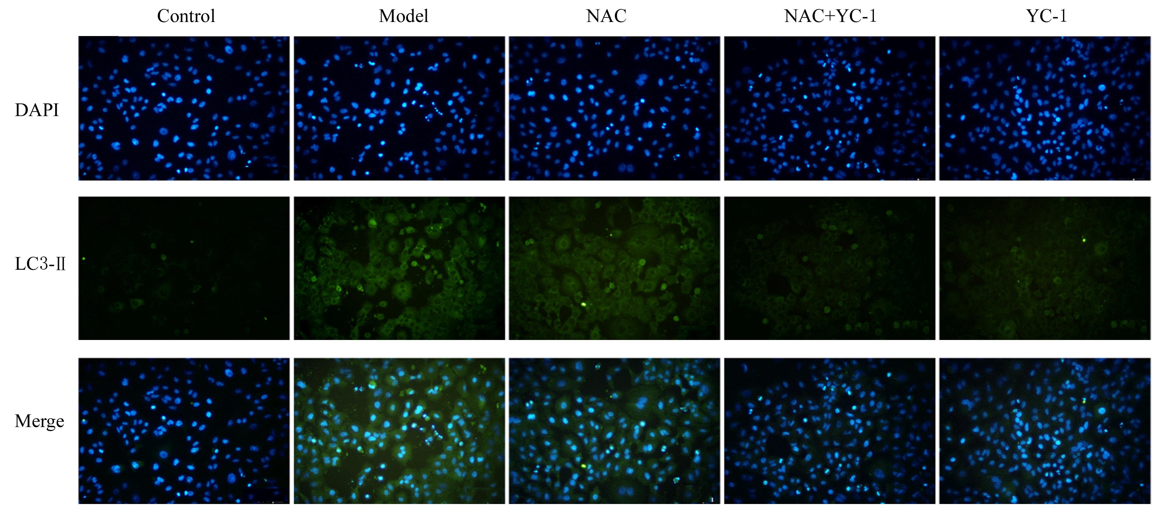
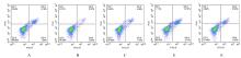
Fig. 9
Apoptotic rates of cells in various groups detected by flow cytometry"
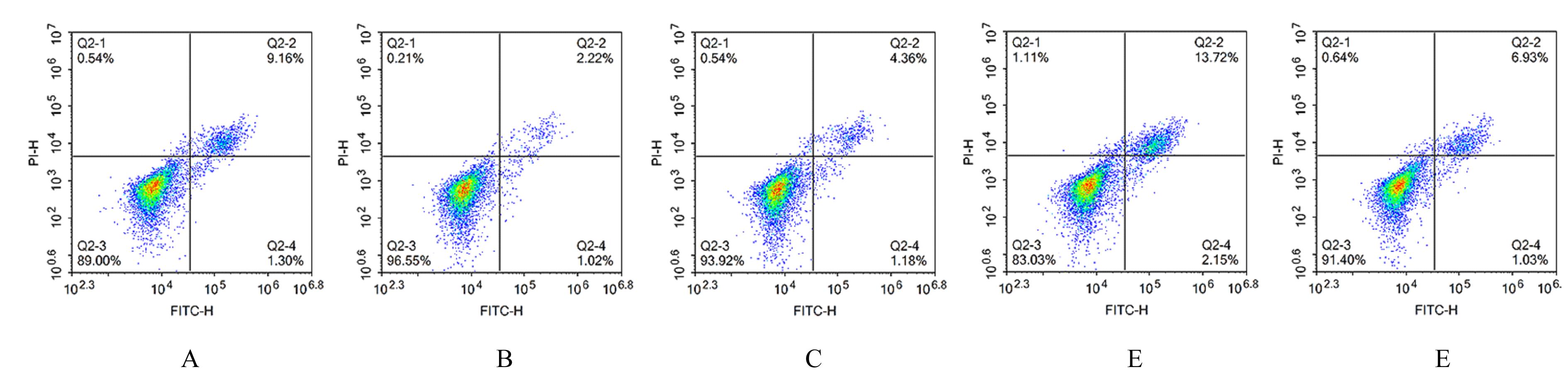

Fig. 10
Apoptotic rates of cells in various groups"
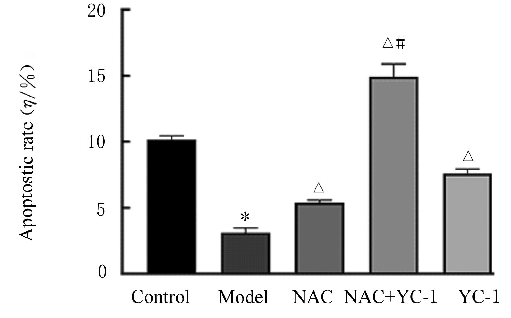
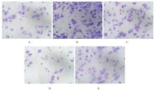
Fig. 11
Invasion of cells in various groups detected by Transwell chamber assay (Crystal violet,×200)"
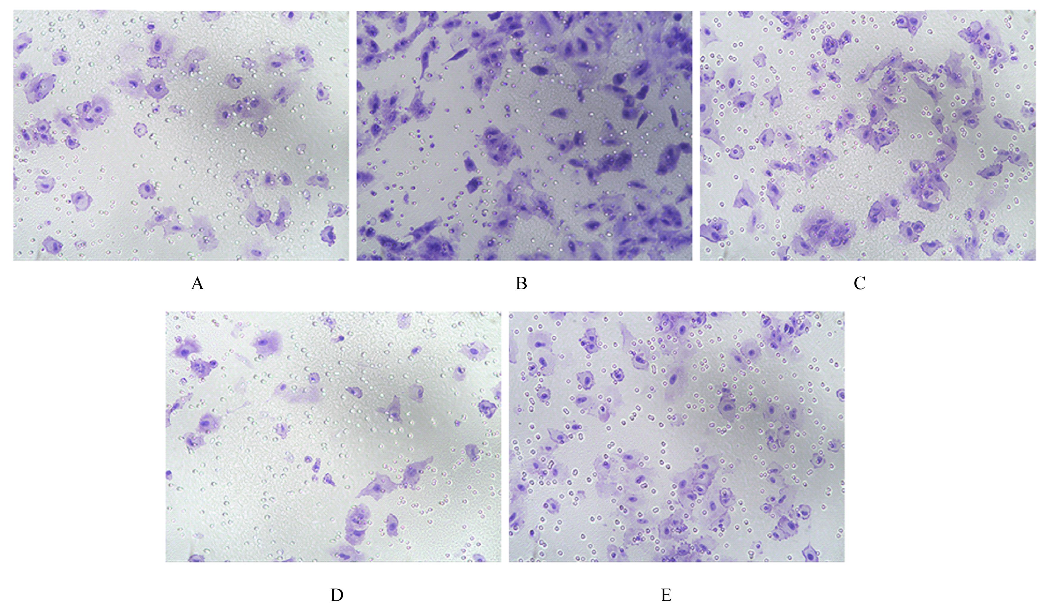

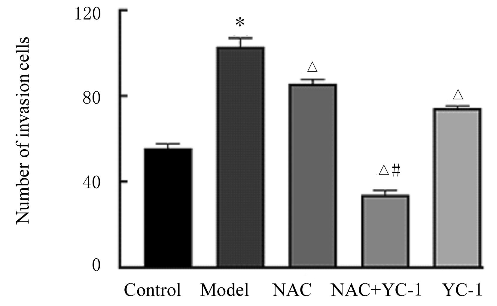
Fig. 12
Numbers of invasion cells in various groups"
| 1 | SCHABATH M B, COTE M L. Cancer progress and priorities: lung cancer[J]. Cancer Epidemiol Biomarkers Prev, 2019, 28(10): 1563-1579. |
| 2 | ALEXANDER M, KIM S Y, CHENG H Y. Update 2020: management of non-small cell lung cancer[J]. Lung, 2020, 198(6): 897-907. |
| 3 | WU F Y, WANG L, ZHOU C C. Lung cancer in China: current and prospect[J]. Curr Opin Oncol, 2021, 33(1): 40-46. |
| 4 | DUMA N, SANTANA-DAVILA R, MOLINA J R. Non-small cell lung cancer: epidemiology, screening, diagnosis, and treatment[J]. Mayo Clin Proc, 2019, 94(8): 1623-1640. |
| 5 | HUA Q, MI B M, XU F,et al.Hypoxia-induced lncRNA-AC020978 promotes proliferation and glycolytic metabolism of non-small cell lung cancer by regulating PKM2/HIF-1α axis[J].Theranostics,2020,10(11):4762-4778. |
| 6 | AJDUKOVIĆ J. HIF-1: a big chapter in the cancer tale[J]. Exp Oncol, 2016, 38(1): 9-12. |
| 7 | ONORATI A V, DYCZYNSKI M, OJHA R, et al. Targeting autophagy in cancer[J].Cancer,2018,124(16): 3307-3318. |
| 8 | SHIDA M, KITAJIMA Y, NAKAMURA J, et al. Impaired mitophagy activates mtROS/HIF-1α interplay and increases cancer aggressiveness in gastric cancer cells under hypoxia[J]. Int J Oncol, 2016, 48(4):1379-1390. |
| 9 | JEONG J K, GURUNATHAN S, KANG M H, et al. Hypoxia-mediated autophagic flux inhibits silver nanoparticle-triggered apoptosis in human lung cancer cells[J]. Sci Rep, 2016, 6: 21688. |
| 10 | YU H L, CHEN B, REN Q. Baicalin relieves hypoxia-aroused H9c2 cell apoptosis by activating Nrf2/HO-1-mediated HIF1α/BNIP3 pathway[J]. Artif Cells Nanomed Biotechnol, 2019, 47(1): 3657-3663. |
| 11 | MAO X W, NANZHANG N, XIAO J, et al. Hypoxia-induced autophagy enhances cisplatin resistance in human bladder cancer cells by targeting hypoxia-inducible factor-1α[J]. J Immunol Res, 2021, 2021: 8887437. |
| 12 | TIRPE A A, GULEI D, CIORTEA S M, et al. Hypoxia: overview on hypoxia-mediated mechanisms with a focus on the role of HIF genes[J]. Int J Mol Sci, 2019, 20(24): 6140. |
| 13 | CUI W, WU F, MA L. Hypoxia associated biomarkers in lung cancer - an update[J]. Eur Rev Med Pharmacol Sci, 2017, 21(3 ): 43-46. |
| 14 | LI Y N, ZHANG Y S, LI X R, et al. Status of hypoxia-inducible factor-1α expression in non-small cell lung cancer[J]. Pharmazie, 2021, 76(9): 404-411. |
| 15 | MOLONEY J N, COTTER T G. ROS signalling in the biology of cancer[J].Semin Cell Dev Biol,2018,80:50-64. |
| 16 | 黄 波, 郭红荣, 丁 洁, 等. 肺腺癌中FPR1的表达及其对细胞迁移、成瘤能力的影响[J]. 临床与实验病理学杂志, 2018, 34(10): 1104-1109. |
| 17 | LI X H, HE S K, MA B Y. Autophagy and autophagy-related proteins in cancer[J].Mol Cancer,2020,19(1):12. |
| 18 | ARIOSA A R, LAHIRI V, LEI Y C,et al. A perspective on the role of autophagy in cancer[J]. Biochim Biophys Acta Mol Basis Dis, 2021, 1867(12): 166262. |
| 19 | TUREK K, JAROCKI M, KULBACKA J, et al. Dualistic role of autophagy in cancer progression[J]. Adv Clin Exp Med, 2021, 30(12): 1303-1314. |
| 20 | DENG M Z, ZHANG W Z, YUAN L L, et al. HIF-1a regulates hypoxia-induced autophagy via translocation of ANKRD37 in colon cancer[J]. Exp Cell Res, 2020, 395(1): 112175. |
| 21 | JOSHI S, KUMAR S, PONNUSAMY M P, et al. Hypoxia-induced oxidative stress promotes MUC4 degradation via autophagy to enhance pancreatic cancer cells survival[J]. Oncogene, 2016, 35(45): 5882-5892. |
| 22 | TANIDA I, UENO T, KOMINAMI E. LC3 and autophagy[J]. Methods Mol Biol, 2008, 445: 77-88. |
| 23 | FAN S J, YUE L Y, WAN W, et al. Inhibition of autophagy by a small molecule through covalent modification of the LC3 protein[J]. Angew Chem Int Ed Engl, 2021, 60(50): 26105-26114. |
| [1] | Wenli JIANG,Lin ZHANG,Junyao LI,Mingyu XU,Jie ZHANG,Chunling DONG. Targeted therapy of osimertinib combined with savolitinib in NSCLC with EGFR-TKI resistance complicated with MET amplification: A case report and literature review [J]. Journal of Jilin University(Medicine Edition), 2023, 49(3): 782-788. |
| [2] | Yunpeng LIU,Zihao LIU,Boming KANG,Zhiguang YANG. Regulatory effect of lncRNA SNHG17 on biological behavior of non-small cell lung cancer cells by targeting AEG1 through miR-384 [J]. Journal of Jilin University(Medicine Edition), 2023, 49(2): 298-307. |
| [3] | Zongnan YU,Ying CUI. Effect of miR-216b-5p on proliferation, migration and invasion of laryngeal cancer TU686 cells and its mechanism [J]. Journal of Jilin University(Medicine Edition), 2023, 49(1): 116-121. |
| [4] | Yun LIU,Linqi ZHU,Shihe SHAO. Effects of circular RNA hsa_circ_0009735 on epithelial mesenchymal transformation, cell cycle, and autophagy of gastric cancer cells [J]. Journal of Jilin University(Medicine Edition), 2022, 48(6): 1498-1509. |
| [5] | Xuechun DU,Baosheng LI,Shuwei QIAO,Yanzhen OU,Zhen LI,Weiyan MENG. Effect of Porphyromonas gingivalis-LPS on expression levels of ferroptosis-related factors in macrophages [J]. Journal of Jilin University(Medicine Edition), 2022, 48(5): 1148-1155. |
| [6] | Qian ZHANG,Jing LI. Inhibitory effect of TLR4 gene overexpression on autophagy of gastric cancer cells and its mechanism [J]. Journal of Jilin University(Medicine Edition), 2022, 48(5): 1238-1246. |
| [7] | Shuyuan YU,Ping WANG,Xinrui WANG,Lanzi GONGGA,Li YANG. Effects of high altitude hypoxia on auditory conduction and peripheral auditory nerve myelin regeneration in plateau migrated rats [J]. Journal of Jilin University(Medicine Edition), 2022, 48(4): 875-882. |
| [8] | Wentao WANG,Xuguang MI,Yang ZHOU,Wenxing PU,Jiaxu GAO,Meng JING,Fankai MENG. Effect of autophagy induced by exosomes derived from bone marrow mesenchymal stem cells on survival of SH-SY5Y cells inhibited by MPP+ and its mechanism [J]. Journal of Jilin University(Medicine Edition), 2022, 48(3): 606-614. |
| [9] | Haixin QU,Erwei YUAN,Weiping GUO,Yajing ZHANG,Wenxia MA,Dan WU. Improvement effect of luteolin on acute respiratory distress syndrome by inhibiting ROS/TXNIP/NLRP3 signaling pathway activation in mice [J]. Journal of Jilin University(Medicine Edition), 2022, 48(3): 676-683. |
| [10] | Yabo MA,Xiaotan YUAN,Xianguo XIE,Xinfeng LIU,Jinrui XU,Yi YANG. Expression levels of Wnt5a protein in ovary tissue of mice at different development stages and its effect on oocyte autophagy [J]. Journal of Jilin University(Medicine Edition), 2022, 48(2): 277-283. |
| [11] | Yinong LIU,Qiang ZHANG,Li XU. Improvement effect of atorvastatin on vascular endothelial dysfunction induced by Ox-LDL/β2GPⅠ/anti-β2GPⅠ complex and its mechanism [J]. Journal of Jilin University(Medicine Edition), 2022, 48(2): 317-323. |
| [12] | Ying DONG,Jianyu GUO,Siyi WANG,Dan GUO,Like WANG,Xu WEN,Lifeng LIU,Meng QU,Chunyan YU,Nannan LIU,Dan WANG,Changjie CHEN. Effect of endoplasmic reticulum stress PERK-eIF2α-ATF4 signaling pathway on delaying transplanted tumor growth in APP/PS1 mice [J]. Journal of Jilin University(Medicine Edition), 2022, 48(2): 324-330. |
| [13] | Yang ZHOU,Xuguang MI,Wenxing PU,Wentao WANG,Meng JING,Fankai MENG. Ameliorative effect of melatonin on oxidative stress of human neuroblastoma SHSY5Y cells induced by hydrogen peroxide and its mechanism [J]. Journal of Jilin University(Medicine Edition), 2022, 48(2): 340-347. |
| [14] | Junjie HOU,Xuguang MI,Xiaonan LI,Xiaonan LI,Ying YANG,Xianzhuo JIANG,Ying ZHOU,Zhiqiang NI,Ningyi JIN,Yanqiu FANG. Bronchopleural fistula complicated in treatment process of non-small cell lung cancer by bevacizumab combined with paclitaxel: A case report and literature review [J]. Journal of Jilin University(Medicine Edition), 2022, 48(2): 513-517. |
| [15] | Yanmin SUN,Junpan HU,Bingyu WANG,Jinying FU. Therapeutic effect of alpinetin in letrozole-induced polycystic ovary syndrome model rats and its mechanism [J]. Journal of Jilin University(Medicine Edition), 2022, 48(1): 129-135. |