| [1] |
Yanjue YE,Ziyi TANG,Yupei TAN,Shiying YANG,Yong LIU,Li YIN.
Effect of azathioprine on ferroptosis in spermatocytes of mice induced by RSL3
[J]. Journal of Jilin University(Medicine Edition), 2024, 50(5): 1217-1226.
|
| [2] |
Yongjing YANG,Tianyang KE,Shixin LIU,Xue WANG,Dequan XU,Tingting LIU,Ling ZHAO.
Synergistic sensitization of apatinib mesylate and radiotherapy on hepatocarcinoma cells invitro
[J]. Journal of Jilin University(Medicine Edition), 2024, 50(4): 1009-1015.
|
| [3] |
Yuanhang JIA,Yixia JIANG,Zhenhua HE,Lin CHEN,Fang ZHOU.
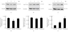
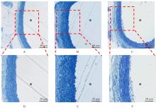
Effect of SENP-1/HIF-1α pathway on vascular endothelial injury in rats with chronic intermittent hypoxia
[J]. Journal of Jilin University(Medicine Edition), 2024, 50(4): 1026-1034.
|
| [4] |
Aimin WANG,Fenglin WANG,Yiming HUANG,Yaqi XU,Wenjing ZHANG,Xianzhu CONG,Weiqiang SU,Suzhen WANG,Mengyao GAO,Shuang LI,Yujia KONG,Fuyan SHI,Enxue TAO.
CatBoost algorithm and Bayesian network model analysis based on risk prediction of cardiovascular and cerebro vascular diseases
[J]. Journal of Jilin University(Medicine Edition), 2024, 50(4): 1044-1054.
|
| [5] |
Shan CAO,Yijia ZHANG,Yang BAI,Fang CHEN,Sha XIE,Qianqian HAN.
Network pharmacological analysis and in vitro experimental verification based on anti-atherosclerosis mechanism of Xiaoban Tongmai Formula
[J]. Journal of Jilin University(Medicine Edition), 2024, 50(4): 925-938.
|
| [6] |
Yingqun NI,Mao YANG,Di YANG,Chenglin GUO,Wenjun ZHU,Yaqin YU,Qin LU,Jinzhi LUO,Chunqin WU,Zhaohui FANG.
Screening of key differentially expressed genes involved in osteogenic differentiation of lower limb vascular smooth muscle cells and validation
[J]. Journal of Jilin University(Medicine Edition), 2024, 50(3): 620-627.
|
| [7] |
Yuwei KANG,Wei YANG,Shijie MA,Wei ZHOU,Fei DENG.
Levels of sICAM-1 and sVCAM-1 and activity of SOD in serum and their relationships with coronary artery calcification in patients with maintenance hemodialysis
[J]. Journal of Jilin University(Medicine Edition), 2024, 50(3): 812-818.
|
| [8] |
Li ZHANG,Binfeng XIA,Huihui HUANG,Ru WANG,Min KONG,Xia YIN.
Research progress in pathophysiological mechanism and clinical diagnosis and treatment of hypertension associated with vascular endothelial growth factor and its receptor inhibitors
[J]. Journal of Jilin University(Medicine Edition), 2024, 50(3): 854-863.
|
| [9] |
Linru WANG,Jing ZHANG,Dongchan ZHAO,Jinjun WANG,Wenxian HU.
Effect of silencing FOXO1 gene on autophagy and apoptosis of human aortic vascular smooth muscle cells
[J]. Journal of Jilin University(Medicine Edition), 2024, 50(2): 431-441.
|
| [10] |
Lan ZHANG,Yongxin WU,Tao ZHANG,Dongwei WANG.
Improvement effect of ligustilide on rats with heart failure by regulating PKD1/HIF-1α/VEGF pathway
[J]. Journal of Jilin University(Medicine Edition), 2024, 50(1): 42-49.
|
| [11] |
Xiuling ZHOU,Deyu CONG,Ye ZHANG,Hongshi ZHANG.
Effect of head acupuncture on neurological function and HIF-1α and VEGFR2 expressions in brain tissue in rats with focal cerebral ischemia and its mechanism
[J]. Journal of Jilin University(Medicine Edition), 2023, 49(6): 1431-1436.
|
| [12] |
Ruipeng ZHANG,Jie LI.
Resistance and regeneration effects of lncRNA GPRC5D-AS1 on muscle atrophy of myocytes in mice induced by dexamethasone and its mechanism
[J]. Journal of Jilin University(Medicine Edition), 2023, 49(6): 1457-1465.
|
| [13] |
Aihua REN,Xinda JU,Aofei LIU,Yongchao LIU,Yanfeng LIU.
Effect of ganoderic acid A on proliferation and apoptosis of human non-small cell lung cancer PC-9 cells and its mechanism
[J]. Journal of Jilin University(Medicine Edition), 2023, 49(6): 1466-1472.
|
| [14] |
Xiaolei XUE,Baomei XU.
Expression of Klotho protein in placenta exosomes in patients with pre-eclampsia and its effect on oxidative stress in vascular endothelial cells
[J]. Journal of Jilin University(Medicine Edition), 2023, 49(6): 1528-1538.
|
| [15] |
Chang GAO,Yan LIU,Haoxiang YANG,Cuicui ZHANG.
Effect of long non-coding RNA MALAT1 on angiogenesis of human brain microvascular endothelial cells induced by oxygen-glucose deprivation/reoxygenation hypoxic
[J]. Journal of Jilin University(Medicine Edition), 2023, 49(4): 832-839.
|
 )
)


