Journal of Jilin University(Medicine Edition) ›› 2023, Vol. 49 ›› Issue (6): 1528-1538.doi: 10.13481/j.1671-587X.20230616
• Research in clinical medicine • Previous Articles Next Articles
Expression of Klotho protein in placenta exosomes in patients with pre-eclampsia and its effect on oxidative stress in vascular endothelial cells
Xiaolei XUE,Baomei XU( )
)
- Department of Obstetrics,Fifth Affiliated Hospital,Xinjiang Medical University,Urumqi 830011,China
-
Received:2022-09-28Online:2023-11-28Published:2023-12-22 -
Contact:Baomei XU E-mail:xubaomei198@163.com
CLC Number:
- R394
Cite this article
Xiaolei XUE,Baomei XU. Expression of Klotho protein in placenta exosomes in patients with pre-eclampsia and its effect on oxidative stress in vascular endothelial cells[J].Journal of Jilin University(Medicine Edition), 2023, 49(6): 1528-1538.
share this article
Tab.1
Clinical data of subjects in two groups"
| Group | Maternal age(year) | Gestational age (week) | Systolic blood pressure(P/mmHg) | Diastolic blood pressure (P/mmHg) | Expression level of Klotho protein [ρB/(ng·L-1)] | Body weight of newborn (m/g) | Placenta weight(m/g) |
|---|---|---|---|---|---|---|---|
| NP | 31.56±0.37 | 38.15±1.46 | 120.06±2.13 | 72.33±1.74 | 947.65±279.39 | 2 141.09±581.22 | 428.74±98.35 |
| PE | 30.20±1.34 | 37.12±2.19 | 158.45±2.57* | 96.26±1.83* | 554.81±178.15** | 1 296.17±426.09* | 315.62±68.57* |
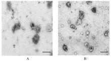
Fig.1
Morphology of placenta Exo of subjects in two groups observed by TEM (Bar=100 μm)"
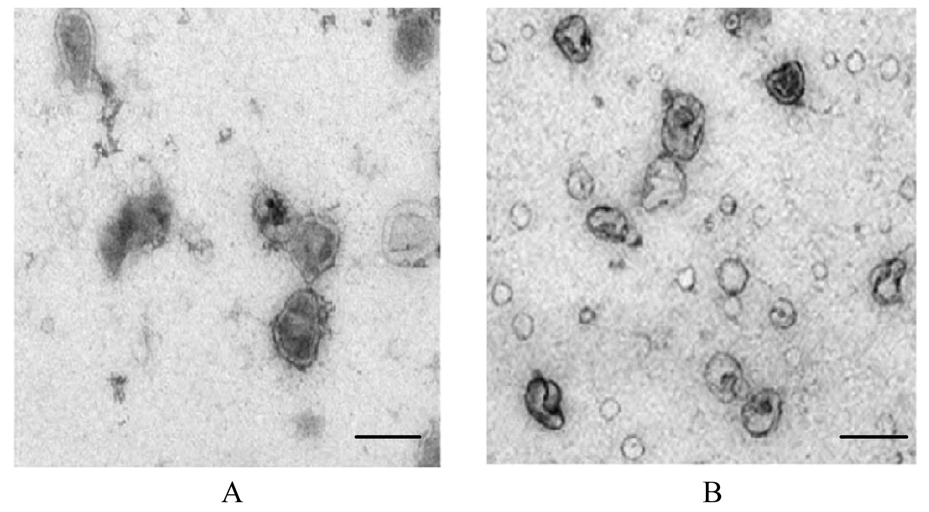
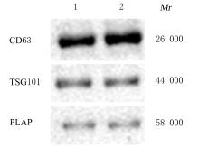
Fig. 2
Expressions of CD63, TSG101, and PLAP proteins in placenta Exo of subjects in two groups detected by Western blotting method"

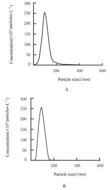
Fig. 3
Particle sizes and concentrations of placenta Exo of subjects in the two groups detected by NTA"
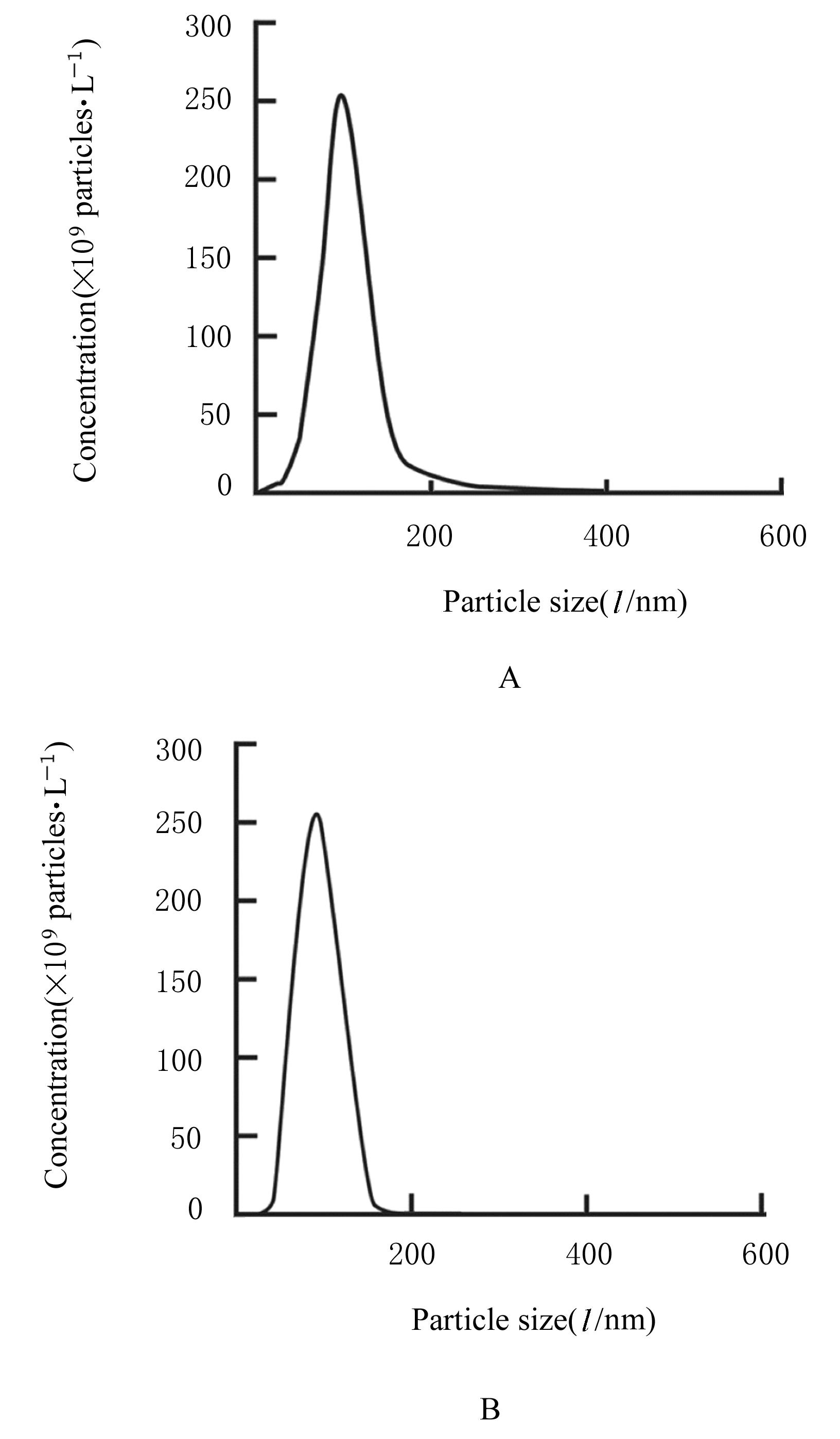
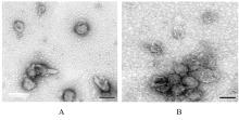
Fig.4
Morphology of Exo in trophoblast cells in two groups observed by TEM (Bar=100 μm)"


Fig. 5
Expressions of CD63 and TSG101 proteins in Exo in trophoblast cells in two groups detected by Western blotting method"
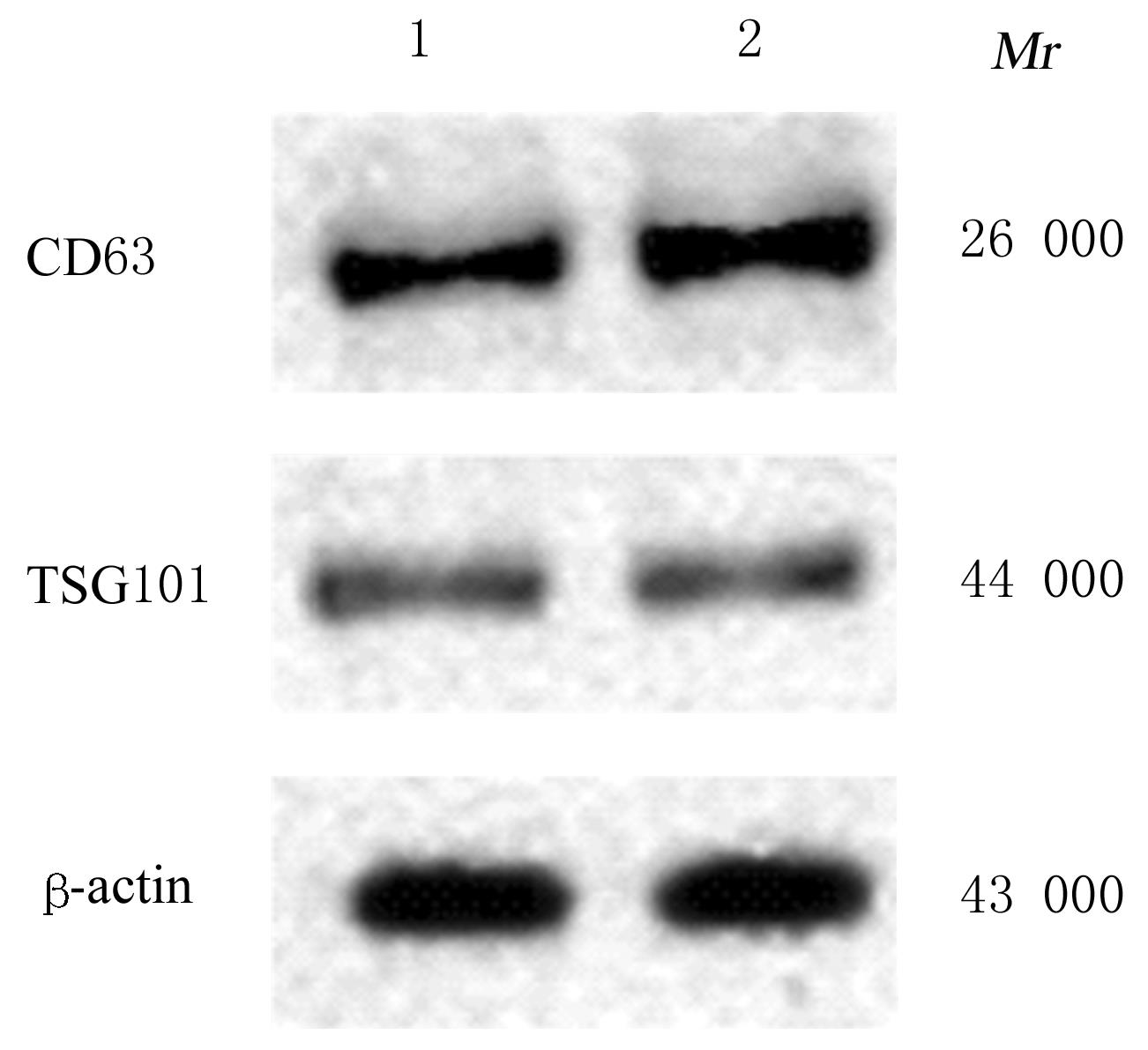

Fig. 6
Particle sizes and concentrations of Exo in trophoblast cells in two groups detected by NTA"
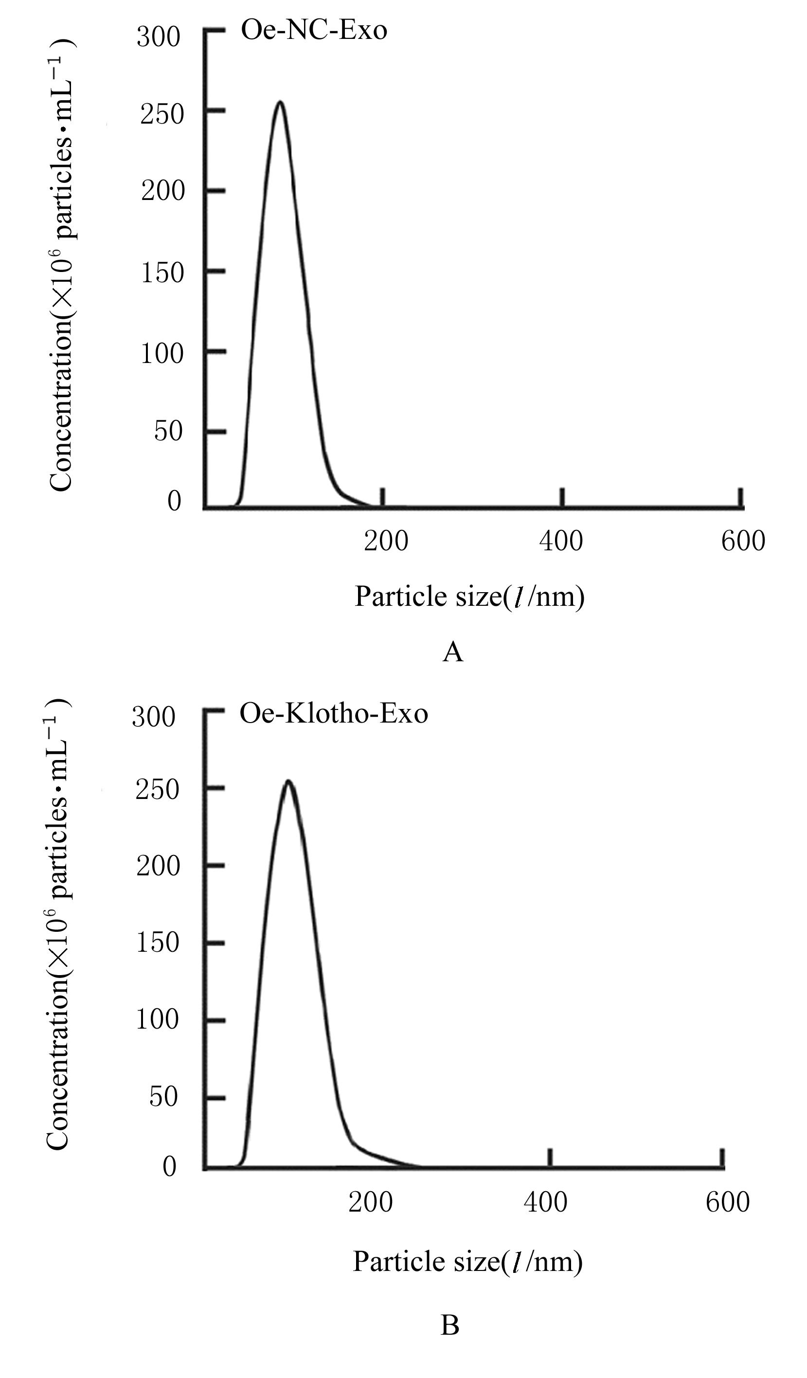

Fig. 7
Uptake of placenta Exo by HUVECs observed under fluorescence microscope (Bar=25 μm)"
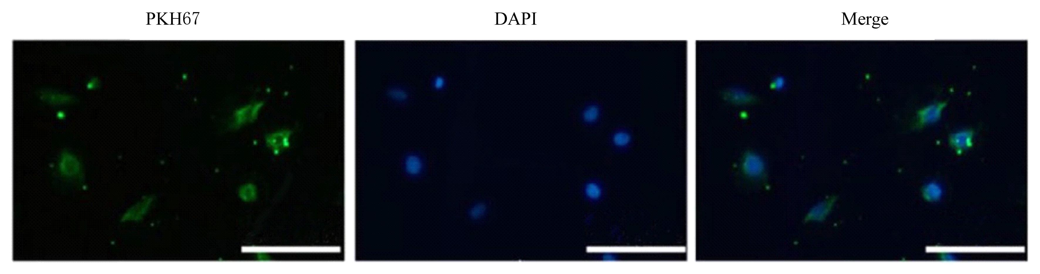
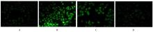
Fig. 8
Level of ROS in HUVECs in various groups detected by DCFDA fluorescence staining(×400)"

Fig. 9
Levels of MDA and activities of SOD in HUVECs in various groups"
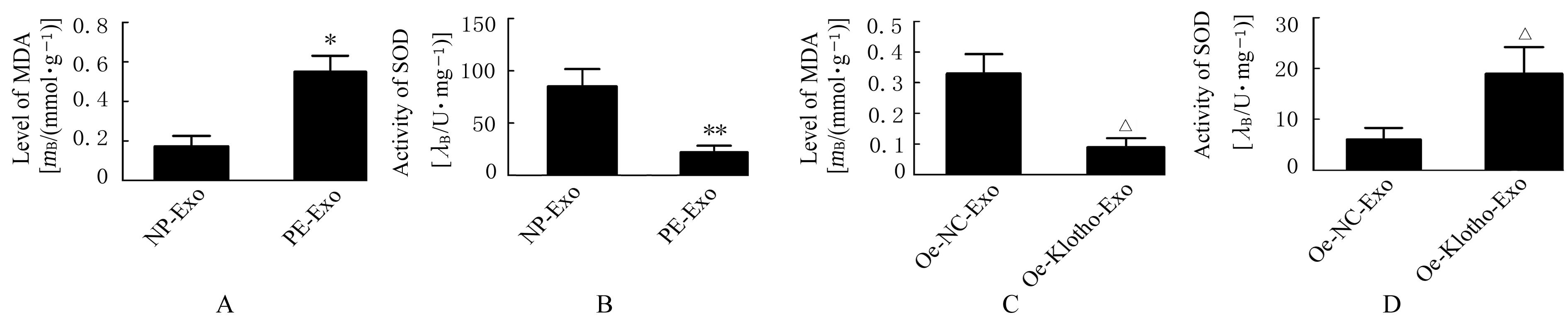

Fig. 10
Expressions of Klotho protein in plancenta Exo of subjects in two groups and expression of eNOS protein in HUVECs in various groups"
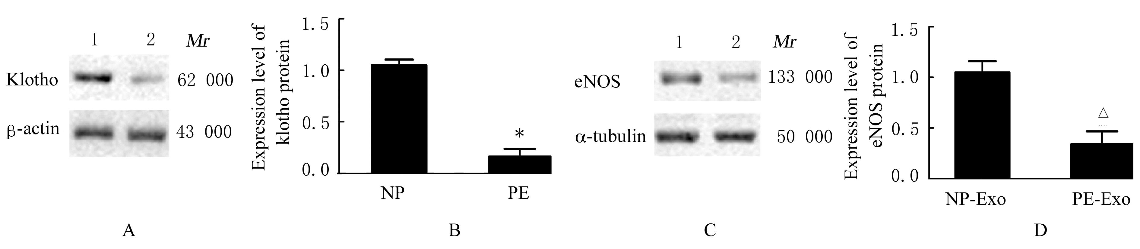

Fig. 11
Electrophoregram (A) and histogram (B) of expressions of Klotho protein in Exo in trophoblast cells in two groups"
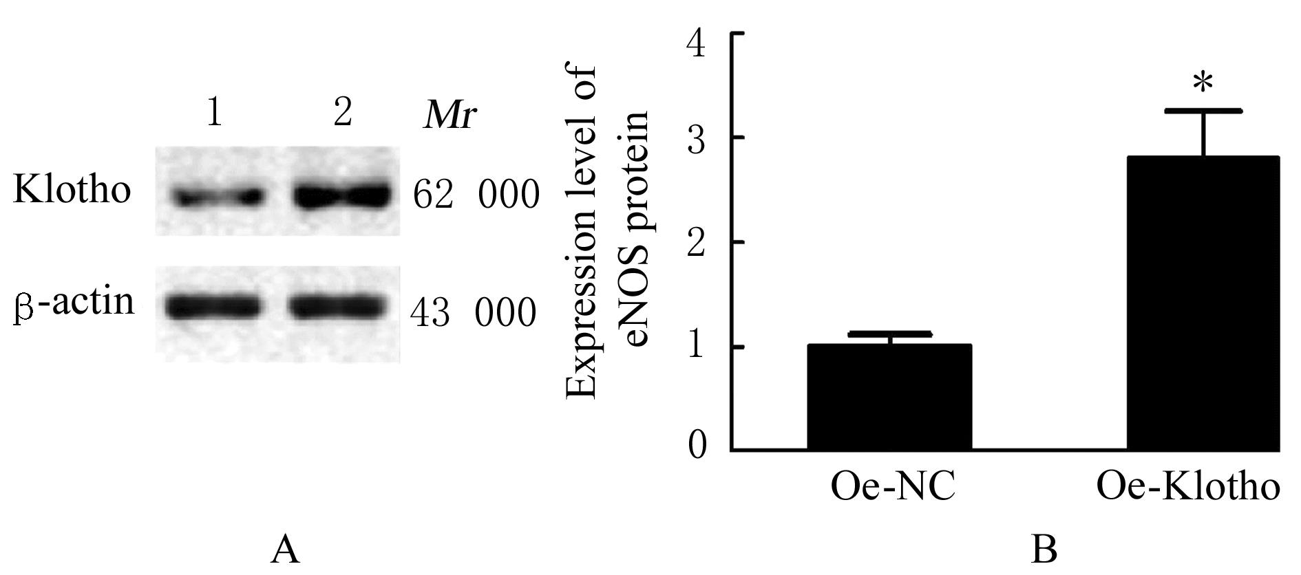
Fig. 12
Expressions of Klotho protein in Exo in trophoblast cells and expressions of eNOS protein in HUVECs in two groups"

| 1 | MAGEE L A, NICOLAIDES K H, VON DADELSZEN P. Preeclampsia[J]. N Engl J Med, 2022, 386(19): 1817-1832. |
| 2 | JUNG E, ROMERO R, YEO L, et al. The etiology of preeclampsia[J]. Am J Obstet Gynecol,2022,226(2S): S844-S866. |
| 3 | GHAFOURIAN M, MAHDAVI R, AKBARI JONOUSH Z, et al. The implications of exosomes in pregnancy: emerging as new diagnostic markers and therapeutics targets[J].Cell Commun Signal,2022,20(1): 51. |
| 4 | MATSUBARA K, MATSUBARA Y, UCHIKURA Y, et al. Pathophysiology of preeclampsia: the role of exosomes[J]. Int J Mol Sci, 2021, 22(5): 2572. |
| 5 | TYURENKOV I N, PERFILOVA V N, NESTEROVA A A, et al. Klotho protein and cardio-vascular system[J].Biochemistry,2021,86(2): 132-145. |
| 6 | RICHTER B, HALLER J, HAFFNER D, et al. Klotho modulates FGF23-mediated NO synthesis and oxidative stress in human coronary artery endothelial cells[J]. Pflugers Arch, 2016, 468(9): 1621-1635. |
| 7 | FAN C F, WANG Y Q, WANG J Y, et al. Clinic significance of markedly decreased α-klothoin women with preeclampsia[J]. Am J Transl Res, 2016, 8(5): 1998-2010. |
| 8 | 谢幸, 孔北华, 段涛. 妇产科学[M]. 9版. 北京: 人民卫生出版社, 2019:83-90. |
| 9 | SHAH K B, CHERNAUSEK S D, TEAGUE A M, et al. Maternal diabetes alters microRNA expression in fetal exosomes, human umbilical vein endothelial cells and placenta[J]. Pediatr Res, 2021, 89(5): 1157-1163. |
| 10 | WANG D, NA Q, SONG G Y, et al. Human umbilical cord mesenchymal stem cell-derived exosome-mediated transfer of microRNA-133b boosts trophoblast cell proliferation, migration and invasion in preeclampsia by restricting SGK1[J].Cell Cycle, 2020,19(15):1869-1883. |
| 11 | AMINI P, AMROVANI M, NASSAJ Z S, et al. Hypertension: potential player in cardiovascular disease incidence in preeclampsia[J]. Cardiovasc Toxicol, 2022, 22(5): 391-403. |
| 12 | KURO-O M, MATSUMURA Y, AIZAWA H, et al. Mutation of the mouse klotho gene leads to a syndrome resembling ageing[J]. Nature, 1997, 390(6655): 45-51. |
| 13 | SHIRAKI-IIDA T, AIZAWA H, MATSUMURA Y, et al. Structure of the mouse klotho gene and its two transcripts encoding membrane and secreted protein[J]. FEBS Lett, 1998, 424(1/2): 6-10. |
| 14 | HONG K, KIM S H, CHA D H, et al. Defective uteroplacental vascular remodeling in preeclampsia: key molecular factors leading to long term cardiovascular disease[J]. Int J Mol Sci, 2021, 22(20): 11202. |
| 15 | UDENZE I C. Association of pre-eclampsia with metabolic syndrome and increased risk of cardiovascular disease in women: a systemic review[J]. Niger J Clin Pract, 2016, 19(4): 431-435. |
| 16 | 宋小刚, 吴 冰, 陈永清. Drp1在糖尿病心肌病中的作用机制研究进展[J]. 解放军医学杂志, 2022, 47(9): 926-931. |
| 17 | SAITO Y, YAMAGISHI T, NAKAMURA T, et al. Klotho protein protects against endothelial dysfunction[J]. Biochem Biophys Res Commun, 1998, 248(2): 324-329. |
| 18 | KANBAY M, DEMIRAY A, AFSAR B, et al. Role of klotho in the development of essential hypertension[J]. Hypertension, 2021, 77(3): 740-750. |
| 19 | 多妍蓓, 赵维纲. 妊娠糖尿病产后发生2型糖尿病的危险因素及早期预测研究进展[J]. 中国实用内科杂志, 2021, 41(11): 990-994. |
| 20 | CECATI M, GIANNUBILO S R, SACCUCCI F,et al. Potential role of placental klotho in the pathogenesis of preeclampsia[J].Cell Biochem Biophys,2016,74(1): 49-57. |
| 21 | MIRANDA J, ROMERO R, KORZENIEWSKI S J, et al. The anti-aging factor α-klotho during human pregnancy and its expression in pregnancies complicated by small-for-gestational-age neonates and/or preeclampsia[J]. J Matern Fetal Neonatal Med, 2014, 27(5): 449-457. |
| 22 | JIA L Y, ZHOU X Y, HUANG X J, et al. Maternal and umbilical cord serum-derived exosomes enhance endothelial cell proliferation and migration[J].FASEB J, 2018, 32(8): 4534-4543. |
| 23 | SHEN L, LI Y J, LI R T, et al. Placenta‑associated serum exosomal miR‑155 derived from patients with preeclampsia inhibits eNOS expression in human umbilical vein endothelial cells[J]. Int J Mol Med, 2018, 41(3): 1731-1739. |
| [1] | Wenting HUI,Tongtong SONG,Min HUANG,Xia CHEN. Effect of hydrogel-based delivery of bFGF on function of NIH3T3 cells after oxygen-glucose deprivation [J]. Journal of Jilin University(Medicine Edition), 2023, 49(6): 1484-1490. |
| [2] | Jiao ZHAGN,Baolian MA,Yonglan ZHANG. Antioxidant capacities of edible plant-exosomes-like nanoparticles in vitro and their protective effects on oxidative damage of PC12 cells induced by hydrogen peroxide [J]. Journal of Jilin University(Medicine Edition), 2023, 49(5): 1117-1124. |
| [3] | Shan LIU,Zhaodong XING,Ping HUANG. Effect of expression of miR-17-5p in exosomes derived from colorectal cancer cells on chemosensitivity of colorectal cancer cells and its mechanism [J]. Journal of Jilin University(Medicine Edition), 2023, 49(4): 975-984. |
| [4] | Hongli CUI,Siqi FAN,Wenfei GUAN,E MENG,Jiatong LIU,Xuetong SUN,Chunxu CAO,Lixin LIU,Yali QI,Fang FANG,Zhicheng WANG. Inhibitory effect of irradiation enhanced by gallic acid-lecithin complex-induced oxidative stress on proliferation of A549 cells [J]. Journal of Jilin University(Medicine Edition), 2023, 49(4): 941-946. |
| [5] | Yintao ZHAO, Yingying YANG, Xiangqin ZHANG, Lu ZHENG, Yawei XU, Haibo YANG, Yuan LIU. Improvement effect of follistatin-like 1 on doxorubicin-induced acute myocardial injury in mice and its mechanism [J]. Journal of Jilin University(Medicine Edition), 2023, 49(3): 565-572. |
| [6] | Jing GUAN,Shen HA,Hao YUAN,Ying CHEN,Pengju LIU,Zhi LIU,Shuang JIANG. Protective effect of Modified Xiao-Xian-Xiong Decoction on liver injury in rats with type 2 diabete mellitus and its mechanism [J]. Journal of Jilin University(Medicine Edition), 2023, 49(3): 608-616. |
| [7] | Yue CHENG,Keke NI,Dewang FU,Yang ZHANG,Hongyang TIAN,Huamao JIANG. Improvement effect of tetramethylpyrazine on oxidative damage of kidney tissue in nephrolithiasis model rats [J]. Journal of Jilin University(Medicine Edition), 2023, 49(1): 67-73. |
| [8] | Jun ZHU,Nan ZHOU,Deming LI. Improvement effect of propofol postconditioning on focal cerebral ischemia-reperfusion injury in rats and its mechanism [J]. Journal of Jilin University(Medicine Edition), 2023, 49(1): 46-54. |
| [9] | Chuan LIU,Huan LI,Dawei WANG. Effects of Dendrobium huoshanense polysaccharide on oxidative stress and inflammation in brain tissue of mice with Parkinson’s disease [J]. Journal of Jilin University(Medicine Edition), 2023, 49(1): 110-115. |
| [10] | Hao LI,Dongge LIU,Shuqi YAN,Shuping REN. Improvement effect of 1,25(OH)2D3 combined with astragalus polysaccharide on insulin resistance of skeletal muscle cells in vitro and its mechanism [J]. Journal of Jilin University(Medicine Edition), 2022, 48(6): 1411-1421. |
| [11] | Tingyu RUAN,Weidi SHI,Xun MA,Jing WANG,Lichao KANG,Dongdong DU,Yuelan YIN. Construction of NDRG1 interference vector and establishment of human brain microvascular endothelial cells stably over-expressing NDRG1 [J]. Journal of Jilin University(Medicine Edition), 2022, 48(6): 1614-1622. |
| [12] | Junxiu LIU,Jia ZHOU,Guangfu LYU,Yuchen WANG,Xuefeng ZHUANG,Jiarui ZHAO,Xiaowei HUANG,Ruili LI. Protective effect of Shenhong Buxue Granule on vascular endothelium of mice with vascular endothelial dysfunction of Qi stagnation and blood stasis type and its mechanism [J]. Journal of Jilin University(Medicine Edition), 2022, 48(6): 1437-1447. |
| [13] | Bingbing WU,Aiping ZHANG,Xinke ZHAO,Yingdong LI,Kai LIU. Protective effect of ultra-filtration extract from Angelica Sinensis Radix and Hedysari Radix on human umbilical vein endothelial cell injury induced by X-ray and its mechanism [J]. Journal of Jilin University(Medicine Edition), 2022, 48(5): 1139-1147. |
| [14] | Xiaochen HUANG,Hao LI,Baohua WANG,Kai LI. Protective effect of lidocaine on PC12 cells in Parkinson’s disease model and its mechanism [J]. Journal of Jilin University(Medicine Edition), 2022, 48(3): 638-647. |
| [15] | Wentao WANG,Xuguang MI,Yang ZHOU,Wenxing PU,Jiaxu GAO,Meng JING,Fankai MENG. Effect of autophagy induced by exosomes derived from bone marrow mesenchymal stem cells on survival of SH-SY5Y cells inhibited by MPP+ and its mechanism [J]. Journal of Jilin University(Medicine Edition), 2022, 48(3): 606-614. |
|
||