Journal of Jilin University(Medicine Edition) ›› 2025, Vol. 51 ›› Issue (1): 191-201.doi: 10.13481/j.1671-587X.20250123
• Research in clinical medicine • Previous Articles
Expressions of autotaxin and lysophosphatidic acid receptor 3 in serum and lung tissue of patients with chronic obstructive pulmonary disease and their significances
Peiqin JIANG1,Zheng ZHANG1,Zhong HUANG2( ),Xianling LU1()
- 1.Department of Pulmonary and Critical Care Medicine,First Affiliated Hospital,Shihezi University,Shihezi 832000,China
2.Emergency Medical Center,First Affiliated Hospital,Shihezi University,Shihezi 832000,China
-
Received:2024-02-10Accepted:2024-04-07Online:2025-01-28Published:2025-03-06 -
Contact:Zhong HUANG,Xianling LU E-mail:10082404@qq.com;luxianlingmary@163.com
CLC Number:
- R563.9
Cite this article
Peiqin JIANG,Zheng ZHANG,Zhong HUANG,Xianling LU. Expressions of autotaxin and lysophosphatidic acid receptor 3 in serum and lung tissue of patients with chronic obstructive pulmonary disease and their significances[J].Journal of Jilin University(Medicine Edition), 2025, 51(1): 191-201.
share this article
Tab.1
Clinical data of subjects in control group and COPD stable group"
| Group | Age (x±s, year) | BMI | FEV1%pred (x±s, η/%) | FEV1/FVC (x±s, η/%) | Percentage of male [n(η/%)] | Percentage of smoking [n(η/%)] |
|---|---|---|---|---|---|---|
| Control | 69.70±7.40 | 24.18±2.30 | 97.10±6.82 | 78.05±3.56 | 19(47.5) | 13(32.5) |
| COPD stable | 69.85±7.32 | 24.91±2.38 | 48.88±6.55 | 51.08±7.47 | 23(57.5) | 18(45.0) |
| t/χ2 | 0.091 | 1.389 | 0.802 | 1.317 | ||
| P | 0.928 | 0.169 | <0.001 | <0.001 | 0.370 | 0.251 |
Tab.2
Correlations between serum ATX levels and clinical indicators of patients in AECOPD group and COPD stable group"
| Index | AECOPD group | COPD stable group | ||
|---|---|---|---|---|
| r | P | r | P | |
| Smoking | 0.052 | 0.749 | 0.107 | 0.512 |
| WBC | 0.110 | 0.499 | 0.384 | 0.014 |
| NEUT% | 0.074 | 0.650 | 0.054 | 0.738 |
| NLR | 0.046 | 0.776 | -0.020 | 0.902 |
| BMI | 0.043 | 0.791 | 0.245 | 0.127 |
| CAT | 0.581 | <0.001 | 0.463 | 0.003 |
| FEV1%pred | - | - | -0.393 | 0.012 |
| FEV1/FVC | - | - | -0.353 | 0.025 |
Tab.3
Clinical data of patients in various groups"
| Group | Age (x±s, year) | BMI (x±s, kg·m-2) | SI (x±s) | FEV1%pred (x±s, η/%) | FEV1/FVC (x±s, η/%) | Percentage of male [n(η/%)] |
|---|---|---|---|---|---|---|
| CS | 64.10±9.88 | 24.43±1.59 | 620.00±311.47 | 77.65±7.58 | 62.85±4.11 | 14(70.0) |
| CNS | 64.20±7.27 | 23.59±1.71 | - | 77.45±8.09 | 63.25±4.23 | 11(55.0) |
| HS | 63.50±8.63 | 24.74±2.02 | 608.00±334.81 | 95.60±9.13*△ | 76.70±3.92*△ | 14(70.0) |
| HNS | 62.45±8.68 | 24.52±2.04 | - | 100.40±11.86*△ | 78.90±4.96*△ | 10(50.0) |
| F/χ2 | 0.172 | 1.501 | 0.014 | 33.025 | 78.503 | 2.686 |
| P | 0.915 | 0.221 | 0.907 | <0.001 | <0.001 | 0.443 |
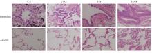
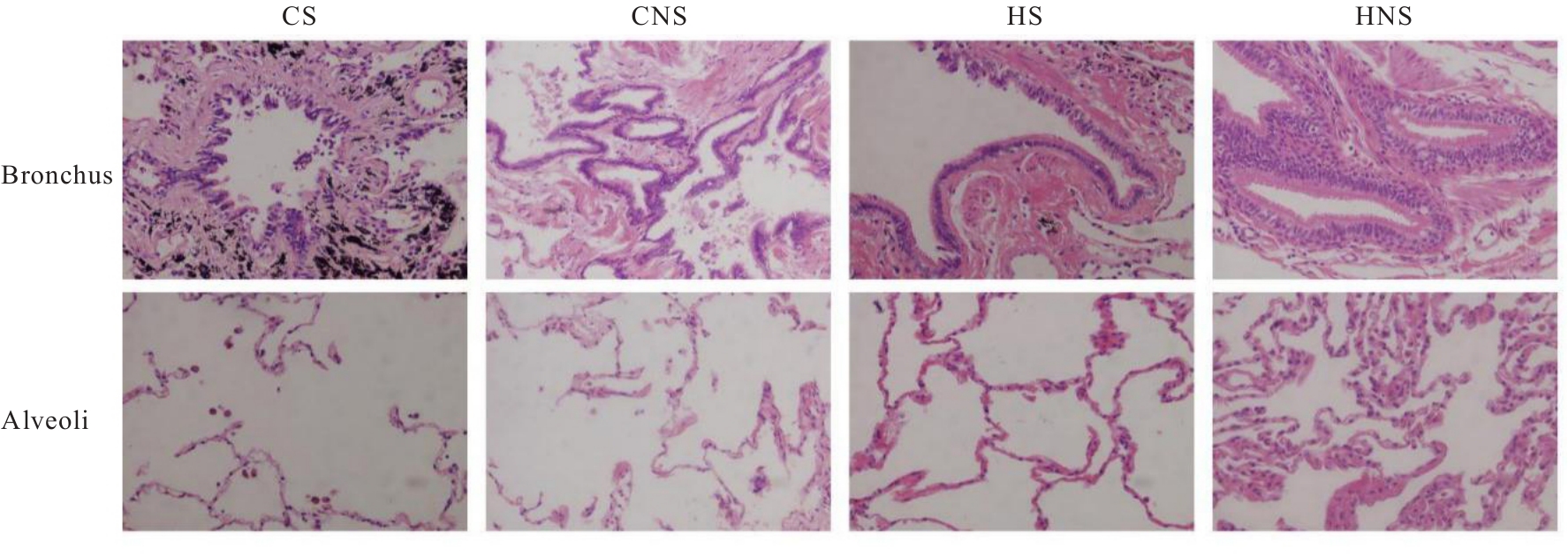
Fig. 1
Pathomorphology of lung tissue of patients in various groups (HE, ×200)"
Tab.4
Bosken scores, MLI, and MAN of lung tissue of patients in various groups"
| Group | Bosken score | MLI(l/mm) | MAN( |
|---|---|---|---|
| CS | 17.75±1.65 | 0.20±0.40 | 31.81±4.92 |
| CNS | 14.35±1.63* | 0.18±0.03 | 33.97±3.55 |
| HS | 6.15±1.23*△ | 0.12±0.04*△ | 53.47±12.64*△ |
| HNS | 2.60±1.10*△# | 0.11±0.03*△ | 56.14±11.96*△ |
| F | 489.311 | 40.068 | 37.022 |
| P | <0.001 | <0.001 | <0.001 |
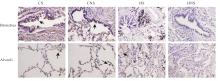
Fig. 2
Expressions of ATX protein in lung tissue of patients in various groups(Immunohistochemistry, ×200)"
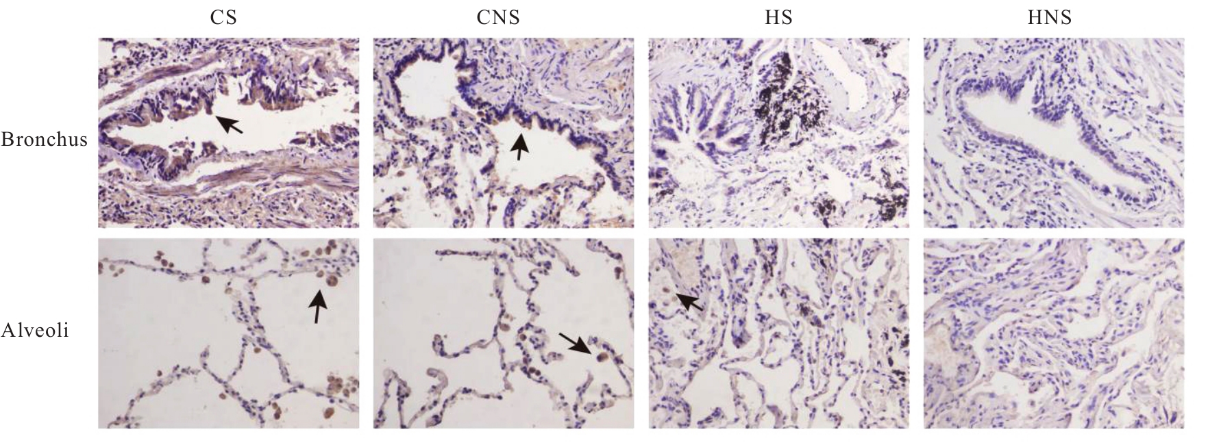
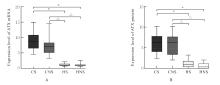
Fig. 3
Expression levels of ATX mRNA and protein in lung tissue of patients in various groups"
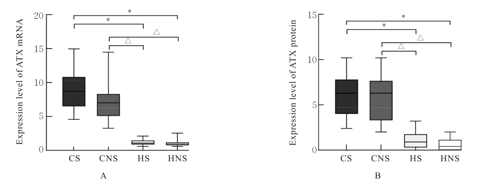
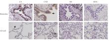
Fig. 4
Expressions of LPA3 protein in lung tissue of patients in various groups (Immunohistochemistry, ×200)"
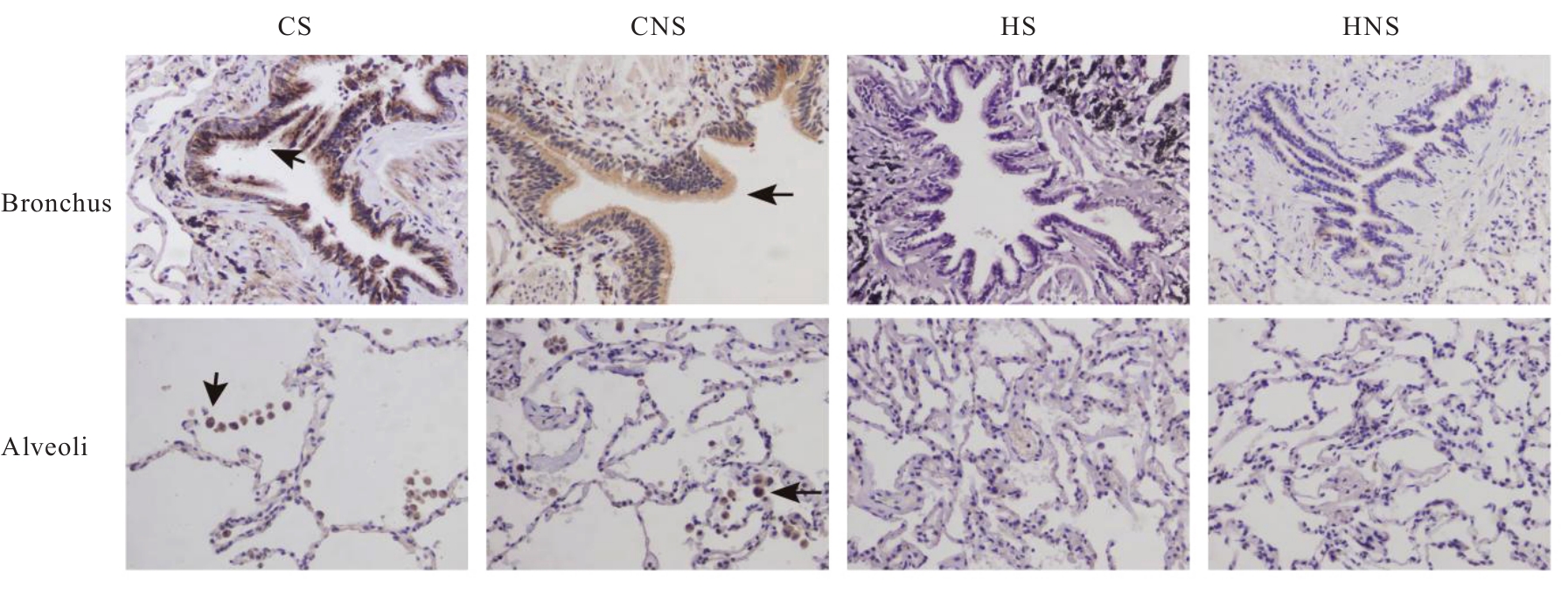
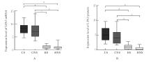
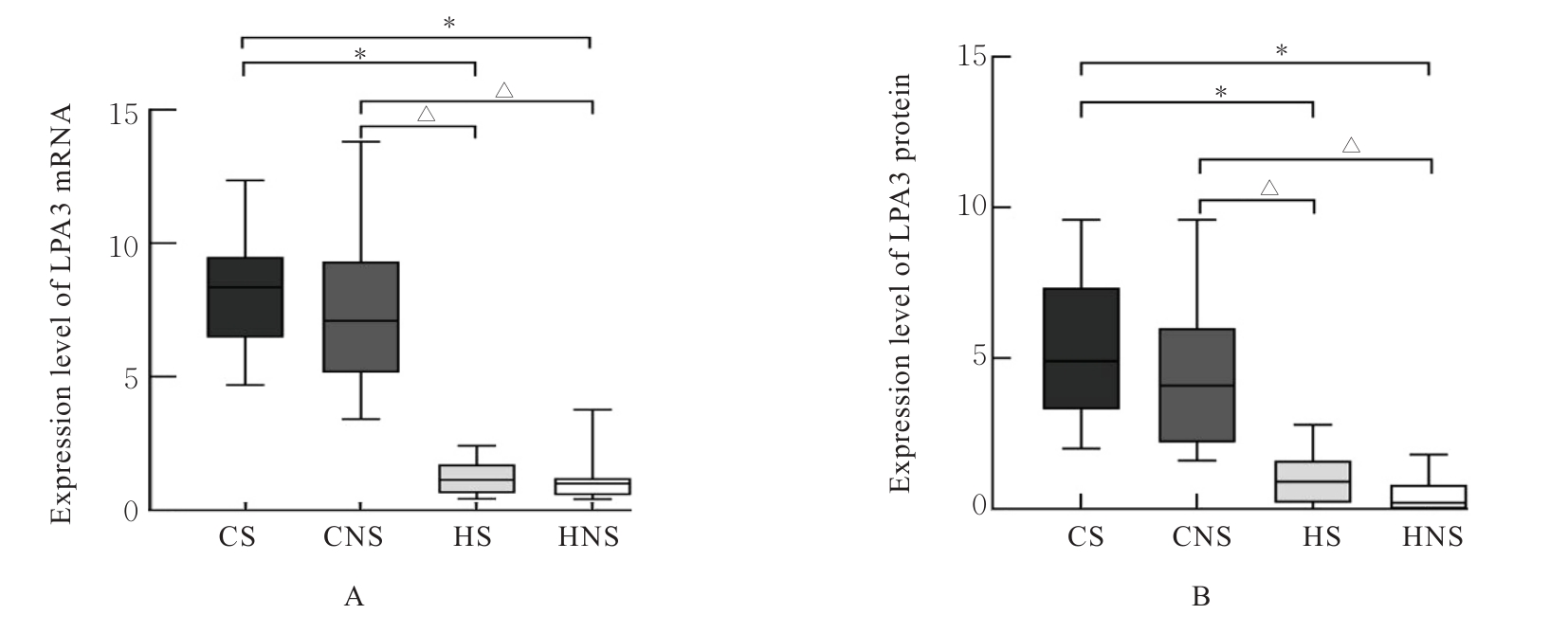
Fig. 5
Expression levels of LPA3 mRNA and protein in lung tissue of patients in various groups"
Tab.5
Correlations between expression levels of ATX and LPA3 proteins and other indicators in lung tissue of COPD patients"
| Index | ATX | LPA3 | ||
|---|---|---|---|---|
| r | P | r | P | |
| FEV1%pred | 0.017 | 0.074 | ||
| FEV1/FVC | 0.019 | 0.090 | ||
| Bosken score | 0.493 | 0.001 | 0.576 | <0.001 |
| MLI | 0.375 | 0.017 | 0.378 | 0.016 |
| MAN | 0.011 | 0.012 | ||
| LPA3 | 0.723 | <0.001 | - | - |
| 1 | GUO P, LI R, PIAO T H, et al. Pathological mechanism and targeted drugs of COPD[J]. Int J Chron Obstruct Pulmon Dis, 2022, 17: 1565-1575. |
| 2 | ZHANG X T, LI M M, YIN N, et al. The expression regulation and biological function of autotaxin[J]. Cells, 2021, 10(4): 939. |
| 3 | KANO K, AOKI J, HLA T. Lysophospholipid mediators in health and disease[J]. Annu Rev Pathol, 2022, 17: 459-483. |
| 4 | ZHAO J, ZHAO Y T. Lysophospholipids in lung inflammatory diseases[J]. Adv Exp Med Biol, 2021, 1303: 373-391. |
| 5 | SOLÍS K H, ROMERO-ÁVILA M T, GUZMÁN-SILVA A, et al. The LPA3 receptor: regulation and activation of signaling pathways[J]. Int J Mol Sci, 2021, 22(13): 6704. |
| 6 | SAATIAN B, ZHAO Y T, HE D H, et al. Transcriptional regulation of lysophosphatidic acid-induced interleukin-8 expression and secretion by p38 MAPK and JNK in human bronchial epithelial cells[J]. Biochem J, 2006, 393(Pt 3): 657-668. |
| 7 | 范 傲, 黄 钟, 段宇清, 等. 慢性阻塞性肺疾病患者血浆中性粒细胞胞外诱捕网和白细胞介素-8及白细胞介素-33的表达水平及其临床意义[J]. 中国呼吸与危重监护杂志, 2022(2): 84-89. |
| 8 | 中华医学会呼吸病学分会慢性阻塞性肺疾病学组, 中国医师协会呼吸医师分会慢性阻塞性肺疾病工作委员会. 慢性阻塞性肺疾病诊治指南(2021年修订版)[J]. 中华结核和呼吸杂志, 2021, 44(3): 170-205. |
| 9 | SHENG H Y, ZHANG Y J, SHI X Q, et al. Functional, ultrastructural, and transcriptomic changes in rat diaphragms with different durations of cigarette smoke exposure[J]. Int J Chron Obstruct Pulmon Dis, 2020, 15: 3135-3145. |
| 10 | ZHENG X R, ZHANG L Y, CHEN J, et al. Dendritic cells and Th17/Treg ratio play critical roles in pathogenic process of chronic obstructive pulmonary disease[J]. Biomedecine Pharmacother, 2018, 108: 1141-1151. |
| 11 | 盛梅梅, 罗专波. 慢性阻塞性肺疾病发病机制的研究现状及进展[J]. 现代医学与健康研究(电子版), 2023, 7(1): 38-41. |
| 12 | 张 地, 张俊杰. Autotaxin表达调控机制及其生物学功能[J]. 中国科学:生命学, 2022, 52(8): 1148-1162. |
| 13 | BRINDLEY D N, TANG X Y, MENG G M, et al. Role of adipose tissue-derived autotaxin, lysophosphatidate signaling, and inflammation in the progression and treatment of breast cancer[J]. Int J Mol Sci, 2020, 21(16): 5938. |
| 14 | BENESCH M G K, MACINTYRE I T K, MCMULLEN T P W, et al. Coming of age for autotaxin and lysophosphatidate signaling: clinical applications for preventing, detecting and targeting tumor-promoting inflammation[J]. Cancers, 2018, 10(3): 73. |
| 15 | BLANQUE R, DESROY N, DUPONT S, et al. Pharmacological profile and efficacy of GLPG1690, a novel ATX inhibitor for COPD treatment[C]//5.1 Airway Pharmacology and Treatment. European Respiratory Society, 2015: PA2129. |
| 16 | LI Q L, WONG W, CHAKRABARTI A, et al. Serum lysophosphatidic acid measurement by liquid chromatography-mass spectrometry in COPD patients[J]. J Am Soc Mass Spectrom, 2021, 32(8): 1987-1997. |
| 17 | 洪静雪, 黄 钟, 张 妤, 等. 慢性阻塞性肺疾病患者血浆溶血磷脂酸和可溶性ST2的表达水平及其临床意义[J]. 西部医学, 2024, 36(1): 57-62. |
| 18 | ZHAO Y T, NATARAJAN V. Lysophosphatidic acid (LPA) and its receptors: role in airway inflammation and remodeling[J]. Biochim Biophys Acta, 2013, 1831(1): 86-92. |
| 19 | JIANG S F, YANG H L, LI M Q. Emerging roles of lysophosphatidic acid in macrophages and inflammatory diseases[J]. Int J Mol Sci, 2023, 24(15): 12524. |
| 20 | 王 蕾, 王桐生, 王雅娟, 等. 巨噬细胞在慢性阻塞性肺疾病发展及治疗中的研究进展[J]. 临床肺科杂志, 2024, 29(2): 293-297. |
| 21 | OIKONOMOU N, MOURATIS M A, TZOUVELEKIS A, et al. Pulmonary autotaxin expression contributes to the pathogenesis of pulmonary fibrosis[J]. Am J Respir Cell Mol Biol, 2012, 47(5): 566-574. |
| 22 | NIKITOPOULOU I, FANIDIS D, NTATSOULIS K, et al. Increased autotaxin levels in severe COVID-19, correlating with IL-6 levels, endothelial dysfunction biomarkers, and impaired functions of dendritic cells[J]. Int J Mol Sci, 2021, 22(18): 10006. |
| 23 | HIKICHI M, MIZUMURA K, MARUOKA S, et al. Pathogenesis of chronic obstructive pulmonary disease (COPD) induced by cigarette smoke[J]. J Thorac Dis, 2019, 11(): S2129-S2140. |
| 24 | ZHAO C Q, SARDELLA A, CHUN J, et al. TNF-alpha promotes LPA1- and LPA3-mediated recruitment of leukocytes in vivo through CXCR2 ligand chemokines[J]. J Lipid Res, 2011, 52(7): 1307-1318. |
| 25 | ZHAO Y, HASSE S, ZHAO C Q, et al. Targeting the autotaxin-Lysophosphatidic acid receptor axis in cardiovascular diseases[J]. Biochem Pharmacol, 2019, 164: 74-81. |
| 26 | LI S, XIONG C Y, ZHANG J J. ATX and LPA receptor 3 are coordinately up-regulated in lipopolysaccharide-stimulated THP-1 cells through PKR and SPK1-mediated pathways[J]. FEBS Lett, 2012, 586(6): 792-797. |
| [1] | Xiaoyu HOU,Ya LI,Yian SONG,Tianhui HE,Jie ZHANG,Jianhui XU. Effect of prostaglandin E2 on discharge activity of warm-sensitive neurons in median preoptic nucleus of hypothalamus in female mice and its mechanism [J]. Journal of Jilin University(Medicine Edition), 2025, 51(1): 17-25. |
| [2] | Xinyue MA,Hui XU,Jiawen DIAO,Aihua JIN,Jishu QUAN. Inhibitory effect of Boschnikia rossica polysaccharides on THP-1 macrophage inflammation and its mechanism [J]. Journal of Jilin University(Medicine Edition), 2024, 50(6): 1499-1511. |
| [3] | Guobin HE,Huan WANG. Effect of knockdown of RIP3 on autophagy, pyroptosis, and ferroptosis of hypoxia/reoxygenation-induced human renal tubular epithelial HK2 cells [J]. Journal of Jilin University(Medicine Edition), 2024, 50(6): 1644-1653. |
| [4] | Weichao WU,Yan GUO,Xiangkai ZHAO,Zhiguang GU,Yijia GUO,Zipeng LAN,Hui HUANG,Lei KUANG,Ming ZHANG,Dongsheng HU,Yongli YANG,Wei WANG,Jinru CHEN. Correlation analysis on occupational acid fog exposure and accelerated biological aging in workers [J]. Journal of Jilin University(Medicine Edition), 2024, 50(6): 1741-1750. |
| [5] | Yuxuan CAO,Wei CHEN,Chengbiao SUN,Na ZHAO,Yan WANG,Mingxin DONG,Na XU,Wensen LIU,Yongmei LI. Damage effect of VSV on vascular endothelial barrier function in vitro and its mechanism [J]. Journal of Jilin University(Medicine Edition), 2024, 50(5): 1275-1285. |
| [6] | Wenhui LIU,Miao YU,Ying GUO,Yupeng LIU,Yang XING,Xinyu HONG,Jiale CUI. Research progress in effect of CXC chemokine receptor 3 on occurrence and development of nervous system diseases [J]. Journal of Jilin University(Medicine Edition), 2024, 50(5): 1474-1480. |
| [7] | Lin CHEN,Limin YAN,Huaijie XING,Min CHEN,Xiaoyan LI,Chaosheng ZENG. Improvement effect of Xuebijing on brain tissue injury and Th17/Treg immune imbalance in cerebrospinal fluid in NMDA receptor encephalitis model mice [J]. Journal of Jilin University(Medicine Edition), 2024, 50(3): 697-707. |
| [8] | Ying ZHANG,Zhaohui WAN,Xianxun JIANG. Effect of over-expression of NDRG1 on resistance of castration-resistant prostate cancer resistant cell line C4-2/ENZA and its mechanism [J]. Journal of Jilin University(Medicine Edition), 2024, 50(3): 708-717. |
| [9] | Li ZHANG,Binfeng XIA,Huihui HUANG,Ru WANG,Min KONG,Xia YIN. Research progress in pathophysiological mechanism and clinical diagnosis and treatment of hypertension associated with vascular endothelial growth factor and its receptor inhibitors [J]. Journal of Jilin University(Medicine Edition), 2024, 50(3): 854-863. |
| [10] | Yingxin RUAN,Junya JIA,Zhanfei WU,Wenya SHANG,Pengyu ZHANG. Effect of NLRP3 inflammatome in renal interstitial fibrosis induced by unilateral ureteral obstruction in rats and its mechanism [J]. Journal of Jilin University(Medicine Edition), 2024, 50(3): 587-595. |
| [11] | Jialiang CHEN,Qi LI,Xiangdong ZHOU,Xiaomei CHEN,Feng LIU,Chang LIU,Youqing ZHONG,Liang LI. Inhibitory effect of nobiletin on airway mucus hypersecretion in model rats with chronic obstructive pulmonary disease and its molecular mechanism [J]. Journal of Jilin University(Medicine Edition), 2024, 50(2): 295-302. |
| [12] | Yu ZHANG,Xianling LU. Expressions of IL-33, IL-8, and NETs in lung tissue of patients with chronic obstructive pulmonary disease and their significances [J]. Journal of Jilin University(Medicine Edition), 2024, 50(2): 498-507. |
| [13] | Zhongyan ZHAO,Zhiyu XU,Chanji WU,Eryi ZHAO,Dan HUANG,Shixiong HUANG. Autoimmune encephalitis with double positive anti-NMDAR and anti-GABABR secondary to herpes simplex virus encephalitis: A case report and literature review [J]. Journal of Jilin University(Medicine Edition), 2024, 50(1): 236-242. |
| [14] | Xinqiang ZHANG,Bo WANG,Huicheng FENG. Effect of local impulse vibration stimulation on proprioception recovery after anterior cruciate ligament reconstruction in rabbits [J]. Journal of Jilin University(Medicine Edition), 2023, 49(6): 1407-1414. |
| [15] | Xiuling ZHOU,Deyu CONG,Ye ZHANG,Hongshi ZHANG. Effect of head acupuncture on neurological function and HIF-1α and VEGFR2 expressions in brain tissue in rats with focal cerebral ischemia and its mechanism [J]. Journal of Jilin University(Medicine Edition), 2023, 49(6): 1431-1436. |
|
||