Journal of Jilin University(Medicine Edition) ›› 2025, Vol. 51 ›› Issue (4): 855-865.doi: 10.13481/j.1671-587X.20250401
• Research in basic medicine • Next Articles
Protective effect of dexmedetomidine on intestinal mucosal injury in rats with enterogenous sepsis and its mechanism
Kun YANG1,Qianyao FU1,Yongqiang SUN1,Kun YANG1,Jun MENG2( )
)
- 1.Department of Anesthesiology,First Affiliated Hospital,Kunming Medical University,Kunming 650000,China
2.Department of Cardiovascular Surgery,People’s Hospital,Wenshan Prefecture,Yunan Province,Wenshan 663000,China
-
Received:2024-09-27Accepted:2024-11-07Online:2025-07-28Published:2025-08-25 -
Contact:Jun MENG E-mail:mengjun888@163.com
CLC Number:
- R614
Cite this article
Kun YANG,Qianyao FU,Yongqiang SUN,Kun YANG,Jun MENG. Protective effect of dexmedetomidine on intestinal mucosal injury in rats with enterogenous sepsis and its mechanism[J].Journal of Jilin University(Medicine Edition), 2025, 51(4): 855-865.
share this article
Tab. 1
Slow wave frequencies and amplitudes of intestinal smooth muscle of rats in various groups"
| Group | Slow wave frequency(f/Hz) | Slow wave amplitude(mV) |
|---|---|---|
| Sham operation | 17.75±1.35 | 0.41±0.03 |
| Model | 9.47±1.05* | 0.14±0.02* |
DEX Low dose | 10.38±0.92 | 0.19±0.01△ |
| Medium dose | 13.29±1.15△# | 0.25±0.03△# |
| High dose | 15.14±1.23△#○ | 0.32±0.02△#○ |
| F | 87.674 | 210.556 |
| P | <0.001 | <0.001 |
Tab. 2
Colony counts of Escherichia coli, Lactobacillus, and Bifidobacterium in cecal tissue of rats in various groups"
| Group | Colony count of Escherichia coli | Colony count of Bifidobacteria | Colony count of Lactobacillus |
|---|---|---|---|
| Sham operation | 5.24±0.25 | 12.35±1.05 | 9.30±0.83 |
| Model | 14.82±0.84* | 5.52±0.34* | 3.75±0.24* |
DEX Low dose | 13.97±0.97 | 6.17±0.53△ | 3.51±0.54 |
| Medium dose | 10.53±0.37△# | 8.37±0.48△# | 5.10±0.38△# |
| High dose | 7.95±0.61△#○ | 9.35±0.76△#○ | 6.32±0.40△#○ |
| F | 365.612 | 161.257 | 207.159 |
| P | <0.001 | <0.001 | <0.001 |
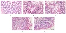
Fig. 1
Pathomophology of small intestinal tissue of rats in various groups detected by HE staining"
Tab.3
Levels of sIgA in supernatant of small intestinal tissue and levels of DAO and D-lactate in serum of rats in various groups detected by ELISA method"
| Group | sIgA [ρB/(mg·L-1)] | DAO [ρB/(mg·L-1)] | D-lactic acid [λB/(U·L-1)] |
|---|---|---|---|
| Sham operation | 29.75±3.51 | 8.18±0.68 | 21.28±2.28 |
| Model | 13.84±1.08* | 14.32±1.27* | 47.36±3.62* |
DEX Low dose | 15.24±1.93 | 13.15±1.10△ | 44.18±4.25 |
| Medium dose | 19.37±2.21△# | 11.29±0.96△# | 36.47±2.98△# |
| High dose | 24.34±1.67△#○ | 9.65±0.81△#○ | 31.23±1.30△#○ |
| F | 87.207 | 64.256 | 115.999 |
| P | <0.001 | <0.001 | <0.001 |
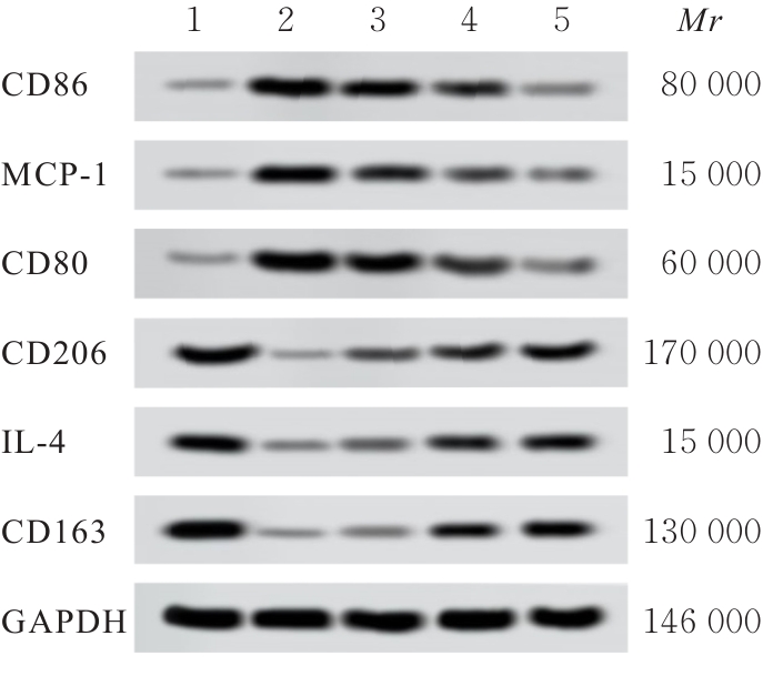
Fig. 2
Electrophoregram of expressions of macrophage polarization-related proteins in small intestine tissue of rats in various groups detected by Western blotting method"
Tab.4
Expression levels of polarization-related protein mRNA in macrophages in small intestine tissue of rats in various groups"
| Group | CD86 mRNA | MCP-1 mRNA | CD80 mRNA | CD206 mRNA | IL-4 mRNA | CD163 mRNA |
|---|---|---|---|---|---|---|
| Sham operation | 1.02±0.09 | 1.03±0.10 | 1.04±0.17 | 1.01±0.11 | 1.03±0.07 | 1.05±0.09 |
| Model | 3.28±0.28* | 4.04±0.32* | 5.94±0.41* | 0.38±0.03* | 0.27±0.02* | 0.51±0.04* |
DEX Low dose | 3.12±0.24 | 3.82±0.35 | 5.41±0.48△ | 0.42±0.07 | 0.33±0.03△ | 0.56±0.05△ |
| Medium dose | 2.68±0.23△# | 3.25±0.30△# | 4.52±0.41△# | 0.58±0.03△# | 0.52±0.04△# | 0.64±0.04△# |
| High dose | 2.24±0.21△#○ | 2.51±0.24△#○ | 3.42±0.32△#○ | 0.76±0.04△#○ | 0.67±0.05△#○ | 0.82±0.07△#○ |
| F | 169.954 | 193.235 | 270.424 | 165.686 | 450.874 | 130.294 |
| P | <0.001 | <0.001 | <0.001 | <0.001 | <0.001 | <0.001 |
Tab.5
Expression levels of polarization-related proteins in macrophages in small intestine tissue of rats in various groups"
| Group | CD86 protein | MCP-1 protein | CD80 protein | CD206 protein | IL-4 protein | CD163 protein |
|---|---|---|---|---|---|---|
| Sham operation | 0.18±0.02 | 0.16±0.01 | 0.17±0.02 | 0.92±0.08 | 0.95±0.08 | 0.93±0.08 |
| Model | 0.91±0.08* | 0.87±0.07* | 0.89±0.07* | 0.12±0.01* | 0.15±0.01* | 0.13±0.01* |
DEX Low dose | 0.82±0.06△ | 0.76±0.08△ | 0.84±0.09 | 0.18±0.01△ | 0.17±0.01 | 0.19±0.02△ |
| Medium dose | 0.65±0.05△# | 0.68±0.06△# | 0.73±0.06△# | 0.35±0.04△# | 0.37±0.03△# | 0.34±0.03△# |
| High dose | 0.26±0.02△#○ | 0.34±0.03△#○ | 0.28±0.02△#○ | 0.68±0.05△#○ | 0.72±0.06△#○ | 0.75±0.08△#○ |
| F | 406.504 | 282.138 | 319.167 | 543.925 | 557.748 | 440.915 |
| P | <0.001 | <0.001 | <0.001 | <0.001 | <0.001 | <0.001 |
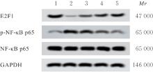
Fig. 3
Electrophoregram of expressions of E2F1, p-NF-κB p65, and NF-κB p65 proteins in small intestine tissue of rats in various groups detected by Western blotting method"
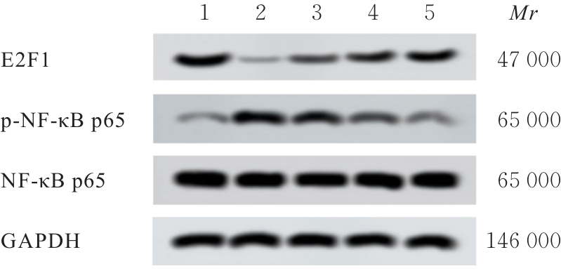
Tab.6
Expression levels of E2F1 protein and ratios of p-NF-κB p65/NF-κB p65 in small intestine tissue of rats in various groups"
| Group | E2F1 protein | Ratio of p-NF-κB p65/NF-κB p65 |
|---|---|---|
| Sham operation | 0.91±0.05 | 0.14±0.01 |
| Model | 0.12±0.02* | 0.73±0.04* |
DEX Low dose | 0.34±0.02△ | 0.65±0.05△ |
| Medium dose | 0.58±0.04△# | 0.33±0.02△# |
| High dose | 0.75±0.05△#○ | 0.28±0.02△#○ |
| F | 673.986 | 637.300 |
| P | <0.001 | <0.001 |
| [1] | YE G J, YE L Q, ZHOU J Q, et al. Challenges in diagnosing community-acquired carbapenem-susceptible Acinetobacter baumannii enterogenic sepsis: a case report[J]. Medicine (Baltimore), 2019, 98(26): e16248. |
| [2] | 房 槟, 曹晓瑞, 闫 昭, 等. MOTS-c通过TLR4对肠源性脓毒症的作用及其机制[J]. 现代生物医学进展, 2020, 20(5): 843-847, 905. |
| [3] | 林耀国, 刘卫明, 魏志亮. 垂体后叶素联合去甲肾上腺素治疗肠源性脓毒性休克患者的效果分析[J]. 医学理论与实践, 2021, 34(2): 240-242. |
| [4] | 刘梦菲, 何 龙, 田丹丹, 等. 艾司氯胺酮复合右美托咪定行无阿片麻醉对乳腺癌改良根治术患者术后恢复质量的影响[J]. 郑州大学学报(医学版), 2023, 58(3):363-366. |
| [5] | 徐 颖, 马四清. 右美托咪定对肠源性脓毒症大鼠肠屏障功能保护及抗凋亡作用的机制研究[J]. 广西医科大学学报, 2021, 38(7): 1313-1318. |
| [6] | 奚高原, 丁成智, 孟 睿, 等. 右美托咪定对食管癌Eca-109细胞LINC00982表达及生物学行为的影响[J]. 郑州大学学报(医学版), 2023, 58(1): 13-18. |
| [7] | 董 岩, 贾依娜尔, 吐尔逊古丽·麦麦提, 等. 抑制AKR1B1表达通过调控巨噬细胞极化改善大鼠脓毒症所致急性肺损伤[J]. 河北医药, 2024, 46(15): 2245-2250. |
| [8] | FOUAD S, HAUTON D, D’ANGIOLELLA V. E2F1: cause and consequence of DNA replication stress[J]. Front Mol Biosci, 2021, 7: 599332. |
| [9] | ZHU L, DOU Z M, WU W, et al. Ghrelin/GHSR axis induced M2 macrophage and alleviated intestinal barrier dysfunction in a sepsis rat model by inactivating E2F1/NF-κB signaling[J]. Can J Gastroenterol Hepatol, 2023, 2023: 1629777. |
| [10] | 李锦灵, 黄树武, 李 舸, 等. 大鼠脓毒症模型的凝血功能研究[J]. 中国实验动物学报, 2018, 26(2): 224-229. |
| [11] | 郑文贺, 闫 超. 地锦草总黄酮对肠源性脓毒症大鼠的肠道保护作用[J]. 福建中医药, 2022, 53(1): 35-39. |
| [12] | 谭 莉, 陈建丽, 倪 佳. 儿童肠源性感染诱发脓毒症及多器官功能障碍临床分析[J]. 贵州医药, 2015, 39(3): 227-228. |
| [13] | 叶森青, 苏 懿, 张云海, 等. 脓毒症中医证型分布、死亡因素分析及AT-Ⅲ联合NT-proBNP、SOFA评分对预后的评估价值[J]. 新中医, 2024, 56(7): 86-91. |
| [14] | 宋 林, 邹 惠, 曾 玲, 等. 脓毒症患儿预后的影响因素分析及pSOFA评分、PCIS评分及早期血乳酸测定的预测价值探讨[J]. 现代生物医学进展, 2023, 23(3): 494-499. |
| [15] | 梁 群, 张 烁. 中医药治疗脓毒症肠屏障损伤的研究进展[J]. 辽宁中医杂志, 2021, 48(3): 203-206. |
| [16] | 孙一凡, 戴林峰, 袁思成, 等. 针刺治疗脓毒症胃肠功能障碍临床研究进展[J]. 中国中医药图书情报杂志, 2020, 44(6): 68-70. |
| [17] | 刘 杰, 费 蕾, 柳 梅. 糖皮质激素对脓毒症休克大鼠肠道功能的保护作用[J]. 内科急危重症杂志, 2016, 22(1): 72-73, 78. |
| [18] | 陈 朴, 赵 静, 吴 琼, 等. 不同营养支持方式对脓毒症患儿肠道屏障功能及肠源性感染指标的影响[J]. 中华医院感染学杂志, 2023, 33(1): 142-146. |
| [19] | ZHANG H L, LU Y, ZHANG Y L, et al. DHA-enriched phosphatidylserine ameliorates cyclophosphamide-induced liver injury via regulating the gut-liver axis[J]. Int Immunopharmacol, 2024, 140: 112895. |
| [20] | WANG C Y, LIU Y X, HE Y Y, et al. Combined effects of TiO2 nanoparticle and fipronil co-exposure on microbiota in mouse intestine[J]. Food Chem Toxicol, 2024, 192: 114931. |
| [21] | LIU Y M, JU M J, PAN S M, et al. Relationship between blood lactate level and the prognosis of patients with diabetic sepsis[J]. Zhonghua Wei Zhong Bing Ji Jiu Yi Xue, 2017, 29(8): 689-693. |
| [22] | GE Z L, CHEN Y, MA L K, et al. Macrophage polarization and its impact on idiopathic pulmonary fibrosis[J]. Front Immunol, 2024, 15: 1444964. |
| [23] | TAO X Y, WANG J L, LIU B, et al. Plasticity and crosstalk of mesenchymal stem cells and macrophages in immunomodulation in sepsis[J]. Front Immunol, 2024, 15: 1338744. |
| [24] | YANG Z B, XIA H, LAI J W, et al. Artesunate alleviates sepsis-induced liver injury by regulating macrophage polarization via the lncRNA MALAT1/PTBP1/IFIH1 axis[J]. Diagn Microbiol Infect Dis, 2024, 110(1): 116383. |
| [25] | SHIMIZU J, MURAO A, LEE Y C, et al. Extracellular CIRP promotes Kupffer cell inflammatory polarization in sepsis[J]. Front Immunol, 2024, 15: 1411930. |
| [26] | ZHANG Q N, DAI J T, LIN Y Z, et al. Isobavachalcone alleviates ischemic stroke by suppressing HDAC1 expression and improving M2 polarization[J]. Brain Res Bull, 2024, 211: 110944. |
| [27] | YI J Z, LI B B, YIN X M, et al. CircMYBL2 facilitates hepatocellular carcinoma progression by regulating E2F1 expression[J]. Oncol Res, 2024, 32(6): 1129-1139. |
| [28] | HUANG Y L, CHEN R, ZHOU J W. E2F1 and NF-κB: key mediators of inflammation-associated cancers and potential therapeutic targets[J]. Curr Cancer Drug Targets, 2016, 16(9): 765-772. |
| [1] | Hongjie LI,Maozhuo LAN,Xiao WANG,Ranran FENG,Yanyan TAO,Jiaqing LIU,Haibai SUN. Effect of BTK inhibitor BGB-3111 combined with bortezomib on apoptosis of human multiple myeloma cells and its mechanism [J]. Journal of Jilin University(Medicine Edition), 2025, 51(3): 599-609. |
| [2] | Xiaoxia HU,Yalong LI,Dongliang YANG,Bazeren LA,Xinyue LIU. Effect of high glucose on polarization of Raw264.7 macrophages in vitro [J]. Journal of Jilin University(Medicine Edition), 2025, 51(2): 403-411. |
| [3] | Tan CHEN,Yan CHEN. Research progress in mechanism of fibrosis regulated by macrophage polarization [J]. Journal of Jilin University(Medicine Edition), 2024, 50(5): 1465-1473. |
| [4] | Shilei GAO,Jiaqiang WANG,Weitao YAO,Zhichao TIAN,Chao LI,Xiaoxiao LIANG,Xin WANG. Effect of miR-761 on epithelial-mesenchymal transition in osteosarcoma MG63 cells by regulating tumor-associated macrophage polarization [J]. Journal of Jilin University(Medicine Edition), 2024, 50(4): 978-988. |
| [5] | Yiyan YU,Zhimin ZHANG,Jiawen CHEN,Xin LIU,Yan LI,Hongyan ZHAO. Research progress in relationship between macrophage polarization and oral diseases [J]. Journal of Jilin University(Medicine Edition), 2024, 50(3): 864-871. |
| [6] | Haitao LI, Qin LI, Fei CAI, Guofu HU, Yunfei TENG. Effect of apigenin on polarization and inflammation of mouse RAW264.7 macrophages and its mechanism [J]. Journal of Jilin University(Medicine Edition), 2023, 49(3): 549-556. |
| [7] | Jing ZHANG,Shaoheng WANG,Pengfei LIU,Lei. GUAN. Effects of continuous infusion of dexmedetomidine on lactate metabolism and postoperative recovery in patients undergoing cytoreductive surgery combined with hyperthermic intraperitoneal chemotherapy [J]. Journal of Jilin University(Medicine Edition), 2022, 48(5): 1305-1313. |
| [8] | Qi LIU,Xin XU,Zhenggen WANG. Effect of calycosin on intestinal mucosal barrier function in cirrhosis rats and its mechanism [J]. Journal of Jilin University(Medicine Edition), 2022, 48(2): 391-398. |
| [9] | Hexin MAO,Linyuan WANG,Ning GUAN,Xiuqiu GAO. Effect of gallic acid on polarization of M1 macrophages [J]. Journal of Jilin University(Medicine Edition), 2021, 47(5): 1139-1145. |
| [10] | YANG Wenqiang, HE Xin, BAI Xue, YU Lu, LI Zongze, ZHANG Jiayue, YANG Jing. Effects of carnosine on oxidative stress and NF-κB signaling pathway in rats with vascular cognitive impairment [J]. Journal of Jilin University(Medicine Edition), 2020, 46(02): 329-334. |
| [11] | HOU Hailong, TANG Ying, QU Xinglong, HUA Shucheng. Clinical effect of dexmedetomidine combined with non-invasive positive pressure ventilation in treatment of patients with AECOPD complicated with pulmonary encephalopathy and evaluation on its safety [J]. Journal of Jilin University(Medicine Edition), 2018, 44(05): 1014-1019. |
| [12] | WANG Haibin, DONG Zhijun, GUO Litao, SHI Jing, DONG Weili. Protective effect of Purendan Superfine Powder on retinopathy of rats with diabetes mellitus induced by streptozotocin and its influence in NF-κB signaling pathway [J]. Journal of Jilin University Medicine Edition, 2018, 44(04): 759-763. |
| [13] | YANG Wenyan, LIU Qiang, SUN Zhijuan, DU Liqing, XU Chang, WANG Yan, LIU Yang, WANG Qin. Effect of melatonin on radiosensitivity of non-small cell lung cancer H1299 cells [J]. Journal of Jilin University Medicine Edition, 2018, 44(03): 532-536. |
| [14] | CAO Xuefeng, ZHAO Liang, LIU Xudong, LI Yan, ZHEN Ruixin, DUAN Fengmei, LIU Yuling. Sedative effect of non-intravenous administration dexmedetomidine in pediatric patients underwent lower abdomen and limb surgery [J]. Journal of Jilin University Medicine Edition, 2018, 44(02): 388-393. |
| [15] | WANG Bo, SHEN Qianqian, ZHANG Hua, FENG Zhiying. Evaluation on postoperative analgesia efficacy of oxycodone combined with dexmedetomidine in patients underwent laparoscopic radical surgery of colon cancer [J]. Journal of Jilin University Medicine Edition, 2017, 43(06): 1231-1236. |
|
||