Journal of Jilin University(Medicine Edition) ›› 2026, Vol. 52 ›› Issue (2): 318-329.doi: 10.13481/j.1671-587X.20260203
• Research in basic medicine • Previous Articles Next Articles
Improvement effect of topical application of exosome loaded hydrogel DCC on peritoneal adhesion in mice and its mechanism
Ning MA1,Shuang LIU1,Chengyao WANG2,Jiajun LU1,Linyu CHEN2,Ziyi WANG2,Weiqin ZHAO2,Weitong WANG1,Pengcheng CHE2,Hong SUN1( )
)
- 1.Hebei Provincial Key Laboratory of Chronic Diseases,School of Basic Medical Sciences,North China University of Science and Technology,Tangshan 063210,China
2.Hebei Provincial Key Laboratory of Rehabilitation Engineering and Regenerative Medicine,School of Nursing and Rehabilitation,North China University of Science and Technology,Tangshan 063000,China
-
Received:2025-04-16Accepted:2025-06-12Online:2026-03-28Published:2026-04-15 -
Contact:Hong SUN E-mail:sunhong@ncst.edu.cn
CLC Number:
- R318.08
Cite this article
Ning MA,Shuang LIU,Chengyao WANG,Jiajun LU,Linyu CHEN,Ziyi WANG,Weiqin ZHAO,Weitong WANG,Pengcheng CHE,Hong SUN. Improvement effect of topical application of exosome loaded hydrogel DCC on peritoneal adhesion in mice and its mechanism[J].Journal of Jilin University(Medicine Edition), 2026, 52(2): 318-329.
share this article
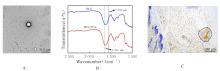
Fig. 1
Exo identification and biocompatibility of DCC/Exo"
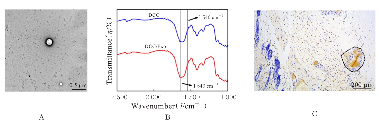
Fig. 2
Gross morphology(A) and adhesion scores (B) of PA tissue of mice in various groups"
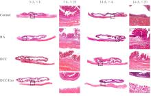
Fig. 3
Pathomorphology of PA tissue of mice in various groups(HE)"
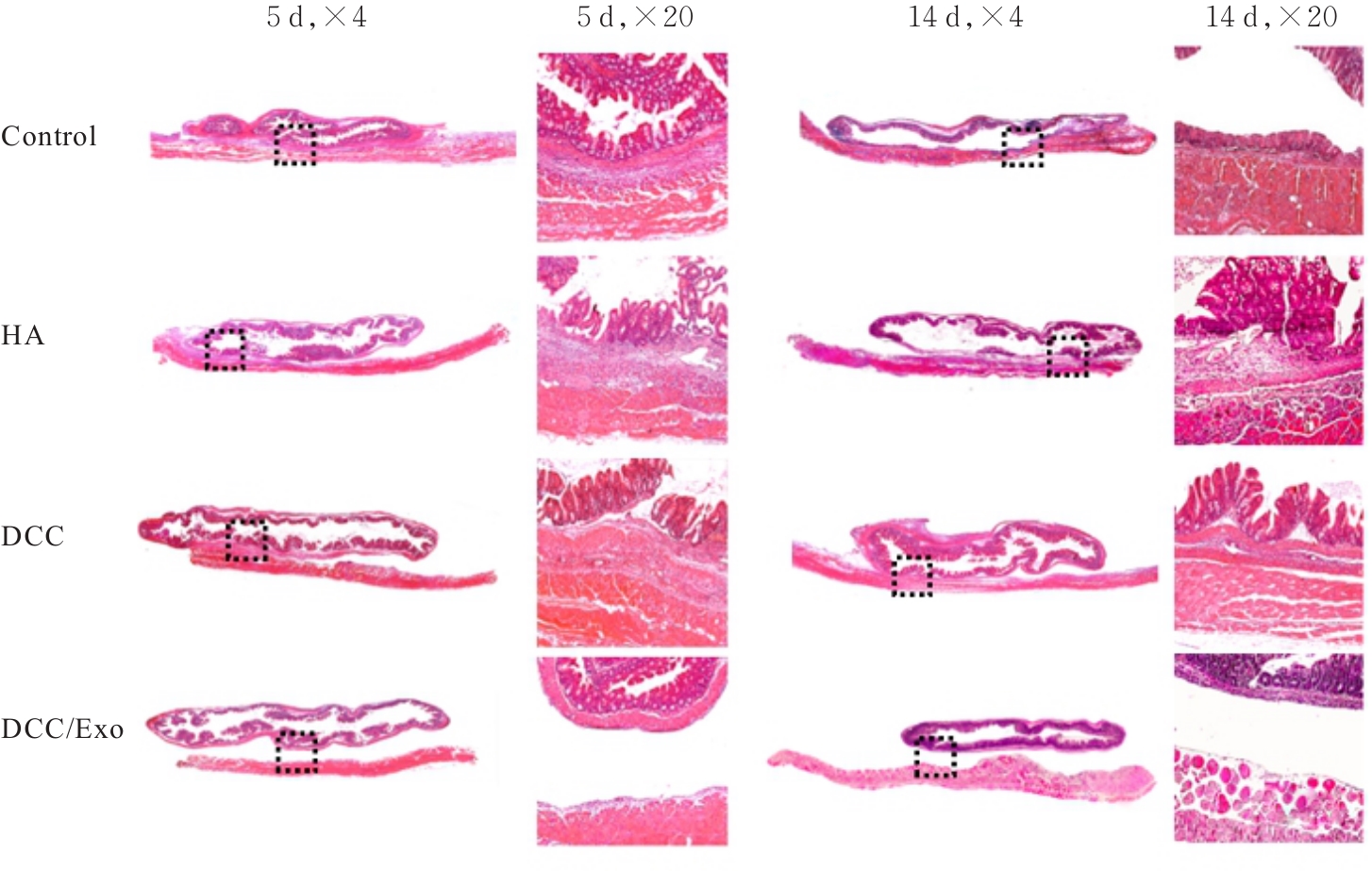

Fig. 4
Expression of MPO in PA tissue of mice in various groups"
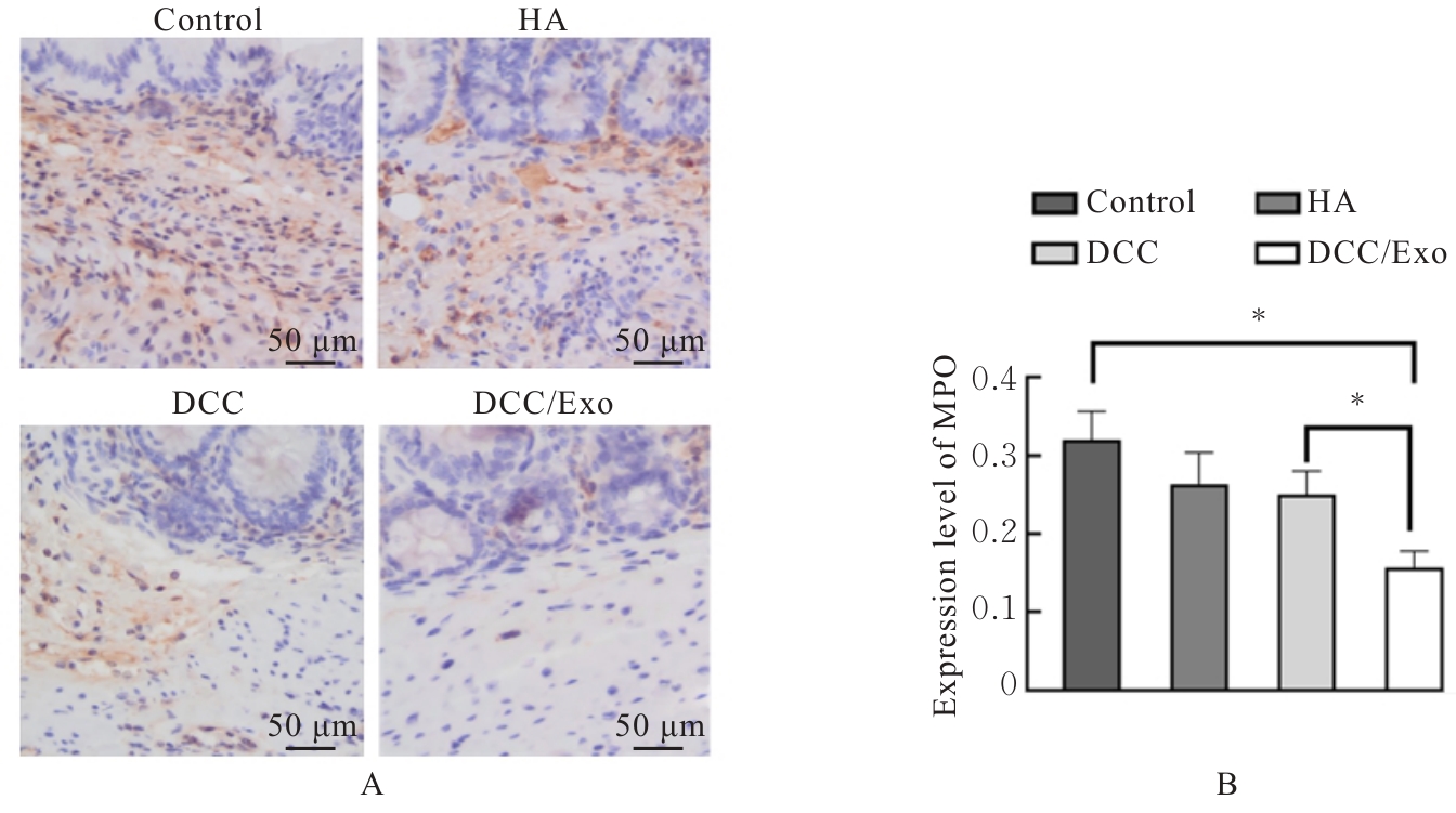
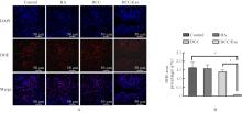
Fig. 5
Expressions of ROS in PA tissue of mice in various groups"
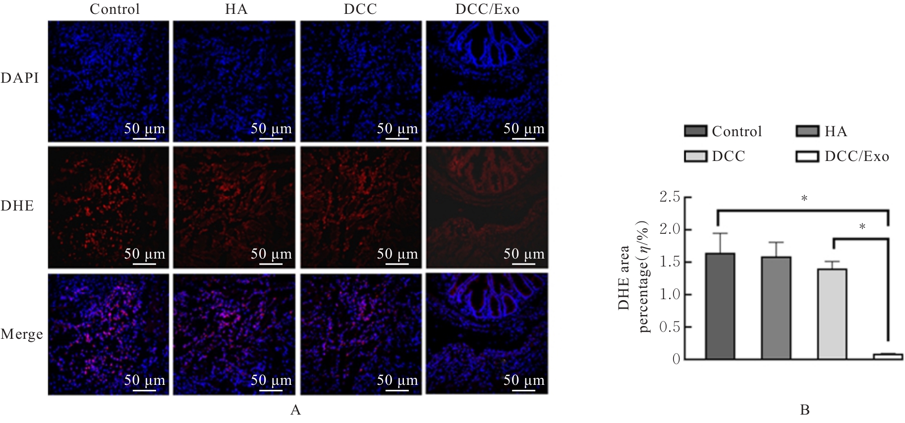
Fig. 6
Expressions of 8-OHdG in PA tissue of mice in various groups"
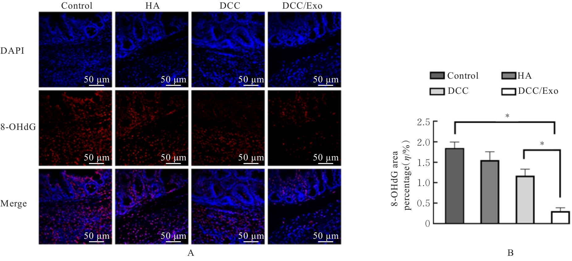
Fig. 7
Expressions of Vimentin and E-cadherin proteins in PA tissue of mice in various groups"
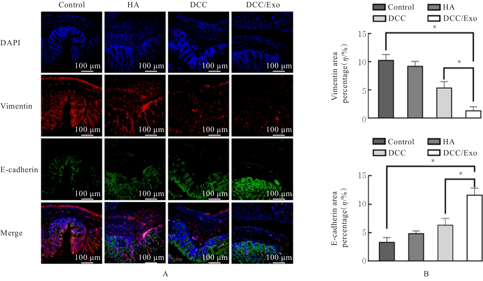
Fig. 8
Mitochondrial injury in PA tissue of mice in various groups"
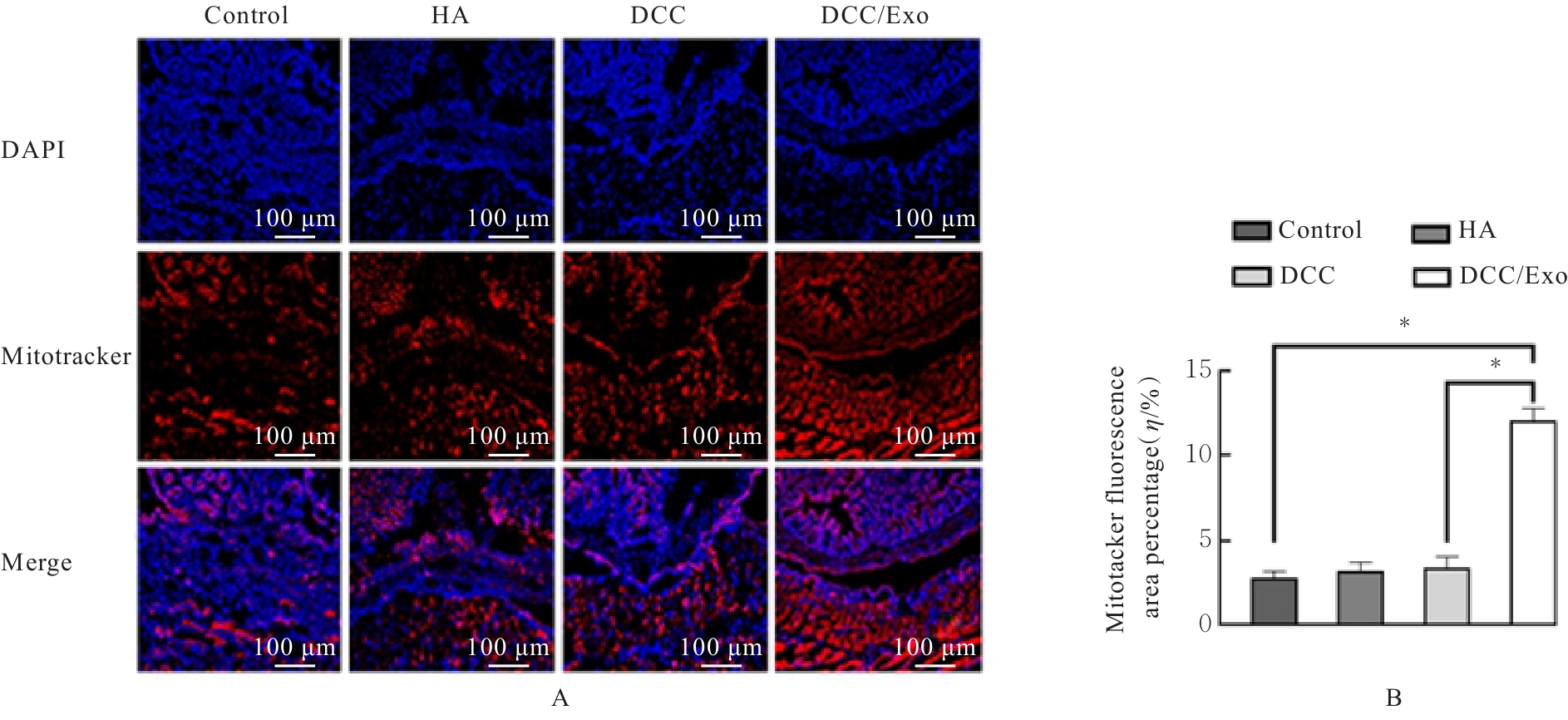
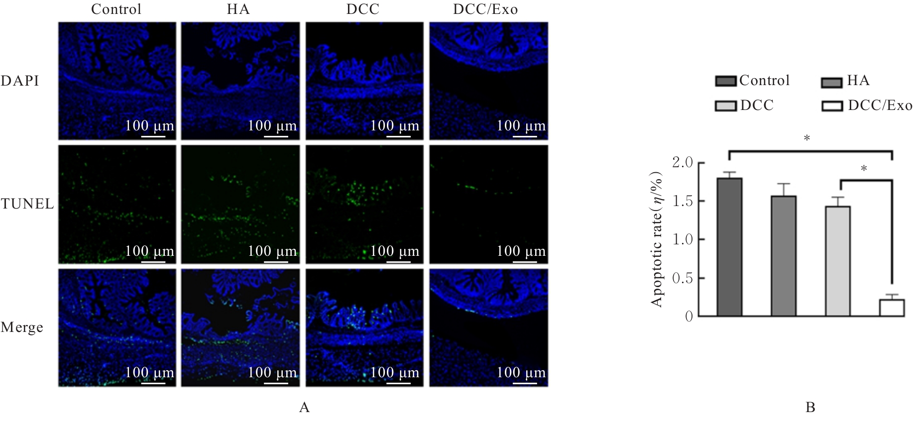
Fig. 9
Apoptosis of cells in PA tissue of mice in various groups"
| [1] | VAKILIAN S, AL-HASHMI S, AL-KINDI J, et al. Avastin-loaded 3D-printed alginate scaffold as an effective antiadhesive barrier to prevent postsurgical adhesion bands formation[J]. Macromol Biosci, 2024, 24(6): e2300530. |
| [2] | LI X Y, CAI J, DUAN X M, et al. Injectable polyamide-amine dendrimer-crosslinked meloxicam-containing poly-γ-glutamic acid hydrogel for prevention of postoperative tissue adhesion through inhibiting inflammatory responses and balancing the fibrinolytic system[J]. J Colloid Interface Sci, 2024, 670: 486-498. |
| [3] | MA Y Y, SUN T H, REN K J, et al. Plasma-activated solutions prevent peritoneal adhesion formation by regulating eNOS expression in mesothelial cells[J]. J Adv Res, 2025, 78: 555-571. |
| [4] | CHEN Y, WANG X, TAO S, et al. Research advances in smart responsive-hydrogel dressings with potential clinical diabetic wound healing properties[J]. Mil Med Res, 2023, 10(1): 37. |
| [5] | 江俊杰, 武 皓, 何 康, 等. 人参多肽温敏水凝胶对大鼠热源性皮肤损伤的修复作用及其机制[J]. 吉林大学学报(医学版), 2025, 51(2): 360-369. |
| [6] | MONDAL J, PILLARISETTI S, JUNNUTHULA V, et al. Hybrid exosomes, exosome-like nanovesicles and engineered exosomes for therapeutic applications[J]. J Control Release, 2023, 353: 1127-1149. |
| [7] | TAN X, ZHANG J, HENG Y Y, et al. Locally delivered hydrogels with controlled release of nanoscale exosomes promote cardiac repair after myocardial infarction[J]. J Control Release, 2024, 368: 303-317. |
| [8] | YU Q Y, SUN H, YUE Z W, et al. Zwitterionic polysaccharide-based hydrogel dressing as a stem cell carrier to accelerate burn wound healing[J]. Adv Healthc Mater, 2023, 12(7): e2202309. |
| [9] | FAN M H, PI J K, ZOU C Y, et al. Hydrogel-exosome system in tissue engineering: a promising therapeutic strategy[J]. Bioact Mater, 2024, 38: 1-30. |
| [10] | ZHANG Z Q, YIN C, SONG X W, et al. A self-fused peptide-loaded hydrogel with injectability and tissue-adhesiveness for preventing postoperative peritoneal adhesions[J]. Mater Today Bio, 2024, 28: 101205. |
| [11] | KRISHNAN M A, ALIMI O A, KUSS M, et al. A dual-layer hydrogel barrier integrating bio-adhesive and anti-adhesive properties prevents postoperative abdominal adhesions[J]. Adv Healthc Mater, 2025, 14(11): 2405238. |
| [12] | ZHANG Y H, YU L, QIU R J, et al. 3D hypoxia-mimicking and anti-synechia hydrogel enabling promoted neovascularization for renal injury repair and regeneration[J]. Mater Today Bio, 2023, 21: 100694. |
| [13] | FOSTER D S, GUO J L, MEANY E, et al. Postoperative adhesions are abrogated by a sustained-release anti-JUN therapeutic in preclinical models[J]. Sci Transl Med, 2025, 17(789): eadp9957. |
| [14] | LI X M, GUO Y F, WANG Z Y, et al. Platelet-rich fibrin promotes mesothelial cell proliferation and peritoneal repair by up-regulating calretinin to prevent postoperative intestinal adhesion[J]. Int J Med Sci, 2025, 22(6): 1254-1268. |
| [15] | JIANG L J, YAO F L, ZHANG E S, et al. Combined treatment of xyloglucan derivative hydrogel and anti-C5a receptor antibody in preventing peritoneal adhesion[J]. Acta Biomater, 2022, 151: 163-173. |
| [16] | GONG P, REN L L, GAO X H, et al. A novel barrier membrane with long-term ROS scavenging function for complete prevention of postoperative adhesion[J]. Mater Des, 2024, 238: 112691. |
| [17] | MASOLA V, BONOMINI M, BORRELLI S, et al. Fibrosis of peritoneal membrane as target of new therapies in peritoneal dialysis[J]. Int J Mol Sci, 2022, 23(9): 4831. |
| [18] | LU Y Q, ELROD J, HERRMANN M, et al. Neutrophil extracellular traps: a crucial factor in post-surgical abdominal adhesion formation[J]. Cells, 2024, 13(11): 991. |
| [19] | HERRO R, GRIMES H L. The diverse roles of neutrophils from protection to pathogenesis[J]. Nat Immunol, 2024, 25(12): 2209-2219. |
| [20] | CHEN J M, AN X Y, XU L, et al. Adhesive nanoparticle-in-microgel system with ROS scavenging capability and hemostatic activity for postoperative adhesion prevention[J]. Small, 2024, 20(27): e2306598. |
| [21] | REN Y W, LI G, LI E M, et al. Luteolin blocks the ROS/PI3K/AKT pathway to inhibit mesothelial-mesenchymal transition and reduce abdominal adhesions[J]. Eur J Pharmacol, 2024, 964: 176272. |
| [22] | LI G, REN Y W, LI E M, et al. Quercetin inhibits mesothelial-mesenchymal transition and alleviates postoperative peritoneal adhesions by blocking the TGF-β1/PI3K/AKT pathway[J]. J Ethnopharmacol, 2024, 319(Pt 2): 117242. |
| [23] | SANDOVAL P, JIMÉNEZ-HEFFERNAN J A, GUERRA-AZCONA G, et al. Mesothelial-to-mesenchymal transition in the pathogenesis of post-surgical peritoneal adhesions[J]. J Pathol, 2016, 239(1): 48-59. |
| [24] | WANG R P, GUO T K, LI J L. Mechanisms of peritoneal mesothelial cells in peritoneal adhesion[J]. Biomolecules, 2022, 12(10): 1498. |
| [25] | WANG Y, SHI Y F, TAO M, et al. Peritoneal fibrosis and epigenetic modulation[J]. Perit Dial Int, 2021, 41(2): 168-178. |
| [1] | Zheng TANG,Wei LIU,Hongmin HU,Qi LAI,Yan SHI,Shengquan LIU,Jun YANG,Chun CHU. Improvement effect of Qishen Yiqi dropping pills on myocardium injury in rats induced by sorafenib and its mechanism [J]. Journal of Jilin University(Medicine Edition), 2026, 52(2): 308-317. |
| [2] | Di LI,Yang SU,Yong ZHANG,Jiaoqi WANG,Xiaofei ZHANG,Xuebing ZHAO. Expression levels of miR-328-3p and miR-671-5p in serum exosomes of Kawasaki disease model mice and their significance [J]. Journal of Jilin University(Medicine Edition), 2026, 52(2): 359-365. |
| [3] | Tian LI,Jiafeng WANG,Zhimin ZHANG. Research progress in application of exosomes in diagnosis and treatment of oral diseases [J]. Journal of Jilin University(Medicine Edition), 2026, 52(2): 543-550. |
| [4] | Ziyi TANG,Shiying YANG,Tianzhen YANG,Wenqiang LIU,Jiangxue ZHONG,Li YIN. Induction effect of pesticide pyraclostrobin on ferroptosis of spermatocytes GC-2 of mice [J]. Journal of Jilin University(Medicine Edition), 2026, 52(1): 18-25. |
| [5] | Huiying YANG,Weihong LIANG,Xinru XU,Haodan SUN,Xin JIAO,Haiping WANG. Research progress in effect of bone marrow mesenchymal stem cells on differentiation induction of cardiomyocyte-like cells, protection of cardiomyocytes, and myocardial-related diseases [J]. Journal of Jilin University(Medicine Edition), 2026, 52(1): 281-289. |
| [6] | Sitong CHEN,Dan YANG,Qingjie LI,Xiaolei TANG,Han WANG,Yangyang LIU,Tiejun LIU. Ameliorative effect of puerarin on non-alcoholic fatty liver disease induced by high-fat diet in mice and its mechanism [J]. Journal of Jilin University(Medicine Edition), 2026, 52(1): 44-55. |
| [7] | Xiaoqian TANG,Shengcong WEN,Zhenya DONG,Jingyi CHEN,Yu CAO,Yunhua ZHANG. Improvement effect of engineered exosomes delivering ANGPTL6 mRNA on liver fibrosis in mice [J]. Journal of Jilin University(Medicine Edition), 2025, 51(6): 1452-1463. |
| [8] | Jing LIU,Yan WANG,Xu HUANG. Promoting effect of overexpressed circRNAs-modified dental pulp stem cell-derived exosomes on angiogenesis of human umbilical vein endothelial cells [J]. Journal of Jilin University(Medicine Edition), 2025, 51(6): 1561-1570. |
| [9] | Yi ZHAO,Bing ZHOU,Huirui QIU,Xuan LI,Xiangli CUI. Improvement effect of lovastatin on hyperlipidemia-induced liver injury in rats and its mechanism [J]. Journal of Jilin University(Medicine Edition), 2025, 51(5): 1155-1164. |
| [10] | Meng XIANG,Bing XU,Peisha WANG,Siqi LIU,Shaohua ZHANG. Effect of exosomes loaded with miR-520a-5p on pregnancy outcomes in fetal mice with intrauterine growth restriction and its mechanism [J]. Journal of Jilin University(Medicine Edition), 2025, 51(5): 1230-1239. |
| [11] | Nan LU,Mingxin DONG,Lei YU,Chengbiao SUN,Yan WANG,Na XU,Wensen LIU,Shumin GE. Transcriptome sequencing-based expression profiling of oxidative stress-related genes and circRNAs in ricin toxin-induced macrophage pyroptosis [J]. Journal of Jilin University(Medicine Edition), 2025, 51(4): 1007-1018. |
| [12] | Xiaoshuang HE,Lina XU,Mei CUI,Yu ZHAO,Bei WANG,Zheng HUANG,Yuchao WANG,Wenyan XIN,Chao WU. Effects of lncRNA DUXAP8 in lung cancer A549 cells-derived exosomes on lung cancer cell growth and its mechnism [J]. Journal of Jilin University(Medicine Edition), 2025, 51(4): 958-967. |
| [13] | DILIXIATI·Dilidaer,Lin JIA. Improvement effect of exosomes derived from human adipose-derived stem cells and human dermal fibroblasts on ultraviolet-induced photoaging skin wrinkles in nude mice [J]. Journal of Jilin University(Medicine Edition), 2025, 51(3): 621-631. |
| [14] | Yue WANG,Ning MA,Jiajun LU,Chengyao WANG,Linyu CHEN,Yuchen REN,Jingwu LI,Hong SUN. Protective effect of novel composite hydrogels on H₂O₂-induced oxidative stress injury in cardiomyocytes [J]. Journal of Jilin University(Medicine Edition), 2025, 51(2): 352-359. |
| [15] | Junjie JIANG,Hao WU,Kang HE,Zhiqiang SAN,Qing YANG,Hui LI,Na LI. Repair effect of ginseng polypeptide thermosensitive hydrogel on heat-induced skin injury in rats and its mechanism [J]. Journal of Jilin University(Medicine Edition), 2025, 51(2): 360-369. |
|