Journal of Jilin University(Medicine Edition) ›› 2026, Vol. 52 ›› Issue (2): 348-358.doi: 10.13481/j.1671-587X.20260206
• Research in basic medicine • Previous Articles Next Articles
Effect of miR-153-3p/PGC-1α axis on apoptosis of oral squamous cell carcinoma cells by modulation of mitochondrial apoptosis pathway
Jie YU,Zongkang WANG,Xun LIU,Yanan YANG,Jin TAN( )
)
- Department of Stomatology,First Affiliated Hospital,Hunan University of Traditional Chinese Medicine,Changsha 410000,China
-
Received:2025-04-14Accepted:2025-07-06Online:2026-03-28Published:2026-04-15 -
Contact:Jin TAN E-mail:woshiyujie2025@163.com
CLC Number:
- R739.8
Cite this article
Jie YU,Zongkang WANG,Xun LIU,Yanan YANG,Jin TAN. Effect of miR-153-3p/PGC-1α axis on apoptosis of oral squamous cell carcinoma cells by modulation of mitochondrial apoptosis pathway[J].Journal of Jilin University(Medicine Edition), 2026, 52(2): 348-358.
share this article
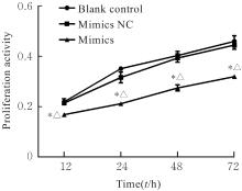
Fig. 1
Proliferation activities of cells in various groups"

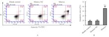
Fig. 2
Apoptotic rates of cells in various groups"
Fig. 3
Mitochondrial membrane potentials of cells in various groups"
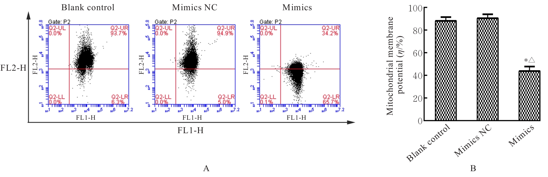
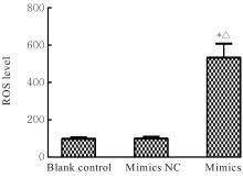
Fig. 4
Levels of ROS in cells in various groups"

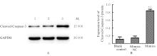
Fig. 5
Electrophoregram(A) and histogram(B) of expressions of Cleaved-Caspase-3 protein in cells in various groups"

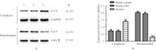
Fig. 6
Electrophoregram(A) and histogram(B) of expressions of Cyt C protein in cell cytoplasm and mitochondria in various groups"
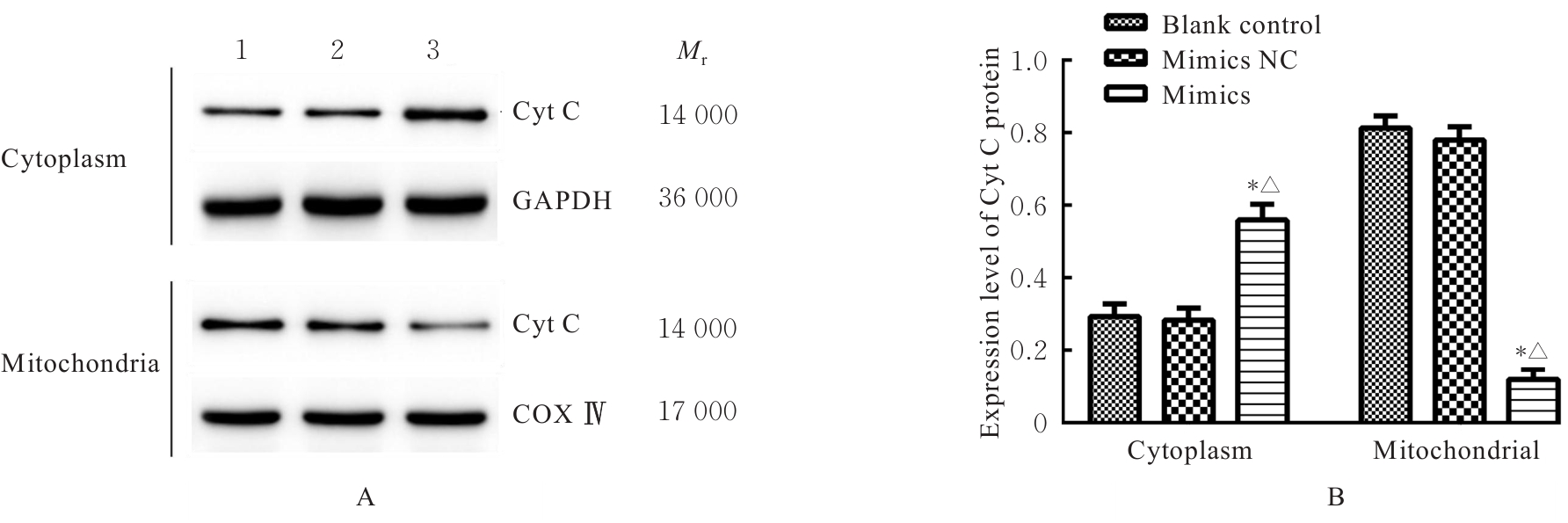
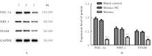
Fig. 7
Electrophoregram(A) and histogram(B) of expressions of PGC-1α, NRF-1 and TFAM proteins in cells in various groups"

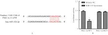
Fig. 8
Targeting regulation relationship between miR-153-3p and PGC-1α"
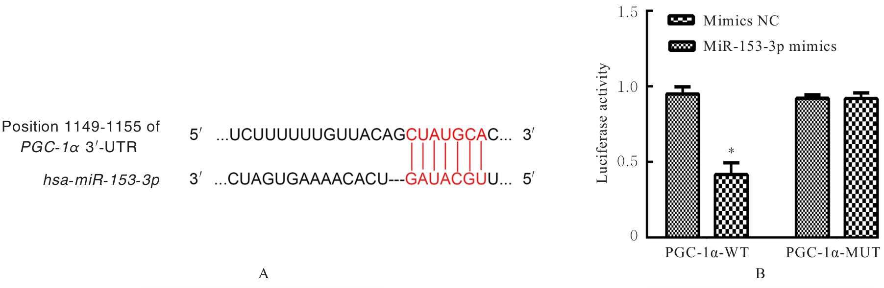
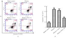
Fig. 9
Apoptotic rates of cells in various groups after co-transfection"
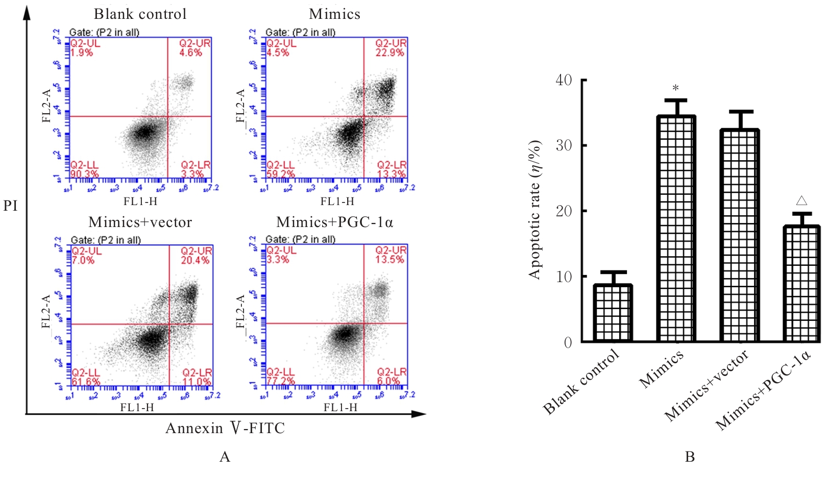

Fig. 10
Levels of ROS in Tca8113 cells in various groups after co-transfection"
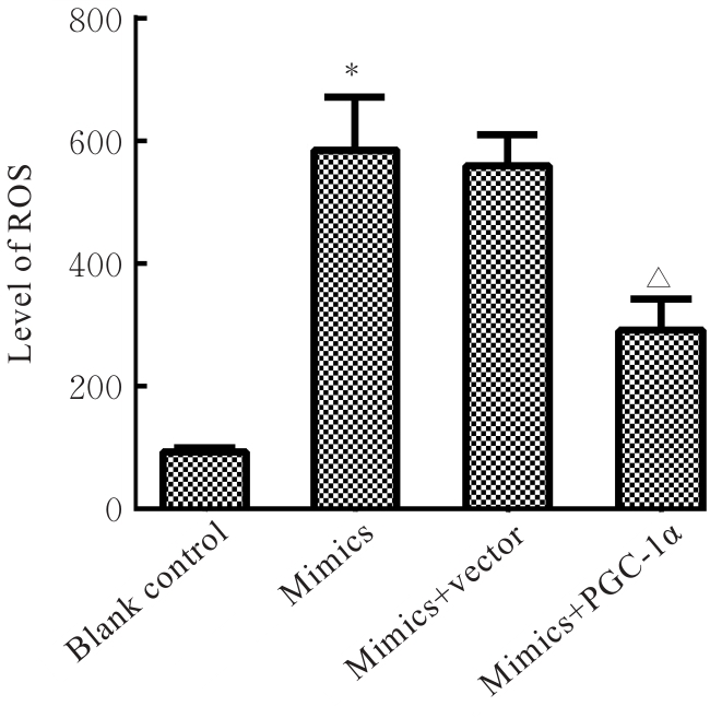
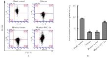
Fig. 11
Mitochondrial membrane potential of cells in various groups after co-transfection"
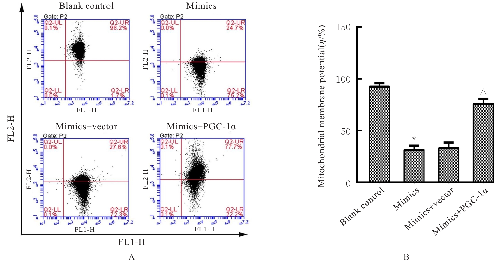
Fig. 12
Electrophoregram(A) and histogram(B) of expressions of PGC-1α/TFAM pathway-related proteins in cells in various groups after co-transfection"

| [1] | BRAY F, FERLAY J, SOERJOMATARAM I, et al. Global cancer statistics 2018: GLOBOCAN estimates of incidence and mortality worldwide for 36 cancers in 185 countries[J]. CA Cancer J Clin, 2018, 68(6): 394-424. |
| [2] | BANKI M, MOOSAVI M S. Umbrella review on cancer stem cell in oral and head and neck squamous cell carcinoma[J]. J Stem Cells Regen Med, 2023, 19(2): 29-33. |
| [3] | ARORA R, CAO C, KUMAR M, et al. Spatial transcriptomics unravels novel signaling patterns at the leading edge of oral squamous cell carcinoma[J]. J Clin Oncol, 2022, 40(16): e18043. |
| [4] | LAN W S, LU H J, CHIU Y W, et al. Clinical outcomes, prognosis, and predictors of salvage surgery in oral cavity squamous cell carcinoma[J]. J Clin Oncol, 2021, 39(15): e18040. |
| [5] | MASAOKA T, SHINOZUKA K, OHARA K, et al. Bioinformatics analysis of dysregulated exosomal microRNAs derived from oral squamous cell carcinoma cells[J]. J Oral Sci, 2021, 63(2): 174-178. |
| [6] | HUA X, LI Y S, PENTAPARTHI S R, et al. Landscape of microRNA regulatory network architecture and functional rerouting in cancer[J]. Cancer Res, 2023, 83(1): 59-73. |
| [7] | 田少辉, 张学浩, 徐江龙, 等. miR-194靶向Bmi-1调控PI3K/Akt信号通路对脑胶质瘤细胞迁移和凋亡的影响[J]. 中国免疫学杂志, 2021, 37(16): 1943-1947. |
| [8] | BABER S, BAYAT M, MOHAMADNIA A, et al. Role of miR153 and miR455-5p expression in oral squamous cell carcinoma isolated from plasma[J]. Asian Pac J Cancer Prev, 2021, 22(1): 157-161. |
| [9] | CHANG A C, LIEN M Y, TSAI M H, et al. WISP-1 promotes epithelial-mesenchymal transition in oral squamous cell carcinoma cells via the miR-153-3p/Snail axis[J]. Cancers, 2019, 11(12): 1903. |
| [10] | ZHANG Y, ZHAO H K, LI Y S. Pleiotropic regulation of PGC-1α in tumor initiation and progression[J]. Antioxid Redox Signal, 2024, 41(7): 557-572. |
| [11] | BRUNO G, PIETRAFESA M, CRISPO F, et al. TRAP1 modulates mitochondrial biogenesis via PGC-1α/TFAM signalling pathway in colorectal cancer cells[J]. J Mol Med, 2024, 102(10): 1285-1296. |
| [12] | 金 铭, 陈金亮, 张冬梅, 等. PGC-1α在肺癌中的研究进展[J]. 国际呼吸杂志, 2021, 41(17): 1355-1360. |
| [13] | 杨 浩, 王馨悦, 罗 瑛, 等. PGC-1α在肿瘤中的作用最新研究进展[J]. 中国细胞生物学学报, 2019, 41(10): 2019-2025. |
| [14] | LI Y, ZHANG H, JIANG X, et al. PGC-1α-mediated mitochondrial biogenesis promotes metastasis in oral squamous cell carcinoma through metabolic reprogramming[J]. J Exp Clin Cancer Res, 2022, 41(1):123. |
| [15] | CHEN L L, MAO X D, HUANG M M, et al. PGC-1α and ERRα in patients with endometrial cancer: a translational study for predicting myometrial invasion[J]. Aging, 2020, 12(17): 16963-16980. |
| [16] | HUANG X Q, RUAN G Y, LIU G F, et al. Immunohistochemical analysis of PGC-1α and ERRα expression reveals their clinical significance in human ovarian cancer[J]. Onco Targets Ther, 2020, 13: 13055-13062. |
| [17] | WANG P, GUO X Y, ZONG W, et al. PGC-1α/SNAI1 axis regulates tumor growth and metastasis by targeting miR-128b in gastric cancer[J]. J Cell Physiol, 2019, 234(10): 17232-17241. |
| [18] | ZHANG Q, LI J, WU S, et al. TFAM overexpression rescues mitochondrial dysfunction and apoptosis in PGC-1α-deficient colorectal cancer cells[J]. Cancer Letters, 2023, 553:215996 |
| [19] | LIU H, ZHANG Y, CHEN X, et al. TFAM-mediated mitochondrial DNA packaging promotes metabolic adaptation in pancreatic ductal adenocarcinoma[J]. Nat Commun, 2022, 13(1):3660. |
| [20] | ZHANG K Z, PETR J. Mitochondrial dynamics: updates and perspectives[J]. Sci Rep, 2024, 14(1): 9936. |
| [21] | TAHERZADEH-FARD E, SAFT C, AKKAD D A, et al. PGC-1alpha downstream transcription factors NRF-1 and TFAM are genetic modifiers of Huntington disease[J]. Mol Neurodegener, 2011, 6(1): 32. |
| [22] | PARK S, LEE J, KIM H, et al. PGC-1α inhibition sensitizes melanoma to ferroptosis via TFAM-dependent mitochondrial dysfunction[J]. Cell Rep, 2023, 42(3):112239. |
| [23] | KALPAGE H A, BAZYLIANSKA V, RECANATI M A, et al. Tissue-specific regulation of cytochrome с by post-translational modifications: respiration, the mitochondrial membrane potential, ROS, and apoptosis[J]. FASEB J, 2019, 33(2): 1540-1553. |
| [24] | SANTUCCI R, SINIBALDI F, COZZA P, et al. Cytochrome с: an extreme multifunctional protein with a key role in cell fate[J]. Int J Biol Macromol, 2019, 136: 1237-1246. |
| [25] | XU W, LI X, ZHANG H, et al. Exosomal miR-153-3p derived from cancer-associated fibroblasts promotes oral squamous cell carcinoma metastasis by regulating mitochondrial quality control[J]. J Extracell Vesicles, 2022, 11(5): e12217. |
| [1] | Moujie LIU,Jing ZHANG,Huihui WU,Juhua XIE. Improvement effect of metformin on myocardial hypertrophy in spontaneously hypertensive rats and its mechanism [J]. Journal of Jilin University(Medicine Edition), 2026, 52(2): 391-397. |
| [2] | Jiawei LI, Adilijiang,Li WU,Yun JIANG. Improvement effect of empagliflozin on ameliorating doxorubicin-induced myocardial injury rat model and its mechanism [J]. Journal of Jilin University(Medicine Edition), 2026, 52(1): 105-115. |
| [3] | Yunshan DING,Haitao DAI,Min CHEN,Xiaohui HAO,Xiao ZHOU,Nan WU. Effect of silencing TRPV2 gene and cannabidiol on biological behaviors of oral squamous cell carcinoma CAL-27 cells [J]. Journal of Jilin University(Medicine Edition), 2026, 52(1): 143-151. |
| [4] | Yaping LI,Chunyan TAN,Lujie ZHAO,Jiayi ZHAO,Ting LI,Xiao YANG,Xiaoyun YANG. Inductive effect of lysophosphatidic acid combined with 6-hydroxydopamine on apoptosis of SH-SY5Y cells and its mechanism [J]. Journal of Jilin University(Medicine Edition), 2026, 52(1): 152-161. |
| [5] | Yuxin LI,Lu YANG,Fengjin LI,Ling QI. Inductive effect of wedelolactone on cuproptosis in human pancreatic cancer PANC-1 cells [J]. Journal of Jilin University(Medicine Edition), 2026, 52(1): 182-191. |
| [6] | Jiaxin WANG,Junwen MAO,Xuan ZHANG. Expression of natural autoantibodies against apoptosis inhibitory genes in plasma of patients with hepatocellular carcinoma and its clinical significance [J]. Journal of Jilin University(Medicine Edition), 2026, 52(1): 228-235. |
| [7] | Huaimin LIANG,Jiacheng JIN,Wenhua CHEN,Yuyao LI,Hangyu WANG,Ke ZHANG,Jinhui WANG. UPLC-Q-TOF/MS and network pharmacology analysis and experimental verification based on potential active ingredients and mechanisms of medicinal Mulberry Leaves in anti-acute kidney injury [J]. Journal of Jilin University(Medicine Edition), 2026, 52(1): 56-69. |
| [8] | Shanshan SUN,Mei LU,Xinfu GAO,LYu Guangyao,Baolei Zhao,LYu Wenwen. Inhibitory effect of Bradykinin 1 receptor antagonist ELN441958 on proliferation of HepG2 cells by regulating Akt/FoxO3a signaling pathway [J]. Journal of Jilin University(Medicine Edition), 2026, 52(1): 70-80. |
| [9] | Xiaohan YAO,Mingchen YAO,Zhiqing WANG,Heyang LI,Yan YAN,Ningjing LEI. Effect of silencing GPR139 gene on proliferation, apoptosis and autophagy of breast cancer cells and its mechanism [J]. Journal of Jilin University(Medicine Edition), 2026, 52(1): 1-9. |
| [10] | Sifan FENG,Yunfeng LI,Jiaying WANG,Fubin MA,Yan WANG. Effects of heme-binding protein 1 gene knockdown on proliferation, migration, and inflammatory response of microglia BV2 and their mechanisms [J]. Journal of Jilin University(Medicine Edition), 2025, 51(6): 1532-1541. |
| [11] | Huiqin SUO,Chenxu JING,Jingming ZHAO,Chikun LI,Yunlu DING,Hongbo CHU,Guangyu CHENG,Qingjie LI,Hongguang JIN. Effect of β-elemene on mitochondrial structure and function of non-small cell lung cancer A549 cells [J]. Journal of Jilin University(Medicine Edition), 2025, 51(5): 1204-1210. |
| [12] | Han XUE,Yuxin FAN,Ting ZHANG,Zhimin LI,Mingge HUO,Xingang GUAN. Preparation of nanodrug PTX2 NPs and its killing effect on human lung cancer A549 cells [J]. Journal of Jilin University(Medicine Edition), 2025, 51(5): 1260-1266. |
| [13] | Xun LU,Chengxin MA,Jianan YANG,Xinxin GUO,Xiaobei XIE,Binghai ZHAO,Hongzhi LI. Research progress in mechanism of podocyte injury and its potential therapeutic strategies for diabetic nephropathy [J]. Journal of Jilin University(Medicine Edition), 2025, 51(5): 1415-1422. |
| [14] | Jiarui LI,Zhenlin YANG,Fan GAO,Jingjing GUO,Jinzi LI. Effect of miR-34a-5p on hippocampal neuron apoptosis in rats with temporal lobe epilepsy and its mechanism [J]. Journal of Jilin University(Medicine Edition), 2025, 51(4): 939-947. |
| [15] | Panxi SUN,Xue QIN,Chongyang ZHANG,Jia LUO,Yong CHEN,Lili WEI. Protective effect of TUG-891 on ischemic stroke induced by ischemia and hypoxia and its mechanism [J]. Journal of Jilin University(Medicine Edition), 2025, 51(4): 968-975. |
|