吉林大学学报(医学版) ›› 2021, Vol. 47 ›› Issue (3): 595-607.doi: 10.13481/j.1671-587X.20210308

• 基础研究 • 上一篇    下一篇

异钩藤碱上调miR-192-5p对TNF-α诱导的人支气管上皮细胞凋亡和炎症因子释放的作用及其机制

侯从岭(),芦晓帆,雷小婷,李彬,赵润杨   

  1. 河南省中医院肺病科,河南 郑州 450002
  • 收稿日期:2020-10-19 出版日期:2021-05-28 发布日期:2021-05-28
  • 通讯作者: 侯从岭 E-mail:xbhhyper@sina.com
  • 作者简介:侯从岭(1981-),男,河南省许昌市人,主治医师,医学硕士,主要从事中医药防治呼吸系统疾病方面的研究。
  • 基金资助:
    河南省中医药管理局国家中医临床研究基地科研专项(2019JDZX049);河南省科技厅攻关项目(192102310423)

Effect of isorhynchophylline on apoptosis and release of inflammatory factors in human bronchial epithelial cells induced by TNF-α via up-regulating miR-192-5p and its mechanism

Congling HOU(),Xiaofan LU,Xiaoting LEI,Bin LI,Runyang ZHAO   

  1. Department of Pulmonary Diseases,Henan Provincial Hospital of Traditional Chinese Medicine,Zhengzhou 450002,China
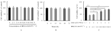
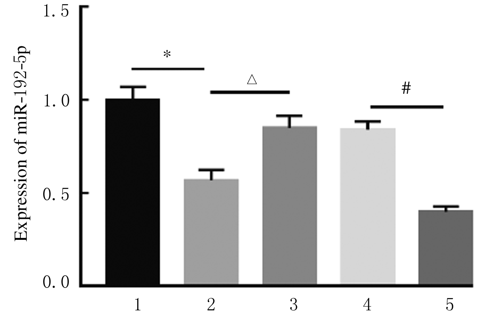
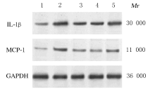
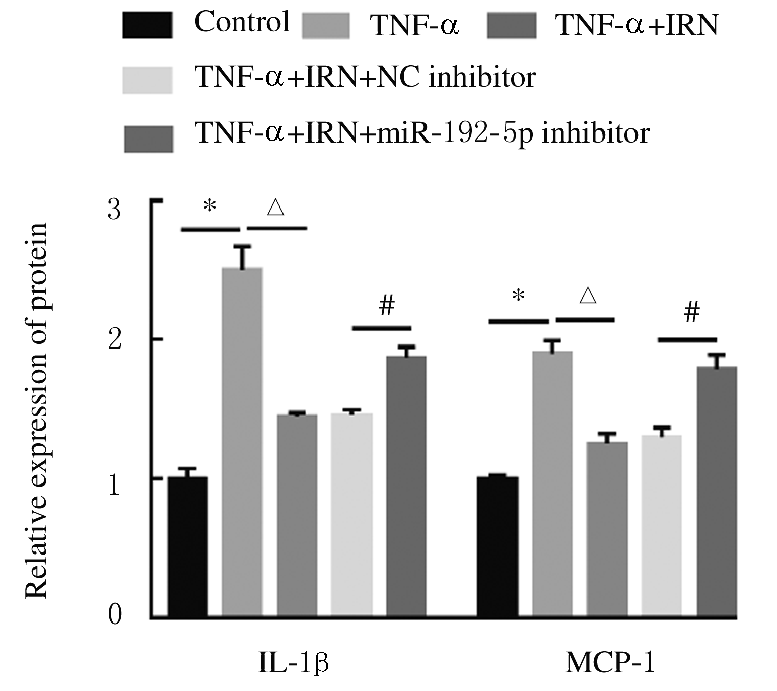
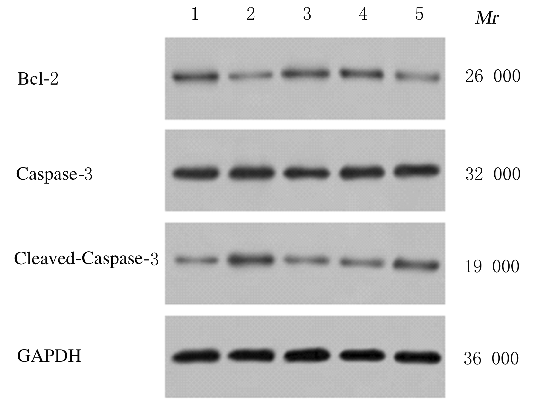
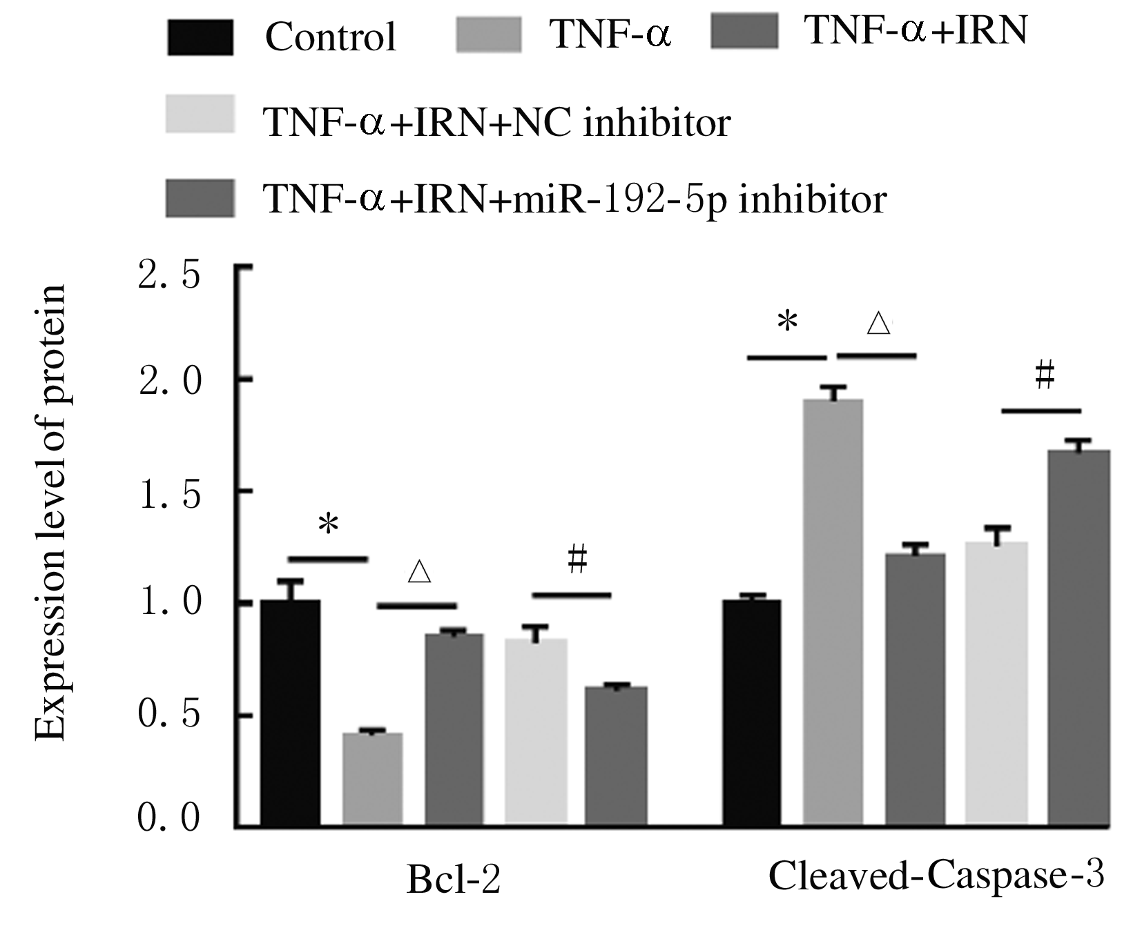
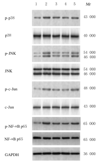
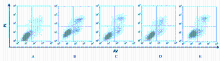
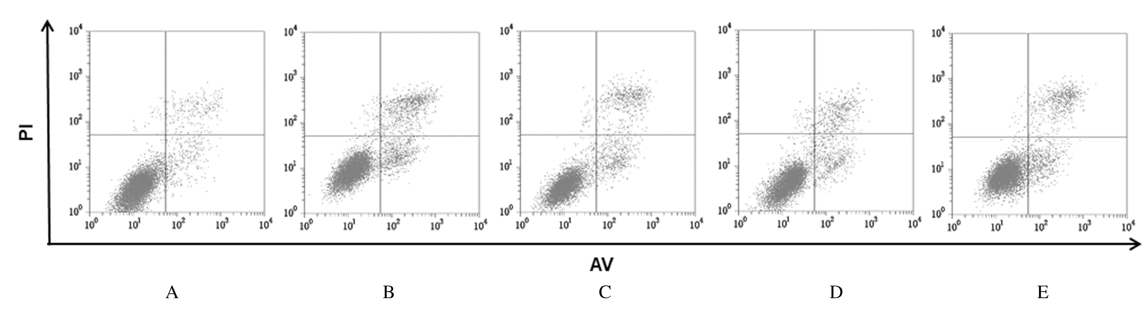
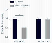
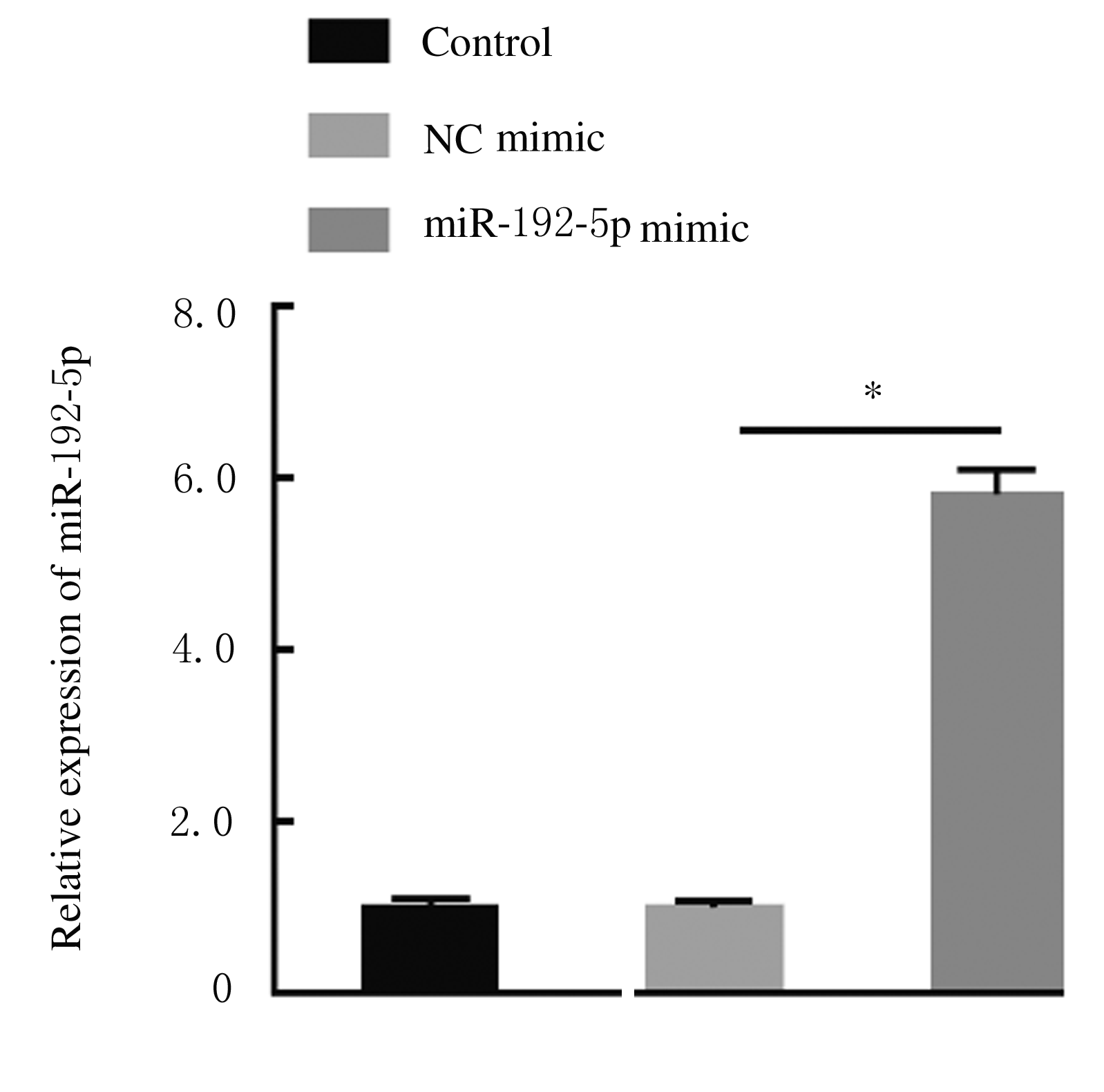
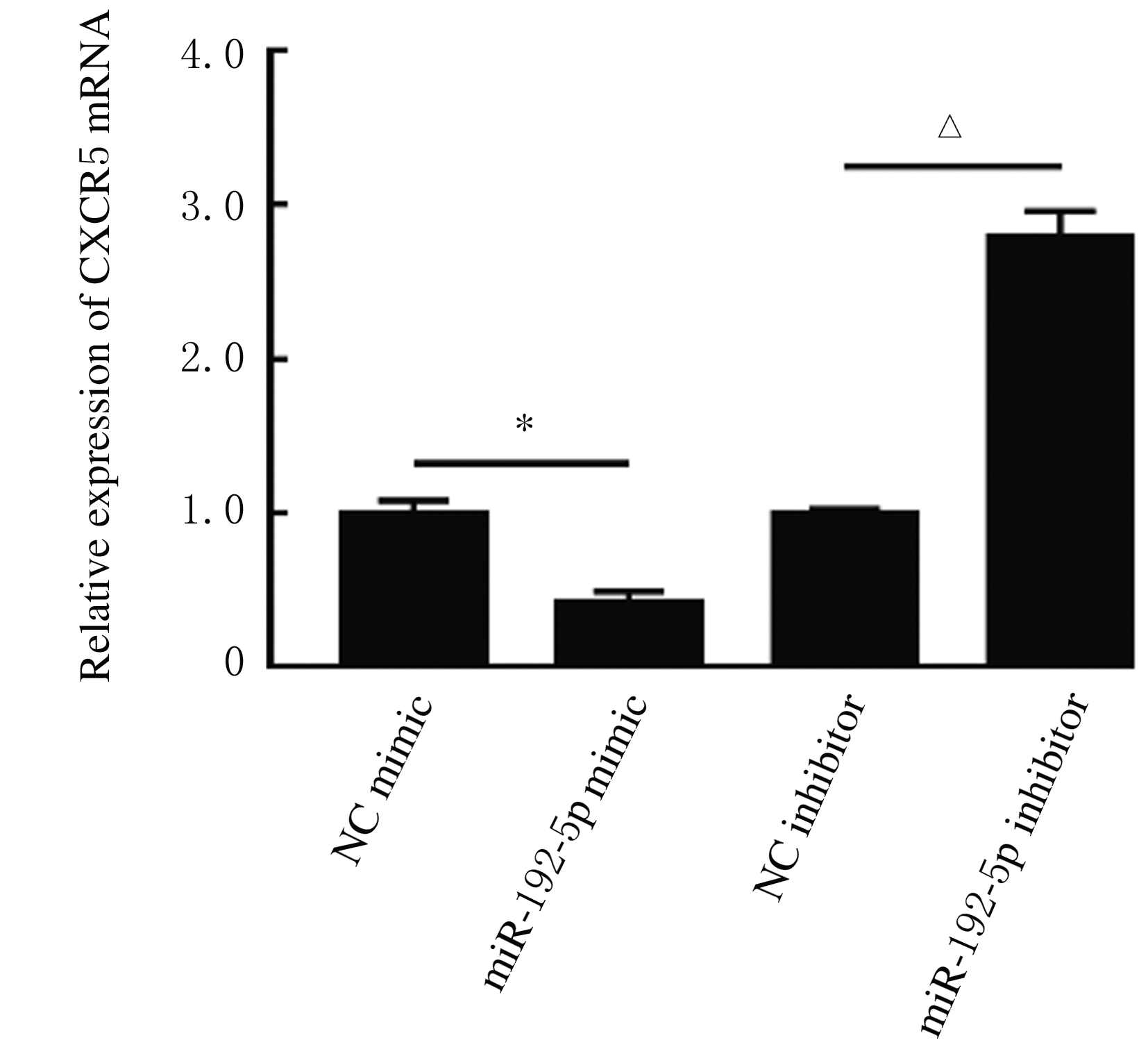
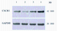
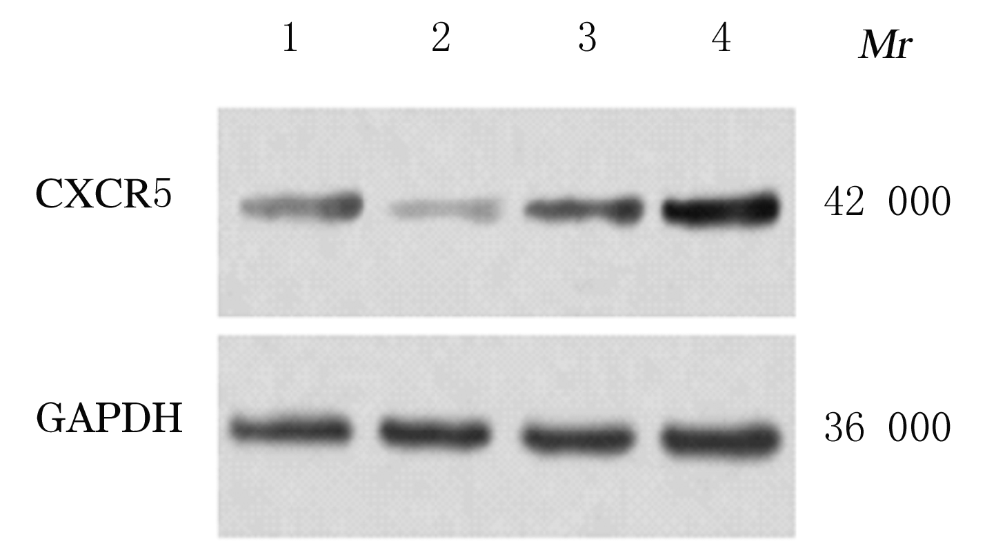
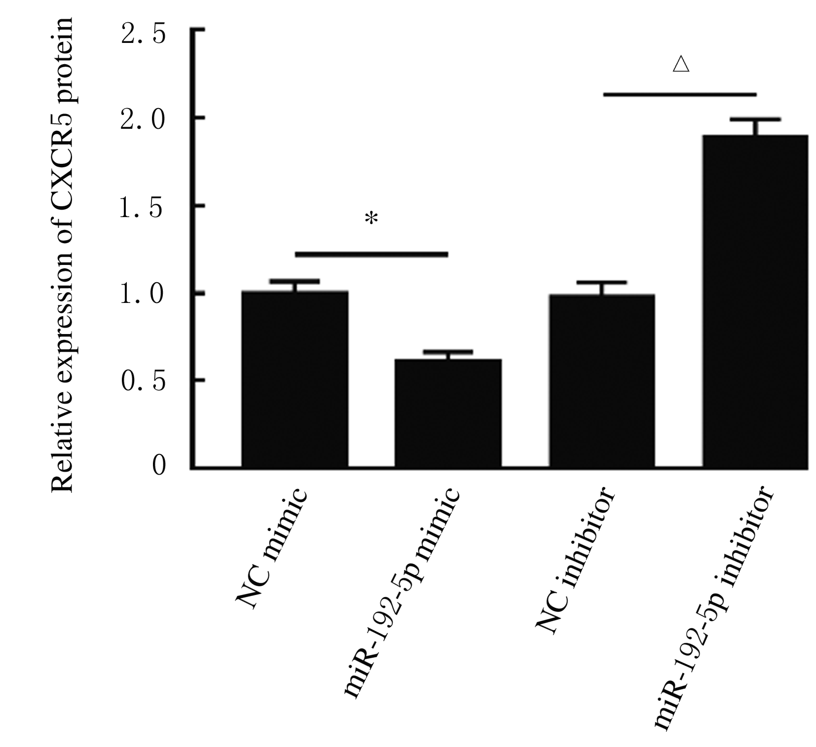
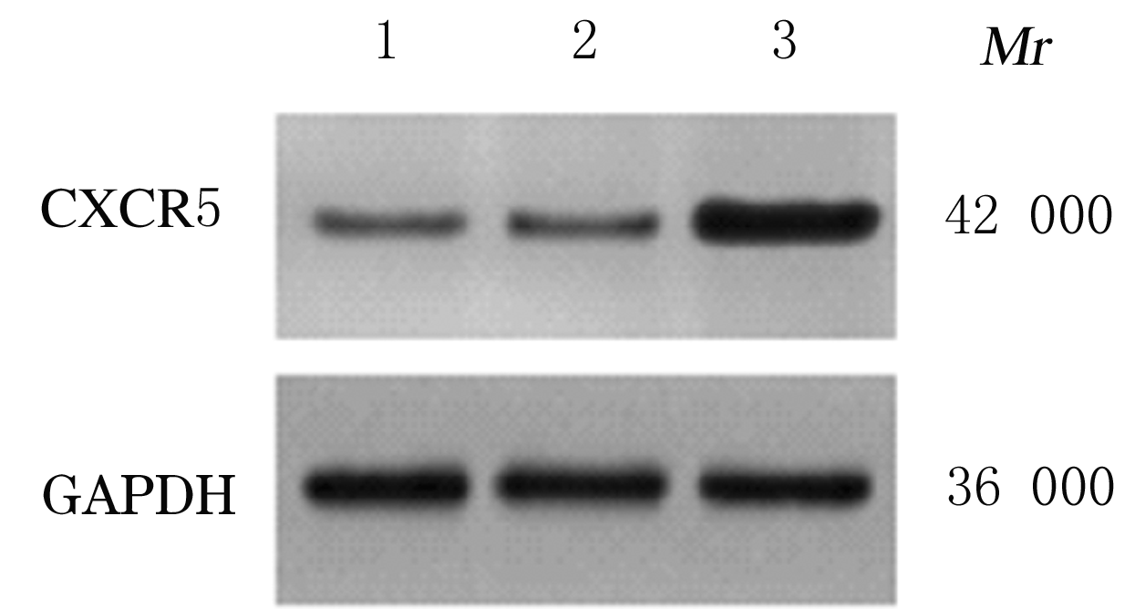
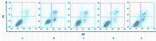
  • Received:2020-10-19 Online:2021-05-28 Published:2021-05-28
  • Contact: Congling HOU E-mail:xbhhyper@sina.com

摘要: 目的

探讨异钩藤碱(IRN)对肿瘤坏死因子α(TNF-α)诱导的人支气管上皮细胞(16HBE)凋亡和炎症因子释放的影响,并阐明其可能的作用机制。

方法

采用不同浓度[(0(对照组)、2.5、5.0、10.0、20.0、30.0和40.0 μmol·L-1]IRN处理16HBE细胞24 h后,采用20 mg·L-1 TNF-α处理细胞18 h。将16HBE细胞分为对照组、TNF-α组(20 mg·L-1)、IRN组(20 μmol·L-1)、TNF-α+IRN组(20 mg·L-1 TNF-α和20 μmol·L-1 IRN)、TNF-α+IRN+miR-192-5p inhibitor组(20 mg·L-1 TNF-α、20 μmol·L-1 IRN和50 nmol·L-1 miR-192-5p inhibitor)以及TNF-α+IRN+pcDNA3.1 CXCR5组[20 mg·L-1 TNF-α、20 μmol·L-1 IRN和2 μmol·L-1 pcDNA3.1-C-X-C趋化因子受体5(CXCR5)]。采用CCK-8法检测各组16HBE细胞活性,实时荧光定量PCR(RT-qPCR)法检测各组16HBE细胞中miR-192-5p和CXCR5 mRNA表达水平,Western blotting法检测各组16HBE细胞中白细胞介素1β(IL-1β)、单核细胞趋化蛋白 1(MCP-1)、Bcl-2、Cleaved-Caspase3、CXCR5以及磷酸化的p38、c-Jun氨基末端激酶(JNK)、c-Jun和核因子-κB(NF-κB)p65蛋白表达水平,ELISA法检测各组16HBE细胞上清液中IL-1β和MCP-1水平,流式细胞术检测各组16HBE细胞凋亡率,TargetScan7.1网站预测靶基因并通过双荧光素酶报告基因实验验证miR-192-5p与CXCR5之间的靶向结合关系。

结果

与对照组比较,不同浓度TNF-α组细胞活性明显降低(P<0.05);与对照组组比较,5.0、10.0、20.0和30.0 μmol·L-1 IRN组细胞活性升高(P<0.05)。与对照组比较,TNF-α组16HBE细胞中miR-192-5p和Bcl-2蛋白表达水平明显降低(P<0.05),IL-1β、MCP-1、Cleaved-Caspase-3、p-p38、p-JNK、p-c-Jun和p-NF-κBp65蛋白表达水平,细胞凋亡率以及上清液中IL-1β和MCP-1水平明显升高(P<0.05);与TNF-α组比较,TNF-α+IRN组16HBE细胞中miR-192-5p和Bcl-2蛋白表达水平明显升高(P<0.05),IL-1β、MCP-1、Cleaved-Caspase-3、p-p38、p-JNK、p-c-Jun和p-NF-κBp65蛋白表达水平,细胞凋亡率以及上清液中IL-1β和MCP-1水平明显降低(P<0.05);与TNF-α+IRN+NC inhibitor组比较,TNF-α+IRN+miR-192-5p inhibitor组16HBE细胞中miR-192-5p和Bcl-2蛋白表达水平明显降低(P<0.05),IL-1β、MCP-1、Cleaved-Caspase-3、p-p38、p-JNK、p-c-Jun和p-NF-κBp65蛋白表达水平,细胞凋亡率以及上清液中IL-1β和MCP-1水平明显升高(P<0.05)。与NC mimic和NC inhibitor组比较,miR-192-5p mimic组16HBE细胞中CXCR5 mRNA和蛋白表达水平明显降低(P<0.05),miR-192-5p inhibitor组16HBE细胞中CXCR5 mRNA和蛋白表达水平明显升高(P<0.05)。与对照组比较,TNF-α组16HBE细胞中CXCR5蛋白表达水平明显升高(P<0.05),细胞活性明显降低(P<0.05),细胞凋亡率明显升高(P<0.05),上清液中IL-1β和MCP-1水平明显升高(P<0.05);与TNF-α组比较,TNF-α+IRN组16HBE细胞中CXCR5蛋白表达水平明显降低(P<0.05),细胞活性明显升高(P<0.05),细胞凋亡率明显降低(P<0.05),上清液中IL-1β和MCP-1水平明显降低(P<0.05);与TNF-α+IRN+ pcDNA3.1组比较,TNF-α+IRN+ pcDNA3.1 CXCR5组16HBE细胞中CXCR5蛋白表达水平明显升高(P<0.05),细胞活性明显降低(P<0.05),细胞凋亡率明显升高(P<0.05),上清液中IL-1β和MCP-1水平明显升高(P<0.05)。

结论

IRN能减轻TNF-α诱导的人支气管上皮细胞凋亡和炎症因子释放,其作用机制可能与miR-192-5p/MAPKs/ NF-κB信号通路有关。

关键词: 异钩藤碱, 慢性阻塞性肺疾病, 支气管上皮细胞, miR-192-5p, C-X-C趋化因子受体5

Abstract: Objective

To observe the effect of isorhynchophylline (IRN) on TNF-α-induced the apoptosis and the release of inflammatory factors in the human bronchial epithelial (16HBE) cells, and to explore its possible mechanism.

Methods

After treated with different concentrations[0(control group),2.5, 5.0, 10.0, 20.0, 30.0 and 40.0 μmol·L-1] of IRN for 24 h, the 16HBE cells were cultured with 20 mg·L-1 TNF-α for 18 h. The 16HBE cells were divided into control group, TNF-α group (20 mg·L-1), IRN group (20 μmol·L-1), TNF-α+IRN group (20 mg·L-1 TNF-α and 20 μmol·L-1 IRN), TNF-α+IRN+miR-192-5p inhibitor group (20 mg·L-1 TNF-α, 20 μmol·L-1 IRN and 50 nmol·L-1 miR-192- 5p inhibitor) and TNF-α+IRN+pcDNA3.1 CXCR5 group [20 mg·L-1 TNF-α, 20 μmol·L-1 IRN and 2 μmol·L-1 pcDNA3.1-C-X-C chomokine receptor type 5(CXCR5)]. CCK-8 method was used to detect the viabilities of 16HBE cells in various groups; Real-time fluorescence quantitative PCR(RT-qPCR) method was used to detect the expression levels of miR-192-5p and CXCR5 mRNA in the 16HBE cells in various groups;Western blotting method was used to detect the expression levels of interleukins-1β (IL-1β)and monocyte chemoattactant protein-1 (MCP-1),B cell lymphoma-2(Bcl-2), Cleaved-Caspase 3, CXCR5, and phosphorylated p38(p-P38), c-Jun N-terminal kinase (JNK), c-Jun, and nuclear factor-κB (NF-κB) p65 proteins in the 16HBE cells in various groups; ELISA method was used to detect the levels of IL-1β and MCP-1 in supernatant of the 16HBE cells in various groups. TargetScan7.1 website was used to predict the target genes and the targeted binding association between miR-192-5p and CXCR5 was verified by dual-luciferase reporter gene assay.

Results

Compared with control group, the viability of the 16HBE cells in TNF-α group was significantly decreased (P<0.05);compared with control group, the viabilities of 16HBE cells in 5.0,10.0,20.0, and 30.0 μmol·L-1 IRN groups were increased (P<0.05). Compared with control group, the expression levels of miR-192-5p and Bcl-2 proteins in the 16HBE cells in TNF-α group were significantly decreased (P<0.05), the expression levels of IL-1β, MCP-1,Cleaved-Caspase-3, p-p38, p-JNK, p-c-Jun and p-NF-κB p65 proteins, the apoptotic rate and the levels of IL-1β and MCP-1 in supernatant of the 16HBE cells were significantly increased (P<0.05); compared with TNF-α group, the expression levels of miR-192-5p and Bcl-2 protein in the 16HBE cells in TNF-α+IRN group were significantly increased (P<0.05), the the expression levels of IL-1β, MCP-1,Cleaved-Caspase-3, p-p38, p-JNK, p-c-Jun and p-NF-κB p65 proteins, the apoptotic rate and the levels of IL-1β and MCP-1 in supernatant of the 16HBE cells were significantly decreased (P<0.05); compared with TNF-α+IRN+NC inhibitor group, the expression levels of miR-192-5p and Bcl-2 protein in the 16HBE cells in TNF-α+IRN+ miR-192-5p inhibitor group were significantly decreased (P<0.05),the expression levels of IL-1β, MCP-1,Cleaved-Caspase-3, p-p38, p-JNK, p-c-Jun and p-NF-κB p65 proteins,and the apoptotic rate and the levels of IL-1β and MCP-1 in supernatant of the 16HBE cells were significantly increased (P<0.05). Compared with NC mimic and NC inhibitor groups, the expression levels of CXCR5 mRNA and protein in the 16HBE cells in miR-192-5p mimic group were significantly decreased (P<0.05), and the expression levels of CXCR5 mRNA and protein in the 16HBE cells in miR-192-5p inhibitor group were significantly increased (P<0.05). Compared with control group, the expression level of CXCR5 protein in the 16HBE cells in TNF-α group was significantly increased (P<0.05), the cell viability was decreased (P<0.05), and the apoptotic rate and the levels of IL-1β and MCP-1 in supernatant of the 16HBE cells were increased (P<0.05);compared with TNF-α group, the expression level of CXCR5 protein in the 16HBE cells in TNF-α+IRN group was significantly decreased (P<0.05),the cell viability was increased (P<0.05),and the apoptotic rate and the levels of IL-1β and MCP-1 were significantly decreased (P<0.05); compared with TNF-α+IRN+ pcDNA3.1 group, the expression level of CXCR5 protein in the 16HBE cells in TNF-α+IRN+ pcDNA3.1 CXCR5 group was significantly increased (P<0.05),the cell viability was decreased (P<0.05),and the apoptotic rate and the levels of IL-1β and MCP-1 in supernatant of the 16HBE cells were increased (P<0.05).

Conclusion

IRN can reduce the TNF-α-induced apoptosis and release of inflammatory factors in the human bronchial epithelial cells, and its mechanism may be related to the miR-192-5p/MAPKs/NF-κB signal pathway.

Key words: isorhynchophylline, chronic obstructive pulmonary disease, human bronchial epithelial cells, miR-192-5p, C-X-C chemokine receptor type 5

中图分类号: 

  • Q71