吉林大学学报(医学版) ›› 2021, Vol. 47 ›› Issue (5): 1077-1085.doi: 10.13481/j.1671-587X.20210501

• 基础研究 •    下一篇

视黄酸通过GSK-3β对大鼠缺氧缺血性脑损伤后海马神经干细胞增殖的调节作用

李思雨1,2,赵敏1,2,杨茂玲1,2,肖农1,2,江伟1,2()   

  1. 1.重庆医科大学附属儿童医院康复科 儿童发育疾病研究教育部重点实验室 国家儿童健康与疾病 临床医学研究中心 儿童发育重大疾病国家国际科技合作基地,重庆 400014
    2.儿科学重庆市重点 实验室 认知发育与学习记忆障碍转化医学重庆市重点实验室,重庆 400014
  • 收稿日期:2021-01-21 出版日期:2021-09-28 发布日期:2021-10-29
  • 通讯作者: 江伟 E-mail:18745297@qq.com
  • 作者简介:李思雨(1995-),女,河南省洛阳市人,在读硕士研究生,主要从事维生素A在缺氧缺血性脑损伤中作用及其机制方面的研究。
  • 基金资助:
    国家自然科学基金面上项目(81571091);重庆市卫健委及科委联合课题(2018ZDXM029)

Regulatory effect of retinoic acid on proliferation of hippocampal neural stem cells after hypoxic-ischemic brain damage in rats via GSK-3β

Siyu LI1,2,Min ZHAO1,2,Maoling YANG1,2,Nong XIAO1,2,Wei JIANG1,2()   

  1. 1.Department of Rehabilitation,Ministry of Education Key Laboratory of Child Development and Disorders,National Clinical Research Center for Child Health and Disorders,China International Science and Technology Cooperation Base of Child Development and Critical Disorders,Children’s Hospital,Chongqing Medical University,Chongqing 400014,China
    2.Chongqing Key Laboratory of Pediatrics,Chongqing Key Laboratory of Translational Medical Research in Cognitive Development and Learning and Memory Disorders,Chongqing 400014,China
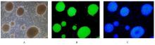
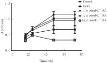
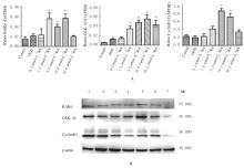
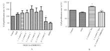
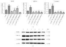
  • Received:2021-01-21 Online:2021-09-28 Published:2021-10-29
  • Contact: Wei JIANG E-mail:18745297@qq.com

摘要: 目的

研究不同浓度视黄酸(RA)对大鼠缺氧缺血性脑损伤(HIBD)后海马神经干细胞增殖的影响,并探讨其可能的作用机制。

方法

体外培养原代海马神经干细胞,并进行免疫荧光鉴定,对神经干细胞施以氧糖剥夺(OGD)损伤,模拟体内HIBD损伤。将细胞分为对照组、OGD组(0 μmol·L-1 RA干预)和不同浓度(0.5、1.0、5.0、10.0和50.0 μmol·L-1) RA干预组,CCK-8法检测OGD后12、24、48和72 h各组细胞增殖活性,实时荧光定量PCR(RT-qPCR)法和Western blotting法检测各组细胞中视黄酸核受体α(RARα)、糖原合酶激酶 3β(GSK-3β)和G1/S-特异性周期蛋白-D1(CyclinD1)mRNA表达水平和蛋白表达量。利用GSK-3β抑制剂CHIR99021处理不同浓度(0、0.5、5.0和50.0 μmol·L-1)RA干预后的海马神经干细胞,将细胞分为对照组,5.0 μmol·L-1 RA干预组,0、0.5、5.0和50.0 μmol·L-1 RA+20 μmol·L-1 CHIR99021干预组,CCK-8法检测OGD后24 h细胞增殖率,RT-qPCR法和Western blotting法检测各组细胞中RARα、GSK-3β和CyclinD1 mRNA表达水平和蛋白表达量。

结果

免疫荧光鉴定,成功提取并培养原代海马神经干细胞。RA干预后的CCK-8法检测,与OGD组比较,1.0和5.0 μmol·L-1 RA干预组在OGD后各时间点细胞增殖活性均明显升高(P<0.05);RT-qPCR法和Western blotting法检测,与OGD组比较,5.0和10.0 μmol·L-1 RA干预组细胞中RARα、GSK-3β和CyclinD1 mRNA表达水平明显升高(P<0.05),1.0、5.0和10.0 μmol·L-1 RA干预组RARα和GSK-3β蛋白表达量增加。加入GSK-3β抑制剂CHIR99021干预后CCK-8法检测,与5.0 μmol·L-1 RA干预组比较,5.0 μmol·L-1 RA+20 μmol·L-1 CHIR99021干预组细胞增殖率明显降低(P<0.01);RT-qPCR法和Western blotting法检测,与5.0 μmol·L-1 RA干预组比较,0和0.5 μmol·L-1 RA+20 μmol·L-1 CHIR99021干预组细胞中RARα mRNA表达水平明显降低(P<0.01),0、0.5、5.0和50.0 μmol·L-1 RA+20 μmol·L-1 CHIR99021干预组细胞中GSK-3β和CyclinD1 mRNA表达水平均明显降低(P<0.05),GSK-3β和CyclinD1蛋白表达量减少。

结论

RA促进HIBD损伤后神经干细胞增殖存在适宜浓度窗,其机制可能是RA通过激活GSK-3β调节CyclinD1的转录,进而促进神经干细胞增殖。

关键词: 视黄酸, 缺氧缺血性脑损伤, 神经干细胞, 细胞增殖, 糖原合酶激酶3β

Abstract: Objective

To investigate the effects of different concentrations of retinoic acid (RA) on the proliferation of hippocampal neural stem cells after hypoxic-ischemic brain damage (HIBD) in the rats, and to investigate its possible mechanism.

Methods

The primary hippocampal neural stem cells were cultured and identified by immunofluorescence. The neural stem cells were injured by oxygen and glucose deprivation (OGD) to simulate the HIBD injury in vivo. The cells were divided into control group, OGD group (0 μmol·L-1 RA intervention) and different concentrations (0.5, 1, 5, 10 and 50 μmol·L-1) of RA intervention groups. The proliferation activities of cells in various groups were measured by CCK-8 method at 12, 24, 48 and 72 h after OGD. Real-time fluorescence quantitative PCR (RT-qPCR) and Western blotting methods were used to detect the expression levels of retinoic acid receptor alpha(RARα),glycogen synthase kinase-3β(GSK-3β) and CyclinD1 mRNA and the protein expression amounts in the hippocampal neural stem cells in various groups. After different concentrations(0, 0.5, 5.0 and 50.0 μmol·L-1) of RA intervention, the hippocampal neural stem cells were treated with GSK-3β inhibitor CHIR99021 and divided into control group, 5 μmol·L-1 RA intervention group, and 0, 0.5, 5.0 and 50.0 μmol·L-1 RA+20 μmol·L-1 CHIR99021 intervention groups. The cell proliferation rates were detected by CCK-8 assay at 24 h after OGD. RT-qPCR and Western blotting methods were used to detect the expression levels of RARα, GSK-3β and CyclinD1 mRNA and the protein expression amounts in the hippocampal neural stem cells in various groups.

Results

The immunofluorescence identification results showed that the primary hippocampal neural stem cells were successfully extracted and cultured.The CCK-8 method detection results after RA intervention showed that the proliferation activities of cells in 1.0 and 5.0 μmol·L-1 RA intervention groups were significantly increased at each time point after OGD compared with OGD group(P<0.05).Compared with OGD group, the expression levels of RARα,GSK-3β,and CyclinD1 mRNA in 5.0 and 10.0 μmol·L-1 RA intervention groups were significantly increased (P<0.05),and the expression amounts of RARα and GSK-3β proteins in 1.0,5.0 and 10.0 μmol·L-1 RA intervention groups were increased.After GSK-3β inhibitor CHIR99021 intervention, compared with 5.0 μmol·L-1 RA intervention group, the cell proliferation rate in 5.0 μmol·L-1 RA+20 μmol·L-1 CHIR99021 intervention group was significantly decreased (P<0.01), the expression levels of RARα mRNA in 0 and 0.5 μmol·L-1 RA+20 μmol·L-1 CHIR99021 intervention groups were significantly decreased (P<0.01),the expression levels of GSK-3β and CyclinD1 mRNA in 0, 0.5, 5.0 and 50.0 μmol·L-1 RA+20 μmol·L-1 CHIR99021 intervention groups were significantly decreased (P<0.05),and the expression amounts of GSK-3β and CyclinD1 were decreased.

Conclusion

There is an appropriate concentration window for RA to promote the proliferation of neural stem cells after HIBD injury,and its mechanism may be that RA regulates the transcription of CyclinD1 through the activation of GSK-3β, thus promotes the proliferation of neural stem cells.

Key words: retinoic acid, hypoxic-ischemic brain damage, neural stem cells, cell proliferation, glycogen synthase kinase 3β

中图分类号: 

  • R493