吉林大学学报(医学版) ›› 2021, Vol. 47 ›› Issue (6): 1415-1421.doi: 10.13481/j.1671-587X.20210610

• 基础研究 • 上一篇    下一篇

实验性种植体周围炎羊模型的建立和炎性因子在模型羊龈沟液及牙龈组织中的表达

杨明1,李含薇1(),吴巍2,宋洋3,贾涵4,林百惠5,赖亚辉5   

  1. 1.北华大学口腔医学院口腔内科学教研室,吉林 吉林 130013
    2.北华大学口腔医学院口外颌面外科教研室,吉林 吉林 130013
    3.北华大学口腔医学院基础教研室,吉林 吉林 130013
    4.北华大学口腔医学院修复学教研室,吉林 吉林 130013
    5.北华大学预防医学院营养学教研室,吉林 吉林 130013
  • 收稿日期:2021-03-26 出版日期:2021-11-28 发布日期:2021-12-14
  • 通讯作者: 李含薇 E-mail:li65575147@163.com
  • 作者简介:杨 明(1988-),男,吉林省集安市人,讲师,医学博士,主要从事牙周病病理基础方面的研究。
  • 基金资助:
    吉林省科技厅科研基金项目(YDZJ202101ZYTS123);吉林省教育厅“十三五”科学技术项目(JJKH20180369KJ)

Establishment of peri-implantitis sheep models and expressions of inflammatory factors in gingival crevicular fluid and gingival tissue of sheep

Ming YANG1,Hanwei LI1(),Wei WU2,Yang SONG3,Han JIA4,Baihui LIN5,Yahui LAI5   

  1. 1.Department of Oral Medicine,School of Stomatology,Beihua University,Jilin 132013,China
    2.Department of Oral and Maxillofacial Surgery,School of Stomatology,Beihua University,Jilin 132013,China
    3.Basic Teaching and Research Section,School of Stomatology,Beihua University,Jilin 132013,China
    4.Department of Prosthodontics,School of Stomatology,Beihua University,Jilin 132013,China
    5.Department of Nutrition,School of Preventive Medicine,Beihua University,Jilin 132013,China
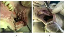
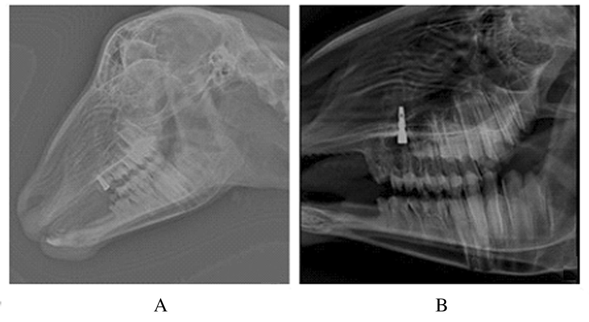
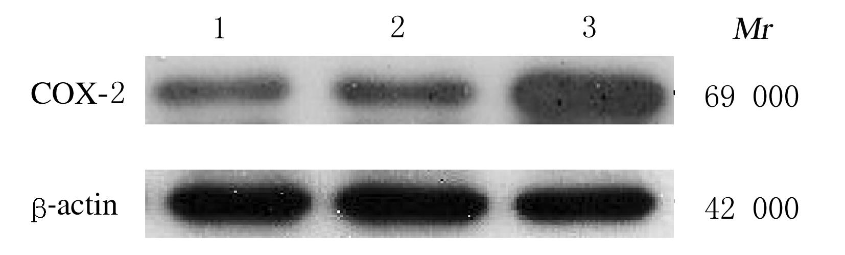
  • Received:2021-03-26 Online:2021-11-28 Published:2021-12-14
  • Contact: Hanwei LI E-mail:li65575147@163.com

摘要: 目的

建立实验性种植体周围炎羊模型,探讨炎性因子白细胞介素17(IL-17)、白细胞介素23(IL-23)、白细胞介素1α(IL-1α)和环氧化酶2(COX-2)在实验性种植体周围炎模型羊龈沟液和牙龈组织中的表达,阐明其在种植体周围炎中的致病作用。

方法

9只20~24个月绵羊随机分为空白组、对照组和模型组,每组3只。空白组羊不进行处理,对照组和模型组羊拔除右上颌双尖牙,即刻植入种植体。3个月待种植体骨结合形成后,模型组羊采用6号丝线环种植体颈部捆绑结扎。空白组羊第0天处死,对照组羊3个月后处死,实验组羊结扎丝线4周后处死。各组羊处死前获取龈沟液,处死后获取牙龈组织标本。观察对照组和模型组羊牙龈组织形态表现,检测对照组和模型组羊牙龈指数(GI)、种植体周围袋深度(PD)和附着丧失(AL),专用动物X光机摄片观察各组羊种植体周围骨密度, ELISA法检测各组羊龈沟液中IL-17和IL-23水平,实时荧光定量PCR(RT-qPCR)法检测各组羊牙龈组织中IL-1α mRNA表达量,Western blotting法检测各组羊牙龈组织中COX-2蛋白表达量。

结果

形态学观察,模型组羊种植体周围牙龈组织可见牙龈红肿、探诊出血、质地变软,有渗出;与对照组比较,模型组羊PD和AL升高(P<0.05)、空白组和对照组羊无肉眼可见的炎症表现。X线片观察,模型组羊可见植体周围阴影,而对照组羊植体周围未见阴影。与空白组比较,对照组和模型组羊龈沟液中IL-23水平明显升高(P<0.05);与对照组比较,模型组羊龈沟液中IL-17水平明显升高(P<0.05);与空白组和对照组比较,模型组羊牙龈组织中IL-1α mRNA和COX-2蛋白表达量明显升高。

结论

采用丝线结扎诱导成功建立实验性种植体周围炎羊模型,炎性因子在模型羊龈沟液和牙龈组织中高表达,其对种植体周围炎的发病可能起到一定的促进作用。

关键词: 种植体周围炎, 即刻种植, 炎性因子, 疾病模型,动物,

Abstract: Objective

To establish the experimental peri-implantitis sheep models and investigate the expressions of inflammatory factors interleukin 17 (IL-17), interleukin 23 (IL-23), interleukin-1α (IL-1α), and cyclooxygenase-2(COX-2) in gingival crevicular fluid and gingival tissue of the experimental peri-implantitis sheep models, and to clarify their pathogenic roles in peri-implantitis.

Methods

Nine sheep were randomly divided into blank group, control group, and model group (n=3).The sheep in blank group were not treated; the right maxillary bicuspid teeth of the sheep in control group and model group were removed, and the sheep received immediate implantation. After three months,when the implant bone bonding formed,the implant neck of the sheep in model group were ligated by the No.6 silk loop. The sheep in blank group were sacrificed at day 0,the sheep in control group were sacrificed after 3 months, and the sheep in model group were sacrificed 4 weeks after the ligation of No.6 silk loop. The gingival crevicular fluid was obtained before sacrifice and the gingival tissue samples were obtained after sacrifice.The morphology of gingival tissue of the sheep in control group and model group was observed.The gingival index(GI),attachment loss (AL),and periimplant pocket(PD) deep of the sheep in control group and model group were detected.The bone densities around the implants of the sheep in various groups were observed by special X-ray machine. The levels of IL-17 and IL-23 in gingival crevicular fluid of the sheep in various groups were determined by ELISA method,the expression amount of IL-1α mRNA in gingival tissue of the sheep in various groups were determined by Real-time fluorecence quantificative PCR(RT-qPCR) method,and the expression amounts of COX-2 protein in gingival crevicular fluid of the sheep in various groups were determined by Western blotting method.

Results

The morphology observation results showed that the gingival tissue around implants of the sheep in model group presented redness and swelling and bleeding on probing, and the texture became soft with exudation.Compared with control group,the PD and AL of the sheep in model group were increased(P<0.05),while there were no significant inflammation of the sheep in blank group and control group. The X-ray detection results showed that the shadow around of implant of the sheep in model group was found, but it was not found in control group.Compared with blank group, the levels of IL-23 in gingival crevicular fluid of the sheep in control group and model group were significantly increased(P<0.05. Compared with blank group and control group, the level of IL-17 in gingival crevicular fluid of the sheep in model group was significantly increased(P<0.05). Compared with blank group and control group, the expression amounts of IL-1α mRNA and COX-2 protein in gingival tissue of the sheep in model group were significantly increased.

Conclusion

The peri-implantitis sheep models are successfully established by using the silk loop ligation. The high expression of inflammatory factors in gingival crevicular fluid and gingival tissue of the model sheep indicates that the inflammatory factors may play a certain role in promoting the development of peri-implantitis.

Key words: peri-implantitis, plant now, inflammatory factors, disease model,animal, sheep

中图分类号: 

  • R783.4