吉林大学学报(医学版) ›› 2022, Vol. 48 ›› Issue (1): 74-81.doi: 10.13481/j.1671-587X.20220110

• 基础研究 • 上一篇    下一篇

Bax抑制因子1过表达对急性心肌梗死大鼠心肌细胞凋亡的影响及其机制

万朝辉1,曾良1(),周辉2   

  1. 1.南华大学附属第二医院急诊科,湖南 衡阳 421001
    2.南华大学附属第三医院急诊科,湖南 衡阳 421900
  • 收稿日期:2021-05-18 出版日期:2022-01-28 发布日期:2022-01-17
  • 通讯作者: 曾良 E-mail:tangs198905@163.com
  • 作者简介:万朝辉(1978-),男,湖南省衡阳市人,医学硕士,主治医师,主要从事急危重症相关诊治方面的研究。
  • 基金资助:
    湖南省教育厅科研项目(19C1618)

Effect of overexpression of Bax inhibitor 1 on cardiomyocyte apoptosis in rats with acute myocardial infarction and its mechanism

Zhaohui WAN1,Liang ZENG1(),Hui ZHOU2   

  1. 1.Department of Emergency,Second Hospital,University of South China,Hengyang 421001,China
    2.Department of Emergency,Third Affiliated Hospital,University of South China,Hengyang 421900,China
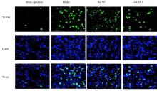
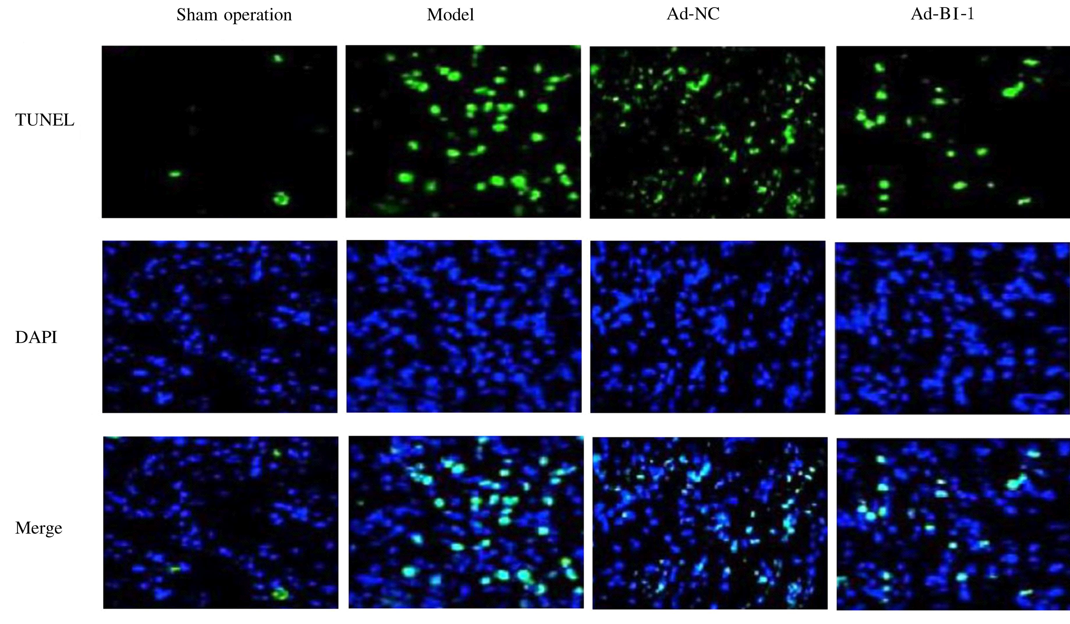
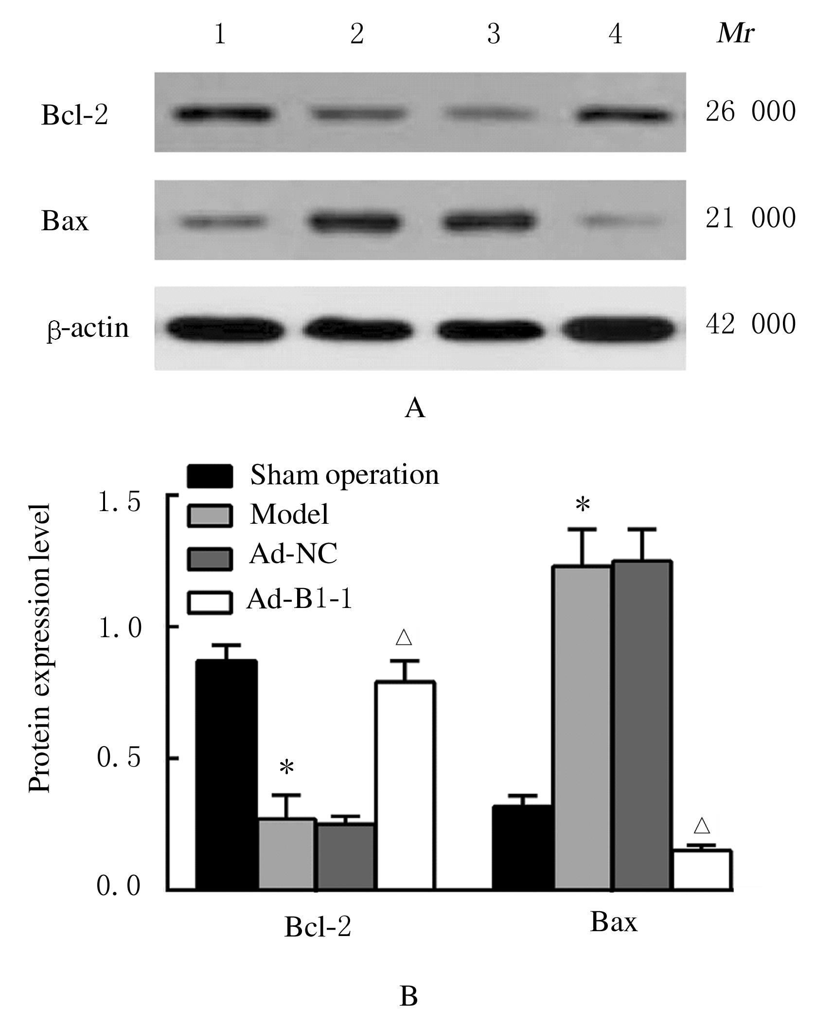
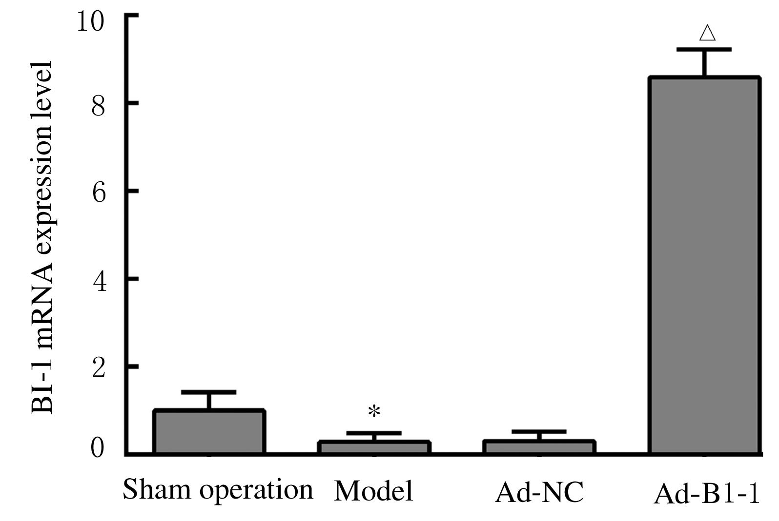
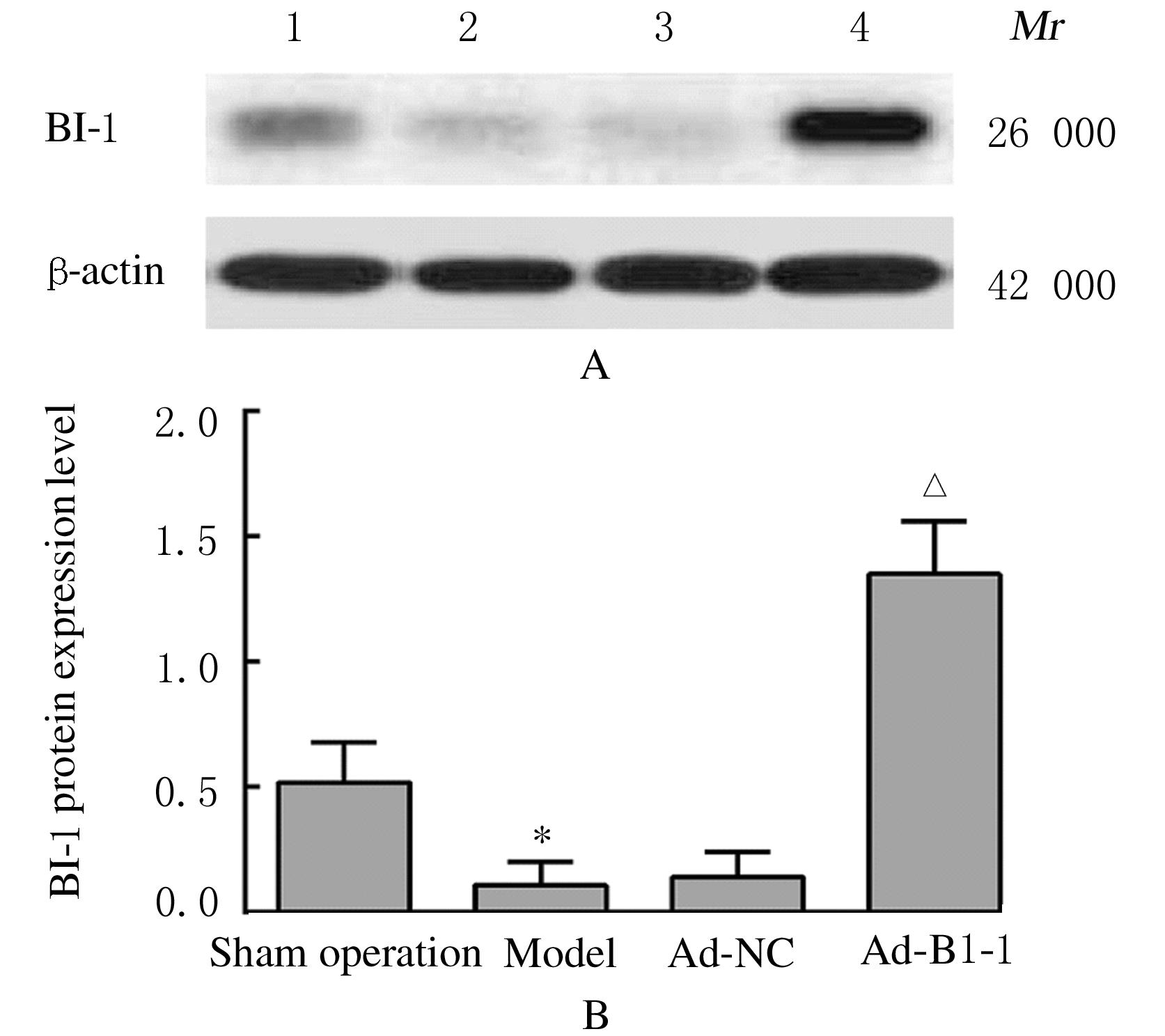
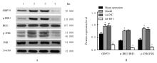
  • Received:2021-05-18 Online:2022-01-28 Published:2022-01-17
  • Contact: Liang ZENG E-mail:tangs198905@163.com

摘要: 目的

探讨过表达B细胞淋巴瘤2(Bcl-2)相关X蛋白(Bax)抑制因子1(BI-1)对急性心肌梗死(AMI)大鼠心肌细胞凋亡的影响,并阐明其作用机制。

方法

60只大鼠随机分为假手术组、模型组、空载腺病毒(Ad-NC)组和BI-1腺病毒(Ad-BI-1)组,每组15只。除假手术组外,其他各组大鼠均采用冠状动脉左前降支结扎法建立AMI大鼠模型,Ad-NC组和Ad-BI-1组大鼠分别于心肌梗死区域注射腺病毒包装的空载质粒和BI-1过表达质粒。术后72 h,检测各组大鼠心功能参数左室射血分数(LVEF)、左室缩短分数(LVFS)、左室舒张末期内径(LVEDD)和左室收缩末期内径(LVESD),TTC染色法检测各组大鼠心肌梗死面积百分率,TUNEL法检测各组大鼠心肌细胞凋亡率,比色法检测各组大鼠心肌组织中含半胱氨酸的天冬氨酸蛋白水解酶3(Caspase-3)活性,实时荧光定量PCR(RT-qPCR)法检测各组大鼠心肌组织中BI-1 mRNA表达水平,Western blotting法检测大鼠心肌组织中BI-1、Bcl-2、Bax和内质网应激相关蛋白葡萄糖调节蛋白78(GRP78)、肌醇依赖酶1(IRE1)、磷酸化IRE1(p-IRE1)、c-Jun氨基末端激酶(JNK)及磷酸化JNK (p-JNK)蛋白表达水平,并计算p-IRE1/IRE1和p-JNK/JNK比值。

结果

与假手术组比较,模型组大鼠LVEF和LVFS明显降低(P<0.05),LVEDD、LVESD、心肌梗死面积百分率、心肌组织中Caspase-3活性和心肌细胞凋亡率明显升高(P<0.05),心肌组织中BI-1 mRNA和蛋白表达水平及Bcl-2蛋白表达水平明显降低(P<0.05),心肌组织中Bax、GRP78蛋白表达水平和p-IRE1/IRE1及p-JNK/JNK比值明显升高(P<0.05);与模型组比较,Ad-BI-1组大鼠LVEF和LVFS明显升高(P<0.05),LVEDD和LVESD明显降低(P<0.05),心肌梗死面积百分率、心肌细胞凋亡率和心肌组织中Caspase-3活性明显降低(P<0.05),心肌组织中BI-1 mRNA和蛋白表达水平及Bcl-2蛋白表达水平明显升高(P<0.05),心肌组织中Bax、GRP78蛋白表达水平和p-IRE1/IRE1及p-JNK/JNK比值明显降低(P<0.05),Ad-NC组大鼠上述各指标差异无统计学意义(P>0.05)。

结论

过表达BI-1通过抑制内质网应激途径降低心肌细胞凋亡水平,从而改善AMI大鼠心功能。

关键词: 急性心肌梗死, 细胞凋亡, Bax抑制因子1, 内质网应激, B细胞淋巴瘤2相关X蛋白

Abstract: Objective

To explore the effect of overexpression of B-cell lymphoma-2(Bcl-2) associated X protein (Bax) inhibitor 1 (BI-1) on the cardiomyocyte apoptosis in the rats with acute myocardial infarction (AMI), and to clarify its mechanism.

Methods

A total of 60 rats were randomly divided into sham operation group, model group, empty adenovirus (Ad-NC) group and BI-1 adenovirus (Ad-BI-1) group; there were 15 rats in each group. Except for sham group, the rats in other groups were ligated in the left anterior descending coronary arteries to establish the AMI rat models. Meanwhile, the rats in Ad-NC group and Ad-BI-1 group were injected with adenovirus-packaged empty plasmid and BI-1 overexpression plasmid in the myocardial infarction area, respectively. The cardiac function parameters left ventricular ejection fraction (LVEF), left ventricular fraction shortening (LVFS), left ventricular end diastolic diameter (LVEDD) and left ventricular end systolic diameter (LVESD) values of the rats were detected 72 h after operation; TTC staining method was used to detect the percentages of myocardial infarction areas of the rats in various groups; TUNEL method was used to detect the apoptotic rates of cardiomyocytes of the rats in various groups; colorimetric method was used to detect the activities of Caspase-3 in myocardium tissue of the rats in various groups; RT-qPCR method was used to detect the expression levels of BI-1 mRNA of the rats in various groups; Western blotting method was used to detect the expression levels of BI-1, Bcl-2, Bax, and endoplasmic reticulum stress related proteins GRP78, IRE1, p-IRE1,JNK,p-JNK proteins in myocardium tissue of the rats in various groups;the ratios of the p-IRE1/IRE1 and p-JNK/JNK were calculated.

Results

Compared with sham operation group, the LVEF and LVFS of the rats in model group were significantly reduced (P<0.05),while the LVEDD and LVESD,the percentage of myocardial infarction area, the Caspase-3 activity in myocardium tissue, and the apoptodtic rates of cardiomocytes were increased (P<0.05), the expression levels of BI-1 mRNA and protein, and the expression levels of Bcl-2 protein in myocardium tissue of the rats in model group were significantly reduced (P<0.05), and the expression levels of Bax, and GRP78 and the ratios of p-IRE1/IRE1 and p-JNK/JNK were significantly increased (P<0.05).Compared with model group, the LVEF and LVFS of the rats in Ad-BI-1 group were significantly increased (P<0.05), while the LVEDD and LVESD were markedly decreased(P<0.05); the percentage of myocardial infarction area,the apoptotic rate of cardiomyocytes,and the Caspase-3 activity in myocardium tissue were decreased (P<0.05); the expression levels of BI-1 mRNA and protein and the expression level of Bcl-2 protein in myocardium tissue of the rats were significantly increased (P<0.05),and the expression levels of Bax and GRP78 and the ratios of p-IRE1/IRE1 and p-JNK/JNK were significantly decreased (P<0.05); the changes of the above indicators in Ad-NC group had no siginficant differences(P>0.05).

Conclusion

Overexpression of BI-1 reduces the level of cardiomyocyte apoptosis by inhibiting the endoplasmic reticulum stress pathway, thereby improving the cardiac function of the AMI rats.

Key words: Acute myocardial infarction, Apoptosis, Bax inhibitor 1, Endoplasmic reticulum stress, B-cell lymphoma-2 associated X protein

中图分类号: 

  • R542.22