吉林大学学报(医学版) ›› 2023, Vol. 49 ›› Issue (3): 557-564.doi: 10.13481/j.1671-587X.20230302

• 基础研究 • 上一篇    下一篇

Cav3.2基因在盆底电刺激治疗小鼠压力性尿失禁中的作用及其机制

刘剑锋,汤剑明,阳莲,张舒飞,洪莉()   

  1. 武汉大学人民医院妇产科,湖北 武汉 430060
  • 收稿日期:2021-06-27 出版日期:2023-05-28 发布日期:2023-06-20
  • 通讯作者: 洪莉 E-mail:drhongli777@163.com
  • 作者简介:刘剑锋(1996-),男,湖北省宜昌市人,在读硕士研究生,主要从事女性盆底功能障碍性疾病和妇科肿瘤方面的研究。
  • 基金资助:
    国家重点研发计划项目(2018YFC2002204);国家自然科学基金项目(81971364);国家自然科学基金青年教师资助项目(82001527)

Effectof Cav3.2 gene in treatment of stress urinary incontinence in mice by pelvic floor electrical stimulation and its mechanism

Jianfeng LIU,Jianming TANG,Lian YANG,Shufei ZHANG,Li HONG()   

  1. Department of Obstetrics and Gynecology,People’s Hospital,Wuhan University,Wuhan 430060,China
  • Received:2021-06-27 Online:2023-05-28 Published:2023-06-20
  • Contact: Li HONG E-mail:drhongli777@163.com

摘要:

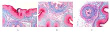
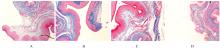
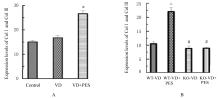
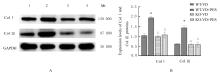
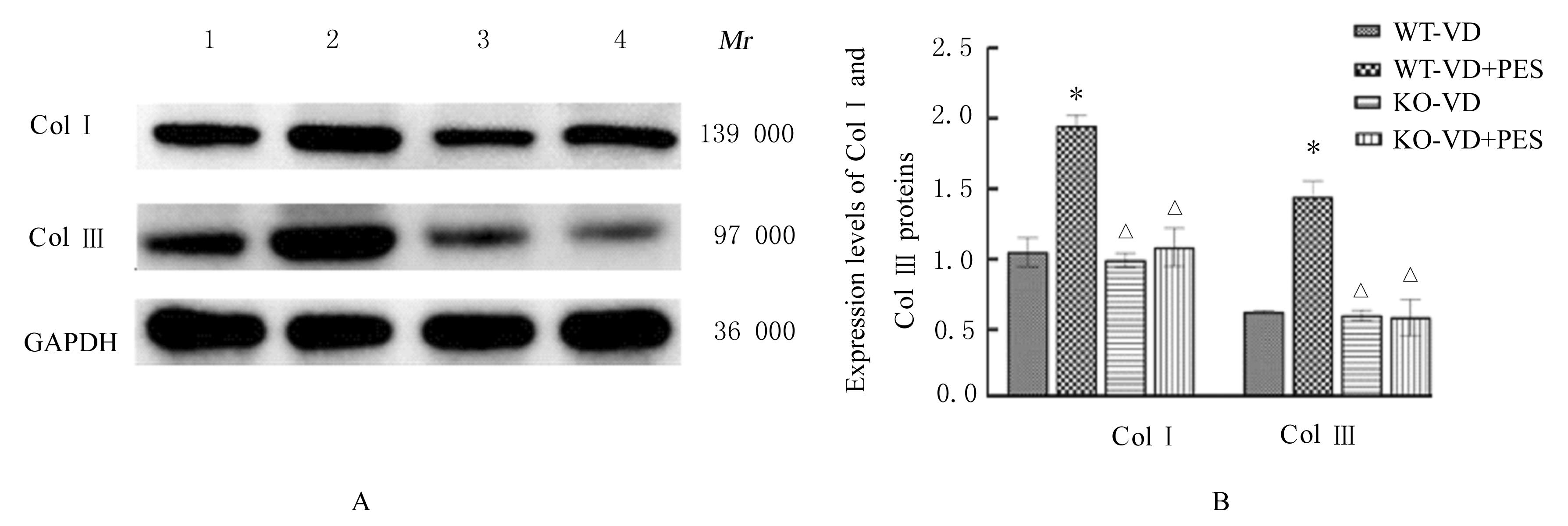
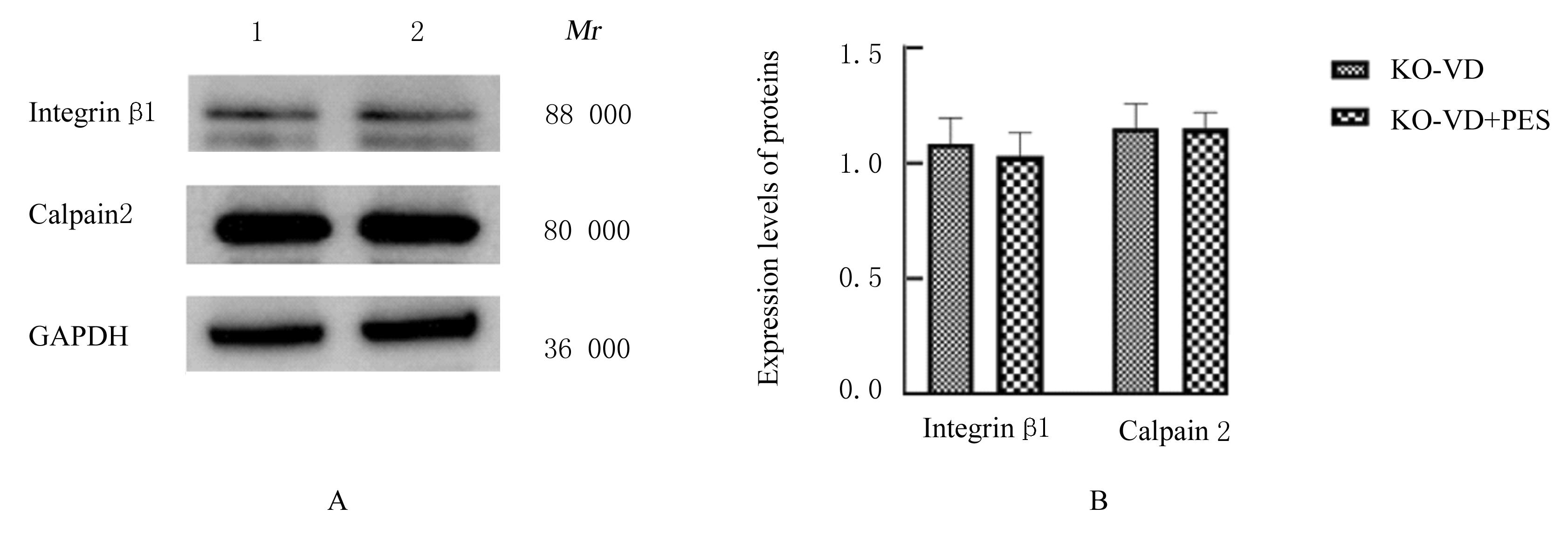
目的 探讨Cav 3.2基因在盆底电刺激(PES)治疗小鼠压力性尿失禁(SUI)中的作用,并阐明其相关机制。 方法 30只C57BL/6小鼠按干预措施随机分为对照组[未进行阴道扩张(VD)造模]、VD组(VD造模)和VD+PES组(VD造模联合PES),每组10只;按照基因型和干预措施将小鼠分为WT-VD组(野生型VD造模)、WT-VD+PES组(野生型VD造模联合PES)、 KO-VD组(Cav 3.2 敲除型VD造模)和KO-VD+PES组(Cav 3.2 敲除型VD造模联合PES)。Masson染色观察各组小鼠尿道和阴道前壁组织中Ⅰ型胶原(ColⅠ)和Ⅲ型胶原(ColⅢ)的胶原纤维沉积情况和胶原纤维表达水平,测定各组小鼠尿动力学参数最大膀胱容积(MBC)和漏尿点压力(LPP),Western blotting法检测各组小鼠尿道和阴道前壁组织中整合素β1、钙蛋白酶2、ColⅠ和ColⅢ蛋白表达水平。 结果 与VD组比较,VD+PES组小鼠尿道和阴道前壁组织中Col Ⅰ和Col Ⅲ胶原纤维表达水平明显升高(P<0.01);与WT-VD组比较, WT-VD+PES 组小鼠尿道和阴道前壁组织中ColⅠ及ColⅢ胶原纤维表达水平明显升高(P<0.01);与WT-VD+PES 组比较,KO-VD组和KO-VD+PES组小鼠尿道和阴道前壁组织中ColⅠ及ColⅢ胶原纤维表达水平明显降低(P<0.01)。与对照组比较,VD组小鼠MBC和LPP均降低(P<0.05);与 VD组比较, VD+PES组小鼠MBC和LPP均升高(P<0.05)。Western blotting法检测,与VD组比较,VD +PES组小鼠尿道和阴道前壁组织中ColⅠ及ColⅢ蛋白表达水平均明显升高(P<0.01);与WT-VD组比较, WT-VD+PES 组小鼠尿道和阴道前壁组织中ColⅠ及ColⅢ蛋白表达水平明显升高(P<0.01);与WT-VD+PES 组比较,KO-VD组和KO-VD+PES组小鼠尿道和阴道前壁组织中ColⅠ及ColⅢ蛋白表达水平明显降低(P<0.01)。与WT-VD组比较,WT-VD+PES组小鼠尿道和阴道前壁组织中整合素β1及钙蛋白酶2蛋白表达水平明显升高(P<0.01)。 结论 PES可改善小鼠尿动力学功能,并通过Cav 3.2基因及其下游整合素β1和钙蛋白酶2促进盆底胶原的生成,促进盆底修复。

关键词: 盆底电刺激, Cav 3.2基因, 整合素β1, 钙蛋白酶2, 小鼠, C57BL/6, 压力性尿失禁

Abstract:

Objective To discuss the effect of Cav 3.2 gene in the treatment of stress urinary incontinence (SUI) of the mice by pelvic floor electrical stimulation (PES), and to clarify its related mechanism. Methods Thirty C57BL/6 mice were randomly divided into control group [without vaginal dilation (VD) modeling], VD group (VD modeling), and VD+PES group (VD modeling combined with PES) according to the intervention measures, and there were 10 mice in each group. The mice were divided into WT-VD group (wild-type VD modeling), WT-VD+PES group (wild-type VD modeling combined with PES), KO-VD group (Cav 3.2 knockout type VD modeling), and KO-VD+PES group (Cav 3.2 knockout type VD modeling combined with PES) according to the genotype and intervention mesures. Masson staining was used to observe the deposition and expression levels of collagen type Ⅰ(Col Ⅰ) and collagen type Ⅲ (Col Ⅲ) collagen fibers in urethral and anterior vaginal wall tissue of the mice in various groups. The maximum bladder capacity(MBC) and leakage point pressure (LPP) of the mice in various groups were detected, and the expression levels of integrins β1, calpain 2, Col Ⅰ, and Col Ⅲ proteins in urethral and anterior vaginal wall tissue of the mice in various groups were detected by Western blotting method. Results Compared with VD group, the expression levels of Col Ⅰ and Col Ⅲ collagen fibers in urethral and anterior vaginal wall tissue of the mice in VD+PES group mice were significantly increased (P<0.01); compared with WT-VD group, the expression levels of Col Ⅰ and Col Ⅲ collagen fibers in urethral and anterior vaginal wall tissue of the mice in WT-VD+PES group were significantly increased (P<0.01); compared with WT-VD+PES group, the expression levels of Col I and Col Ⅲ collagen fibers in urethral and anterior vaginal wall tissue of the mice in KO-VD and KO-VD+PES groups were significantly decreased (P<0.01). Compared with control group, the MBC and LPP of the mice in VD group were decreased (P<0.05); compared with VD group, the MBC and LPP of the mice in VD+PES group were increased (P<0.05). The Western blotting results showed that compared with VD group, the expression levels of Col Ⅰ and Col Ⅲ proteins in urethral and anterior vaginal wall tissue of the mice in VD+PES group were significantly increased (P<0.01); compared with WT-VD group, the expression levels of Col Ⅰ and Col Ⅲ proteins in urethral and anterior vaginal wall tissue of the mice in WT-VD+PES group were increased (P<0.01); compared with WT-VD+PES group, the expression levels of Col Ⅰ and Col Ⅲ proteins in urethral and anterior vaginal wall tissues of the mice in KO-VD and KO-VD+PES groups were significantly decreased(P<0.01). Compared with WT-VD group, the expression levels of integrin β1 and calpain 2 proteins in urethral and anterior vaginal wall tissue of the mice in WT-VD+PES group were increased significantly (P<0.01). Conclusion PES can improve the urodynamic function of the mice and promote the generation of pelvic floor collagen and promote the repairment of pelvic floor through the Cav 3.2 gene and its downstream integrin β 1 and calpain 2.

Key words: Pelvic electrical stimulation, Cav3.2 gene, Integrin β1, Calpain 2, Mice, C57BL/6, Stress urinary incontinence

中图分类号: 

  • R714.6