吉林大学学报(医学版) ›› 2023, Vol. 49 ›› Issue (4): 975-984.doi: 10.13481/j.1671-587X.20230419

• 基础研究 • 上一篇    下一篇

结直肠癌细胞源性外泌体中miR-17-5p的表达对结直肠癌细胞化疗敏感性的影响及其机制

刘珊1,邢兆东2,黄平1()   

  1. 1.海南省人民医院肛肠外科, 海南 海口 570100
    2.中国医学科学院肿瘤医院胰胃外科, 北京 100029
  • 收稿日期:2022-08-22 出版日期:2023-07-28 发布日期:2023-07-26
  • 通讯作者: 黄平 E-mail:13876380118@163.com
  • 作者简介:刘 珊(1985-),女,山东省菏泽市人,主治医师,医学硕士,主要从事普通外科基础和临床方面的研究。
  • 基金资助:
    海南省卫健委2021年度省卫生健康行业科研项目(21A200467)

Effect of expression of miR-17-5p in exosomes derived from colorectal cancer cells on chemosensitivity of colorectal cancer cells and its mechanism

Shan LIU1,Zhaodong XING2,Ping HUANG1()   

  1. 1.Department of Anorectal Surgery,Hainan Provincial People’s Hospital,Haikou 570100,China
    2.Department of Pancreatic and Gastric Surgery,Cancer Hospital,Chinese Academy of Medical Sciences,Beijing 100029,China
  • Received:2022-08-22 Online:2023-07-28 Published:2023-07-26
  • Contact: Ping HUANG E-mail:13876380118@163.com

摘要:

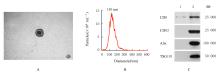
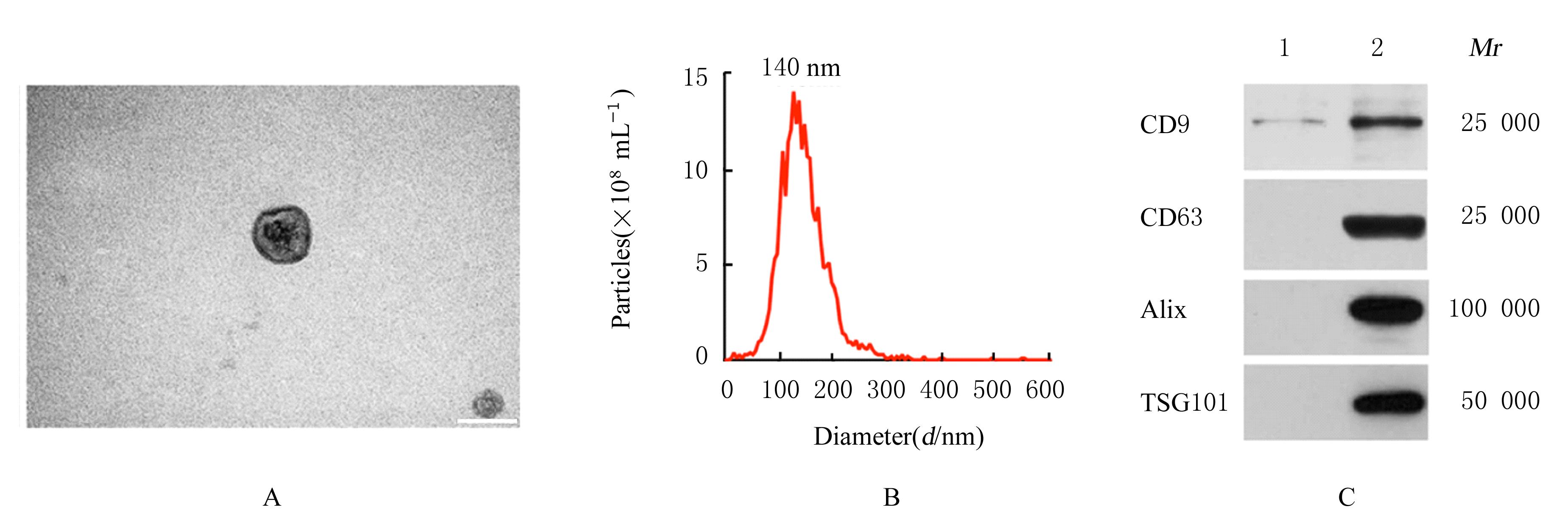
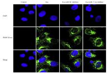
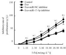
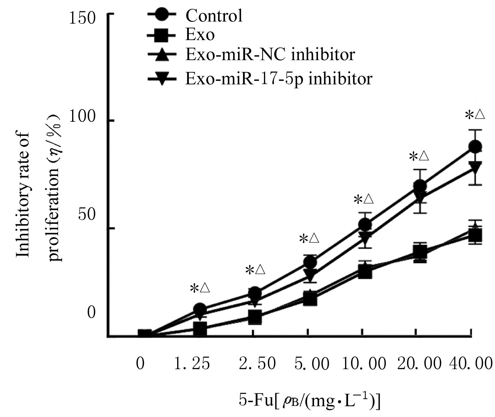
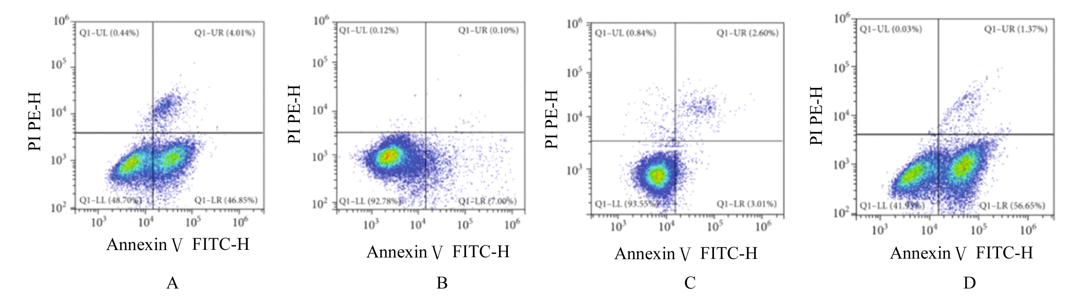
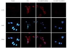
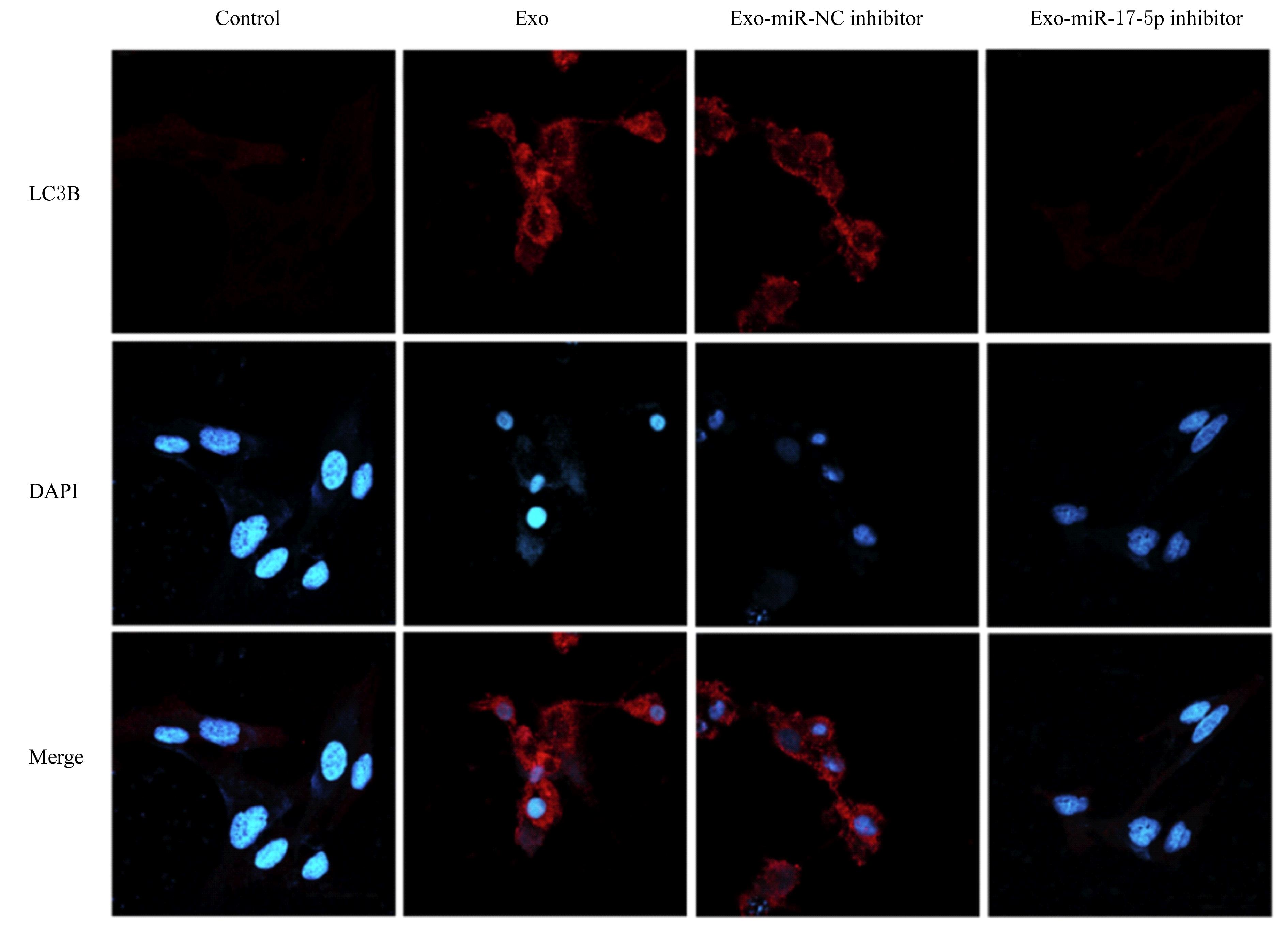
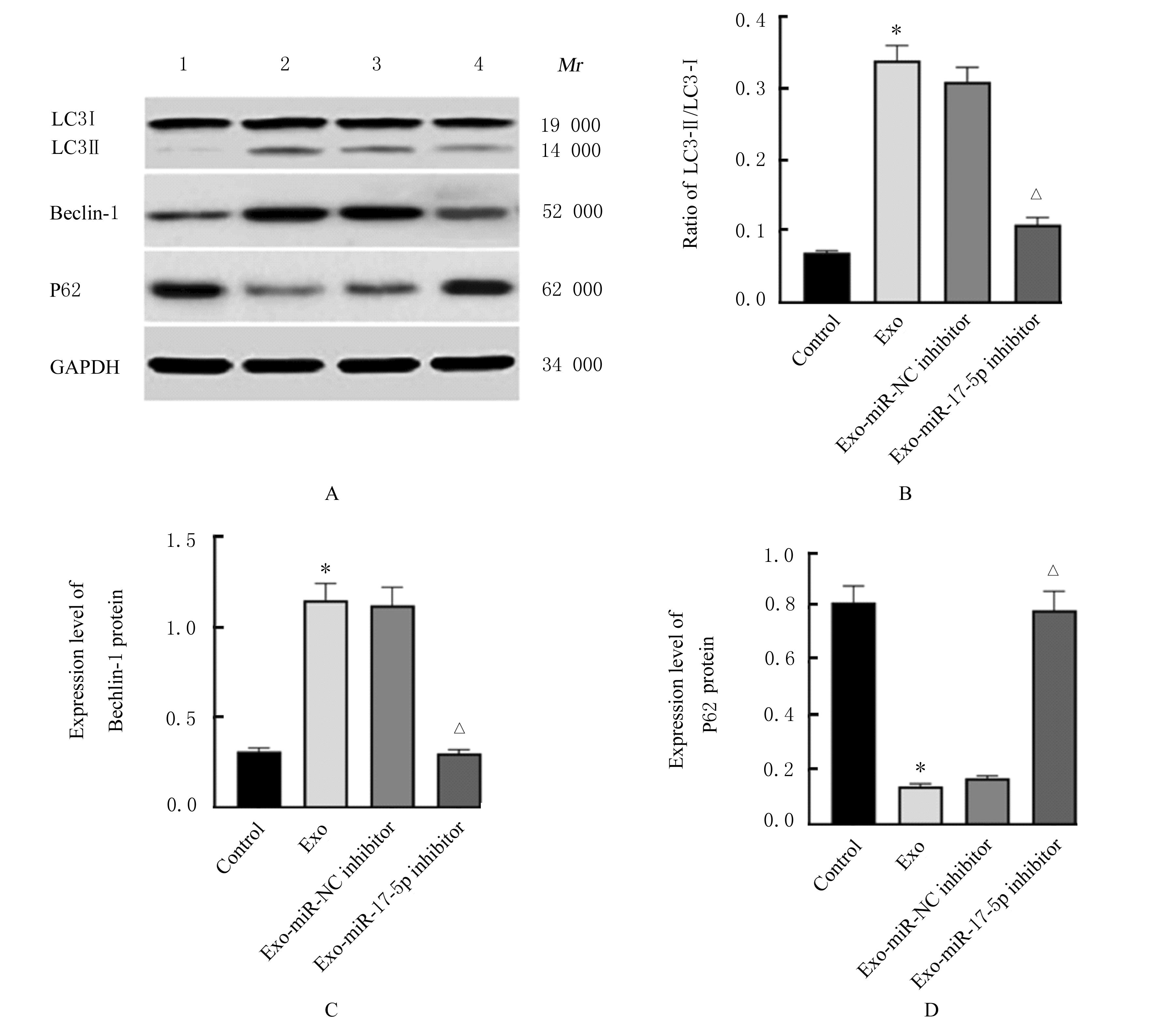
目的 分析结直肠癌细胞源性外泌体(Exo)中微小RNA-17-5p(miR-17-5p)表达对结直肠癌细胞化疗敏感性的影响,阐明其可能的作用机制。 方法 实时荧光定量PCR(RT-qPCR)法检测人结直肠癌HCT116细胞、CT26细胞、LoVo细胞、HT29细胞、SW620细胞、SW480细胞和人正常结肠直肠黏膜上皮HIEC细胞中miR-17-5p表达水平。CT26细胞分为对照组、miR-NC inhibitor组和miR-17-5p inhibitor组,提取各组细胞Exo,透射电子显微镜观察Exo形态表现,纳米粒子跟踪分析法检测粒径分布情况,Western blotting法检测Exo中标志蛋白CD9、CD63、凋亡诱导因子6互作蛋白(Alix)和肿瘤易感基因101(TSG101)蛋白表达水平,RT-qPCR法检测Exo中miR-17-5p表达水平。CT26细胞分为对照组、Exo组、Exo-miR-NC inhibitor组和Exo-miR-17-5p inhibitor组,分别以不同转染组CT26细胞源性Exo处理CT26细胞,Exo绿色荧光标记PKH67染料示踪法观察各组CT26细胞摄取Exo情况,MTT法检测1.25、2.50、5.00、10.00、20.00和40.00 mg·L-1 5-氟尿嘧啶(5-Fu)处理后各组CT26细胞增殖抑制率,流式细胞术检测各组CT26细胞凋亡率,细胞免疫荧光染色检测各组CT26细胞中微管相关蛋白轻链3B(LC3B)荧光强度,Western blotting法检测各组CT26细胞中微管相关蛋白轻链3 Ⅱ(LC3-Ⅱ)、微管相关蛋白轻链3Ⅰ(LC3-Ⅰ)、自噬效应蛋白Beclin-1和P62蛋白表达水平,并计算LC3-Ⅱ/LC3-Ⅰ比值。 结果 人结直肠癌HCT116、CT26、LoVo、HT29、SW620和SW480细胞中miR-17-5p表达水平明显高于HIEC细胞(P<0.05);分离到的颗粒物呈典型球形囊泡,粒径峰值约140 nm,且CD9、CD63、Alix和TSG101蛋白均明显表达,表明成功分离到Exo。与对照组比较, miR-17-5p inhibitor组Exo中miR-17-5p表达水平明显降低(P<0.05)。与对照组比较,Exo组、Exo-miR-NC inhibitor组和Exo-miR-17-5p inhibitor组CT26细胞周围均可见明显的PKH67染色,显示CT26细胞可摄取Exo。与对照组比较,经1.25、2.50、5.00、10.00、20.00和40.00 mg·L-15-Fu处理后Exo组CT26细胞增殖抑制率明显降低(P<0.05),各组CT26细胞凋亡率明显降低(P<0.05),细胞中LC3B荧光强度明显增强(P<0.05),LC3-Ⅱ/LC3-Ⅰ比值和Beclin-1蛋白表达水平明显升高(P<0.05),P62蛋白表达水平明显降低(P<0.05);与Exo组比较,经1.25、2.50、5.00、10.00、20.00和40.00 mg·L-1 5-Fu处理后Exo-miR-17-5p inhibitor组CT26细胞增殖抑制率和细胞凋亡率明显升高(P<0.05),细胞中LC3B荧光强度减弱(P<0.05),LC3-Ⅱ/LC3-Ⅰ比值和Beclin-1蛋白表达水平明显降低(P<0.05),P62蛋白表达水平明显升高(P<0.05)。 结论 抑制结直肠癌细胞源性Exo中miR-17-5p的表达可提高结直肠癌细胞的化疗敏感性,其作用机制可能与抑制细胞自噬水平有关。

关键词: 结直肠肿瘤, 外泌体, 微小RNA-17-5p, 化疗敏感性, 细胞自噬

Abstract:

Objective To analyze the effect of expression of microRNA-17-5p(miR-17-5p) in the colorectal cancer cell-derived exosomes (Exo) on the chemotherapy sensitivity of colorectal cancer cells,and to clarify its possible mechanism. Methods Real-time fluorescence quantitative PCR (qRT-PCR) method was used to detect the expression levels of miR-17-5p in the human colorectal cancer HCT116 cells, CT26 cells, LoVo cells, HT29 cells, SW620 cells, SW480 cells, and human normal colorectal mucosal epithelial HIEC cells.The CT26 cells were divided into control group,miR-NC inhibitor group, and miR-17-5p inhibitor group, and the exosomes were extracted;the morphology of the Exo was observed under transmission electron microscope; the distribution of particle size was detected by nanoparticle tracking analysis; the expressions levels of marker proteins CD9, CD63, apoptosis-inducing factor 6 interacting protein (Alix),and tumor susceptibility gene 101 (TSG101) in the Exo were detected by Western blotting method; the expression level of miR-17-5p in the Exo was detected by RT-qPCR method. The CT26 cells were divided into control group, Exo group, Exo-miR-NC inhibitor group, and Exo-miR-17-5p inhibitor group. The CT26 cells were treated with CT26 cell-derived Exo in different transfection groups, and the uptake of Exo by CT26 cells were observed by Exo green fluorescence marker PKH67 dye trace method; MTT assay was used to detect the inhibitiory rates of proliferation of the CT26 cells after treated with 1.25, 2.50, 5.00, 10.00, 20.00, and 40.00 mg·L-1 5-fluorouracil(5-FU); flow cytometry was used to detect the apoptotic rates of the CT26 cells in various groups; cell immunofluorescence staining was used to observe the expression intensities of microtubule-associated protein 1B light chain 3 (LC3B) in the CT26 cells in various groups; Western blotting method was used to detect the expression levels of autophagy-related proteins microtubule-associated protein light chain 3Ⅱ (LC3-Ⅱ), microtubule-associated protein light chain 3Ⅰ (LC3-Ⅰ), autophagy effector protein Beclin-1, and P62 proteins in CT26 cells in various groups,and the ratio of LC3-Ⅱ/LC3-Ⅰ was calculated. Results The expression levels of miR-17-5p in the human colorectal cancer HCT116, CT26, LoVo, HT29, SW620, and SW480 cells were significantly higher than that in the HIEC cells (P<0.05); the isolated particles showed typical spherical vesicles with a peak particle size at about 140 nm, and the CD9, CD63, Alix and TSG101 proteins were all significantly expressed, indicating that Exo was successfully isolated.Compared with control group,the expression level of miR-17-5p in the Exo in miR-17-5p inhibitor group was significantly decreased (P<0.05); compared with control group, obvious PKH67 staining could be observed around the CT26 cells in Exo group, Exo-miR-NC inhibitor group, and Exo-miR-17-5p inhibitor group, indicating that the CT26 cells could take in the Exo. Compared with control group, the inhibitory rate of proliferation and the apoptotic rate of the CT26 cells in Exo group were decreased after treated with 1.25, 2.50, 5.00, 10.00, 20.00,and 40.00 mg·L-1 5-FU (P<0.05), the intracellular LC3B fluorescence intensity was increased (P<0.05), the ratio of LC3-Ⅱ/LC3-Ⅰ and the expression level of Beclin-1 protein were increased (P<0.05), while the expression level of P62 protein was decreased(P<0.05); compared with Exo group, the inhibitory rate of proliferation and apoptotic rate of the CT26 cells in Exo-miR-17-5p inhibitor group were increased after treated with 1.25, 2.50, 5.00, 10.00, 20.00,and 40.00 mg·L-1 5-FU(P<0.05), the intracellular LC3B fluorescence intensity was decreased (P<0.05), the ratio of LC3-Ⅱ/LC3-Ⅰand the expression level of Beclin-1 protein were decreased (P<0.05),and the expression level of P62 protein was increased (P<0.05). Conclusion The inhibition of expression of miR-17-5p in Exo derived from colorectal cancer cells can improve the chemosensitivity of the colorectal cancer cells, and the mechanism may be related to the inhibition of cell autophagy levels.

Key words: Colorectal neoplasm, Exosomes, MicroRNA-17-5p, Chemosensitivity, Cell autophagy

中图分类号: 

  • R735.34