吉林大学学报(医学版) ›› 2023, Vol. 49 ›› Issue (6): 1642-1648.doi: 10.13481/j.1671-587X.20230632

• 方法学 • 上一篇    下一篇

胎鼠神经干细胞原代培养及传代条件优化

朱文豪,刘天一,何川,张晓宇,辛强,王海峰()   

  1. 吉林大学第一医院神经创伤外科,吉林 长春 130021
  • 收稿日期:2023-01-06 出版日期:2023-11-28 发布日期:2023-12-22
  • 通讯作者: 王海峰 E-mail:hfwang@jlu.edu.cn
  • 作者简介:朱文豪(1995-),男,浙江省丽水市人,在读博士研究生,主要从事脑损伤疾病中干细胞和组织工程方面的研究。
  • 基金资助:
    国家自然科学基金面上项目(81871555);吉林省财政厅卫生人才专项项目(JLSWSRCZX2021-028)

Primary culture and optimization of subculture conditions of neural stem cells from fetal rats

Wenhao ZHU,Tianyi LIU,Chuan HE,Xiaoyu ZHANG,Qiang XIN,Haifeng WANG()   

  1. Department of Neurotrauma Surgery,First Hospital,Jilin University,Changchun 130021,China
  • Received:2023-01-06 Online:2023-11-28 Published:2023-12-22
  • Contact: Haifeng WANG E-mail:hfwang@jlu.edu.cn

摘要:

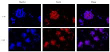
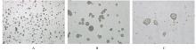
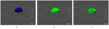
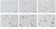
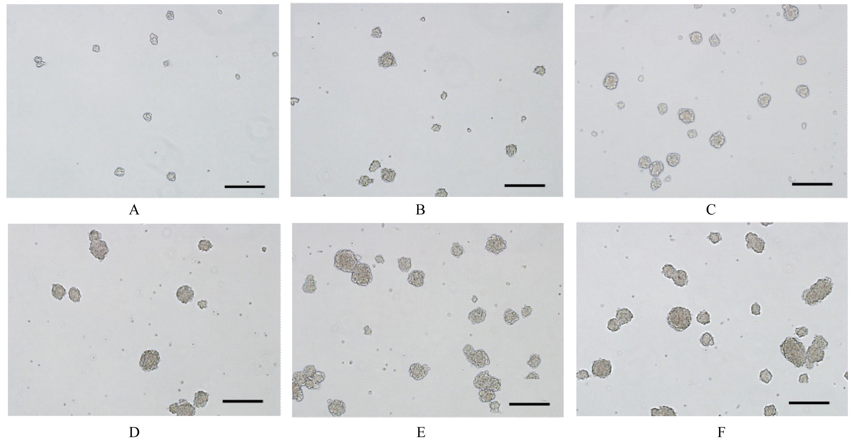
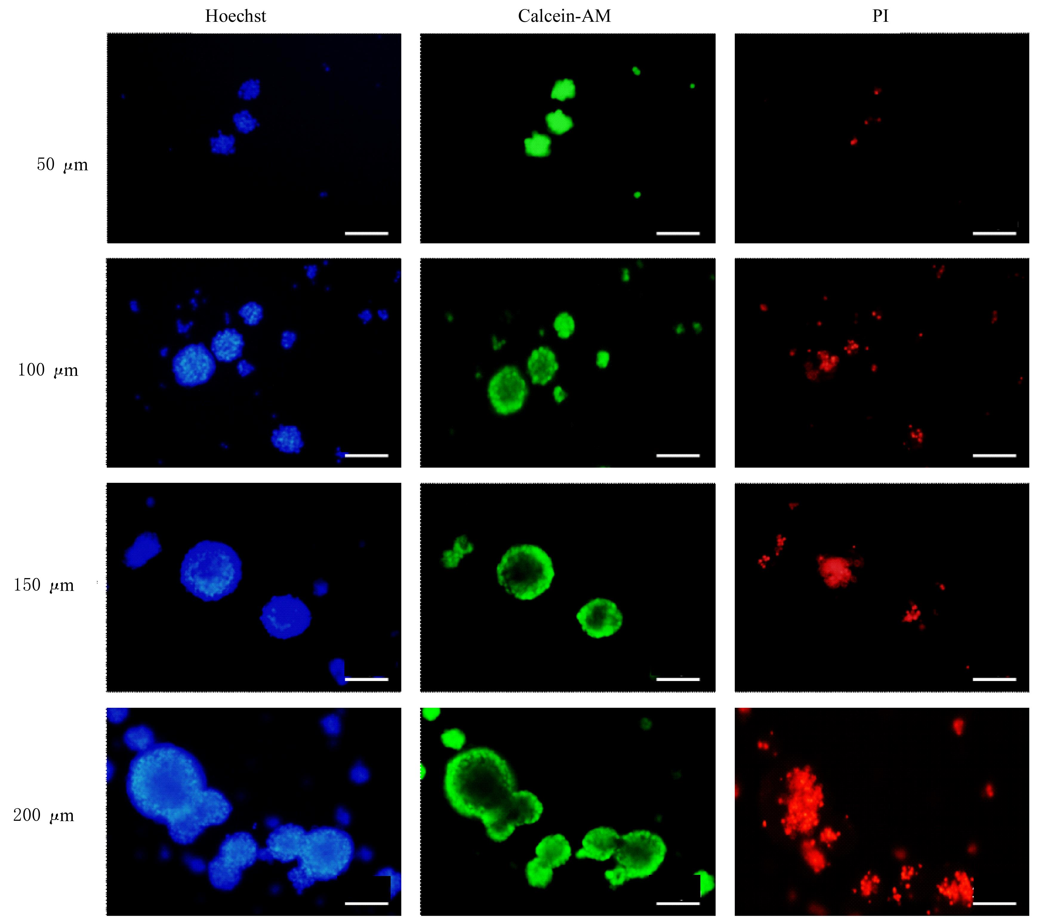
目的 探讨胎鼠神经干细胞(NSCs)原代培养方法和活性评价体系,确定NSCs的最佳传代条件。 方法 选取孕11~14 d SD大鼠,提取胎鼠原代NSCs,采用巢蛋白免疫荧光染色鉴定NSCs。将NSCs以1.0×10⁴、2.0×10⁴、6.0×10⁴、1.0×10⁵、1.6×10⁵和2.0×10⁵ mL-1的细胞密度进行传代培养48 h后观察神经球的形态表现,测定神经球直径。活死细胞染色法检测不同直径神经球中NSCs存活率。 结果 NSCs呈巢蛋白阳性,培养的细胞为神经干细胞。NSCs呈聚集生长和牢固细胞间黏附聚集模式,形成神经球,平均直径为(152.72±47.52)μm,球与球之间少有单独的细胞散在。与2.0×10⁴ mL-1传代密度比较,6.0×104 mL-1 和1.0×10⁵ mL-1 传代密度的神经球直径增大(P<0.05)。1.0×10⁵ mL-1 、1.6×10⁵ mL-1 和2.0×10⁵ mL-1 传代密度的神经球直径比较差异无统计学意义(P>0.05)。与0~40 μm、40~60 μm、60~80 μm和80~100 μm直径神经球比较,100~200 μm直径神经球中NSCs存活率降低(P<0.05)。 结论 以1.0×10⁵ mL-1的密度进行传代时神经球直径为80~100 μm,可有效提高NSCs传代培养效率和活性。

关键词: 神经干细胞, 胎鼠, 原代培养, 传代条件, 神经球

Abstract:

Objective To discuss the method of primary culture and activity evaluation system of the neural stem cells (NSCs) of the fetal rats, and to confirm the optimal subculture conditions of the NSCs. Methods The SD rats with 11-14 d gestation were chosen,and the primary NSCs of the fetal rats were extracted. Nestin immunofluorescence staining was used to identify the NSCs. The NSCs were subcultured at the cell densities of 1.0×104,2.0×104,6.0×104,1.0×105, 1.6×10⁵ and 2.0×10⁵ mL-1, then the morpholgy of the neurospheres was observed 48 h after passage and the diameter of the neurospheres was calculated. The survival rates of NSCs in the neurospheres with different diameters were detected by live-dead cell staining. Results The expression of nestin in the NSCs was positive, indicating that the cultured cells were the NSCs. The NSCs showed aggregation growth and strong cell-cell adhesion pattern forming neurospheres with an average diameter of (152.72±47.52) μm, and there were few single cells scattered between the neurospheres. Compared with 2.0×10⁴ mL-1 subculture density,the diameters of the neurospheres at the subculture densities of differences 6.0×10⁴ mL-1 and 1.0×10⁵ mL-1 were increased (P<0.05). There were no significant differences in the diameters of the neurospheres at the subculture densities of 1.0×10⁵ mL-1, 1.6×10⁵ mL-1, and 2.0×10⁵ mL-1P>0.05). Compared with neurospheres with diameters of 0—40 μm, 40—60 μm, 60—80 μm,and 80—100 μm, the survival rate of NSCs in the neurospheres with the diameters of 100—200 μm was decreased (P<0.05). Conclusion The neurospheres with the diameters of 80—100 μm when subcultured at the density of 1.0×10⁵ mL-1 can effectively improve the efficiency and activity of subculture of the NSCs.

Key words: Neural stem cell, Fetal rat, Primary culture, Subculture condition, Neurosphere

中图分类号: 

  • Q254