吉林大学学报(医学版) ›› 2023, Vol. 49 ›› Issue (5): 1202-1209.doi: 10.13481/j.1671-587X.20230513

• 基础研究 • 上一篇    

丹皮酚对人骨肉瘤MG-63细胞增殖、迁移和CXCR4/STAT3通路的影响

吴琪1(),陈建锋2,李浩2,文峰2   

  1. 1.湖北中医药大学临床技能实训中心针骨实验室,湖北 武汉 430000
    2.湖北中医药大学附属医院 骨科,湖北 武汉 430000
  • 收稿日期:2022-11-27 出版日期:2023-09-28 发布日期:2023-10-26
  • 通讯作者: 吴琪 E-mail:wuqi9278@126.com
  • 作者简介:吴 琪(1982-),女,湖北省咸宁市人,高级实验师,医学博士,主要从事骨关节疾病中医药防治方面的研究。
  • 基金资助:
    湖北省教育厅科研计划重点项目(D20212004)

Effect of paeonol on proliferation, migration, and CXCR4/STAT3 pathway of human osteosarcoma MG-63 cells

Qi WU1(),Jianfeng CHEN2,Hao LI2,Feng WEN2   

  1. 1.Needle Bone Laboratory,Clinical Skills Training Center,Hubei University of Traditional Chinese Medicine,Wuhan 430000,China
    2.Department of Orthopaedics,Affiliated Hospital,Hubei University of Traditional Chinese Medicine,Wuhan 430000,China
  • Received:2022-11-27 Online:2023-09-28 Published:2023-10-26
  • Contact: Qi WU E-mail:wuqi9278@126.com

摘要:

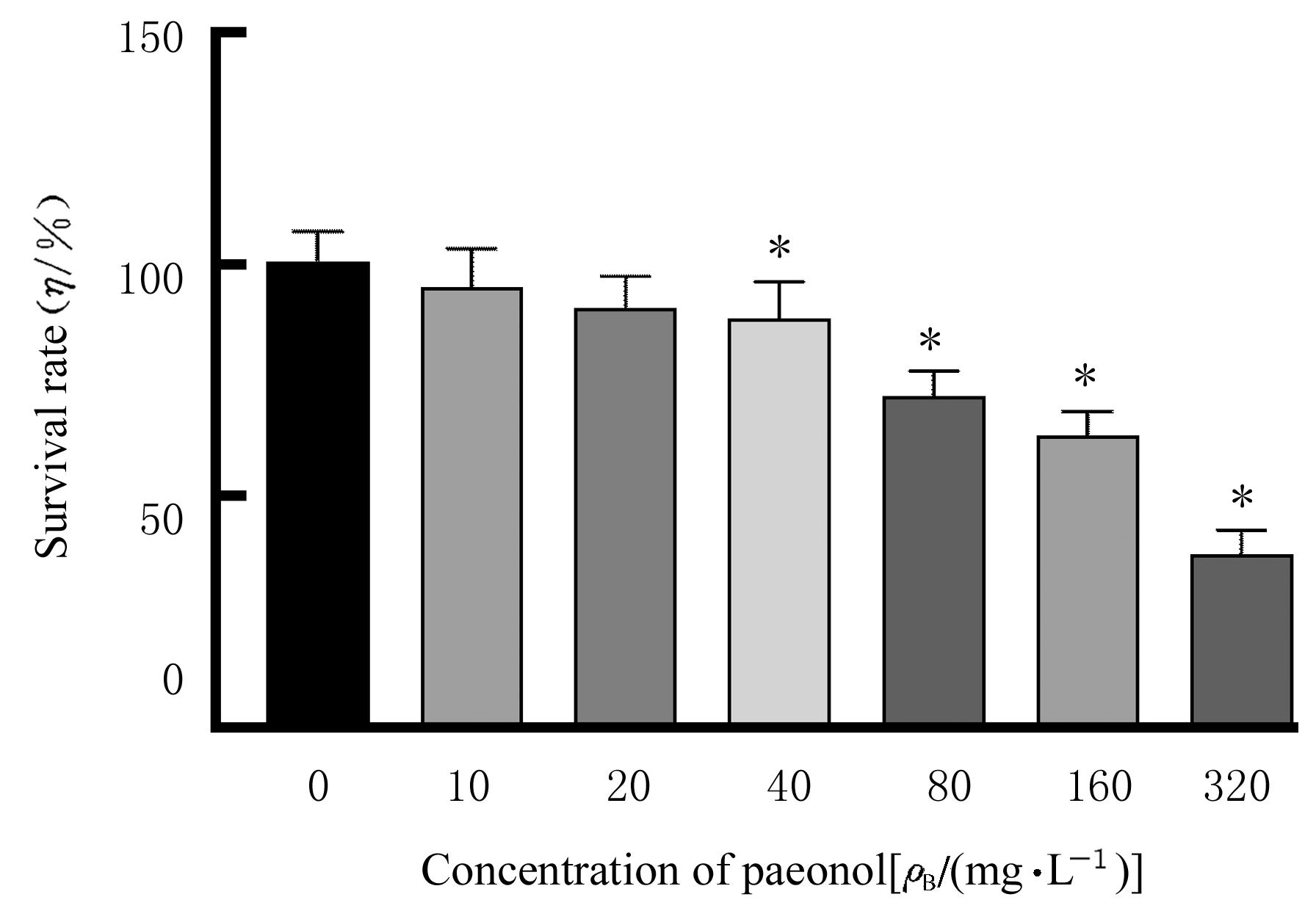
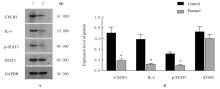
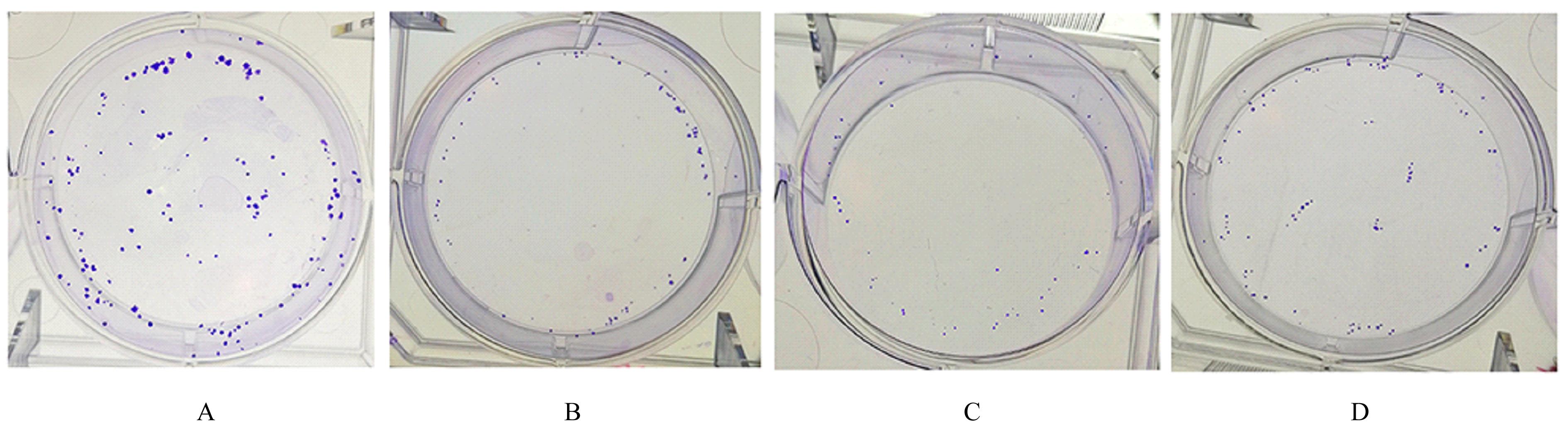
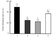
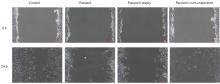
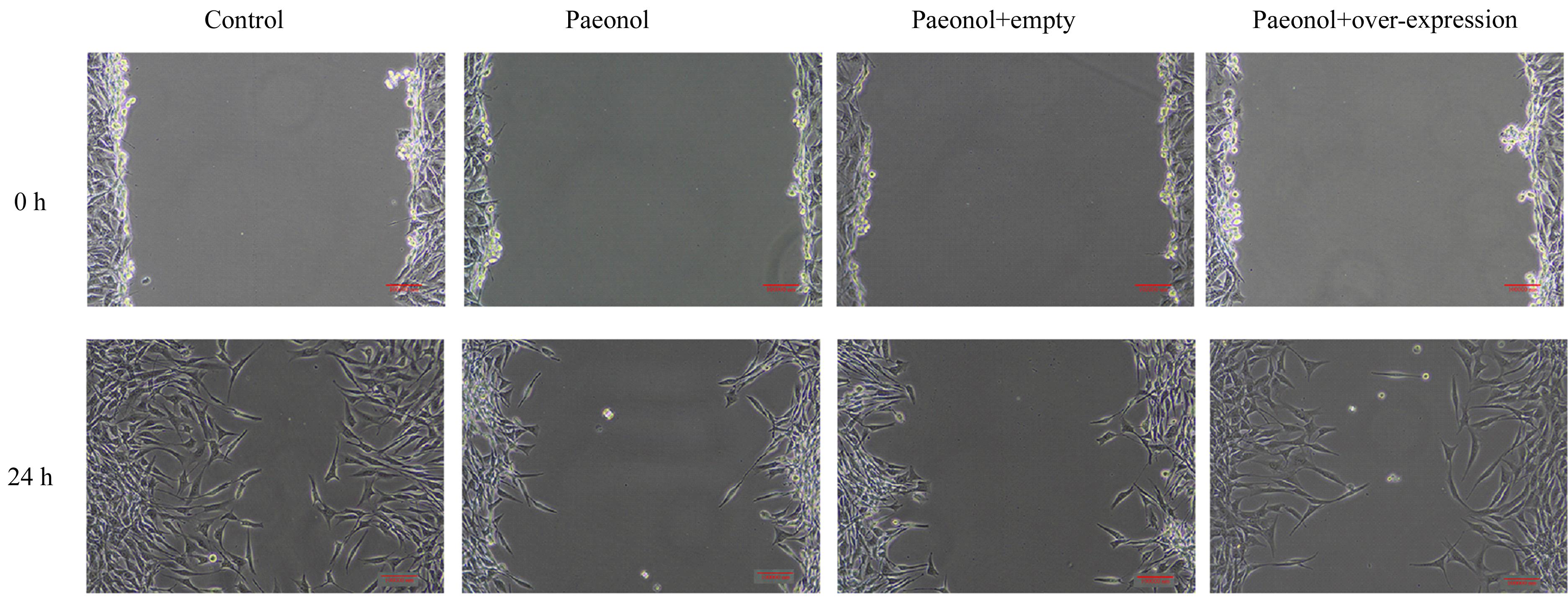
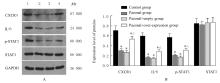
目的 探讨丹皮酚对人骨肉瘤(OS)MG-63细胞增殖、迁移和趋化因子受体4(CXCR4)/信号传导与转录激活因子3(STAT3)通路的影响,并阐明其可能的作用机制。 方法 体外培养MG-63细胞,分为对照组(不给予药物干预)和不同浓度丹皮酚组(给予10、20、40、80、160和320 mg·L-1丹皮酚)。采用CCK-8法检测各组细胞存活率并选择半数抑制浓度(IC50)值作为后续实验药物浓度。将MG-63细胞分为对照组和丹皮酚(215.8 mg·L-1)组,采用Western blotting法检测各组细胞中CXCR4、白细胞介素6(IL-6)、磷酸化STAT3(p-STAT3)和STAT3蛋白表达水平。将MG-63细胞分为对照组(不给予药物干预)、丹皮酚组(给予215.8 mg·L-1丹皮酚)、丹皮酚+空载组(给予215.8 mg·L-1丹皮酚+空载质粒)和丹皮酚+过表达组(给予215.8 mg·L-1丹皮酚+CXCR4过表达质粒)。采用细胞划痕实验检测各组细胞划痕愈合率,克隆形成实验检测各组细胞克隆形成率,Western blotting法检测各组细胞中CXCR4、IL-6、p-STAT3和STAT3蛋白表达水平。 结果 与对照组比较,40、80、160和320 mg·L-1丹皮酚组MG-63细胞存活率降低(P<0.05),丹皮酚IC50值为215.8 mg·L-1。与对照组比较,丹皮酚(215.8 mg·L-1)组MG-63细胞中CXCR4、IL-6和p-STAT3蛋白表达水平降低(P<0.05)。与对照组比较,丹皮酚组和丹皮酚+空载组MG-63细胞划痕愈合率及细胞克隆形成率降低(P<0.05),细胞中CXCR4、IL-6和p-STAT3蛋白表达水平降低(P<0.05);与丹皮酚组比较,丹皮酚+空载组MG-63细胞划痕愈合率、细胞克隆形成率和细胞中CXCR4、IL-6、p-STAT3及STAT3蛋白表达水平差异无统计学意义(P>0.05);与丹皮酚组比较,丹皮酚+过表达组MG-63细胞划痕愈合率和细胞克隆形成率升高(P<0.05),细胞中CXCR4、IL-6和p-STAT3蛋白表达水平升高(P<0.05)。 结论 丹皮酚能抑制MG-63细胞的增殖、迁移和克隆形成,其机制可能与调控CXCR4/STAT3信号通路有关。

关键词: 丹皮酚, 人骨肉瘤细胞, 细胞增殖, 趋化因子受体4, 信号传导与转录激活因子3

Abstract:

Objective To discuss the effect of paeonol on the proliferation, migration and chemokinereceptor4 (CXCR4)/signal transducer and activators of transcription3 (STAT3) pathway of the human osteosarcoma(OS ) MG-63 cells, and to clarify its possible mechanism. Methods The MG-63 cells were cultured in vitro and divided into control group (without drug intervention) and different concentrations of paeonol groups (given 10, 20, 40, 80, 160,and 320 mg·L-1 paeonol, respectively).The survival rates of the MG-63 cells in various groups were detected by CCK-8 method and the half inhibitory concentration (IC50) value was selected as the drug concentration for subsequent experiment. The MG-63 cells were divided into control group(without drug treatment) and paeonol group(given 215.8 mg·L-1 paeonal).The expression levels of CXCR4, interleukin 6 (IL-6),and phosphorylated STAT3 (p-STAT3),and STAT3 proteins in the MG-63 cells in various groups were detected by Western blotting method.The MG-63 cells were divided into control group, paeonol group, paeonol+empty group (given 215.8 mg·L-1 paeonol+empty plasmid), and paeonol+overexpression group (given 215.8 mg·L-1 paeonol+CXCR4 over-expression plasmid). The scratch healing rates of the MG-63 cells in various groups were detected by cell scratch experiment, the clone formation rates the MG-63 cells in various groups were detected by cloning formation experiment, and the expression levels of CXCR4, IL-6, p-STAT3, and STAT3 proteins in the MG-63 cells in various groups were detected by Western blotting method. Results Compared with control group, the survival rates of the MG-63 cells treated in 40, 80, 160, and 320 mg·L-1 paeonol groups were decreased (P<0.05), and the IC50 value of paeonol was 215.8 mg·L-1. Compared with control group, the expression levels of CXCR4, IL-6, and p-STAT3 proteins in the MG-63 cells in paeonol group were decreased (P<0.05). Compared with control group, the scratch healing rates and clone formation rates of the MG-63 cells in paeonol group and paeonol+empty group were decreased (P<0.05), and the expression levels of CXCR4, IL-6,and p-STAT3 proteins in the MG-63 cells were decreased (P<0.05); compared with paeonol group,the scratch healing rate, clone formation rate, expression levels of CXCR4, IL-6, p-STAT3,and STAT3 proteins in the MG-63 cells in paeonol+empty group had no significant differences(P>0.05); compared with paeonol group, the scratch healing rate and clone formation rate of the MG-63 cells in paeonol+overexpression group were increased (P<0.05), and the expression levels of CXCR4, IL-6 and p-STAT3 proteins were increased (P<0.05). Conclusion Paeonol can inhibit the proliferation, migration,and clonogenic ability of the MG-63 cells, and its mechanism may be related to the regulation of CXCR4/STAT3 signal pathway.

Key words: Paeonol, Human osteosarcoma cells, Cell proliferation, Chemokinereceptor 4, Signal transducer and activators of transcription 3

中图分类号: 

  • R273