吉林大学学报(医学版) ›› 2024, Vol. 50 ›› Issue (1): 128-135.doi: 10.13481/j.1671-587X.20240116

• 基础研究 • 上一篇    

载脂蛋白C1表达对人肝癌HepG2细胞增殖和凋亡的影响及其机制

宋慧娟1,徐振华2,何东宁3()   

  1. 1.锦州医科大学生命科学研究院,辽宁 锦州 121000
    2.联勤保障部队三亚康复疗养中心检验病理科,海南 三亚 572000
    3.锦州医科大学附属第三医院肿瘤科,辽宁 锦州 121000
  • 收稿日期:2023-03-17 出版日期:2024-01-28 发布日期:2024-01-31
  • 通讯作者: 何东宁 E-mail:dongning129@sohu.com
  • 作者简介:宋慧娟(1978-),女,辽宁省锦州市人,高级实验师,医学博士,主要从事肿瘤分子生物学方面的研究。
  • 基金资助:
    辽宁省科技厅博士启动基金项目(2022-BS-316)

Effect of apolipoprotein C1 expression on proliferation and apoptosis of human liver cancer HepG2 cells and its mechanism

Huijuan SONG1,Zhenhua XU2,Dongning HE3()   

  1. 1.Institute of Life Science,Jinzhou Medical University,Jinzhou 121000,China
    2.Department of Pathology,Sanya Rehabilitation and Recuperation Center,Joint Logistics Support Force,Sanya 572000,China
    3.Department of Oncology,Third Affiliated Hospital,Jinzhou Medical University,Jinzhou 121000,China
  • Received:2023-03-17 Online:2024-01-28 Published:2024-01-31
  • Contact: Dongning HE E-mail:dongning129@sohu.com

摘要:

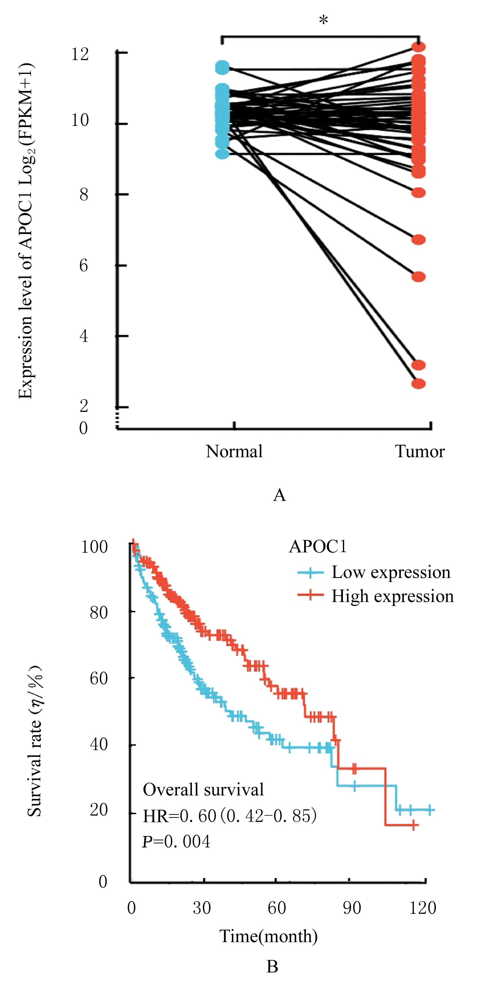
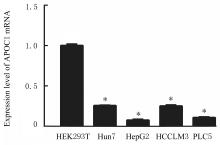
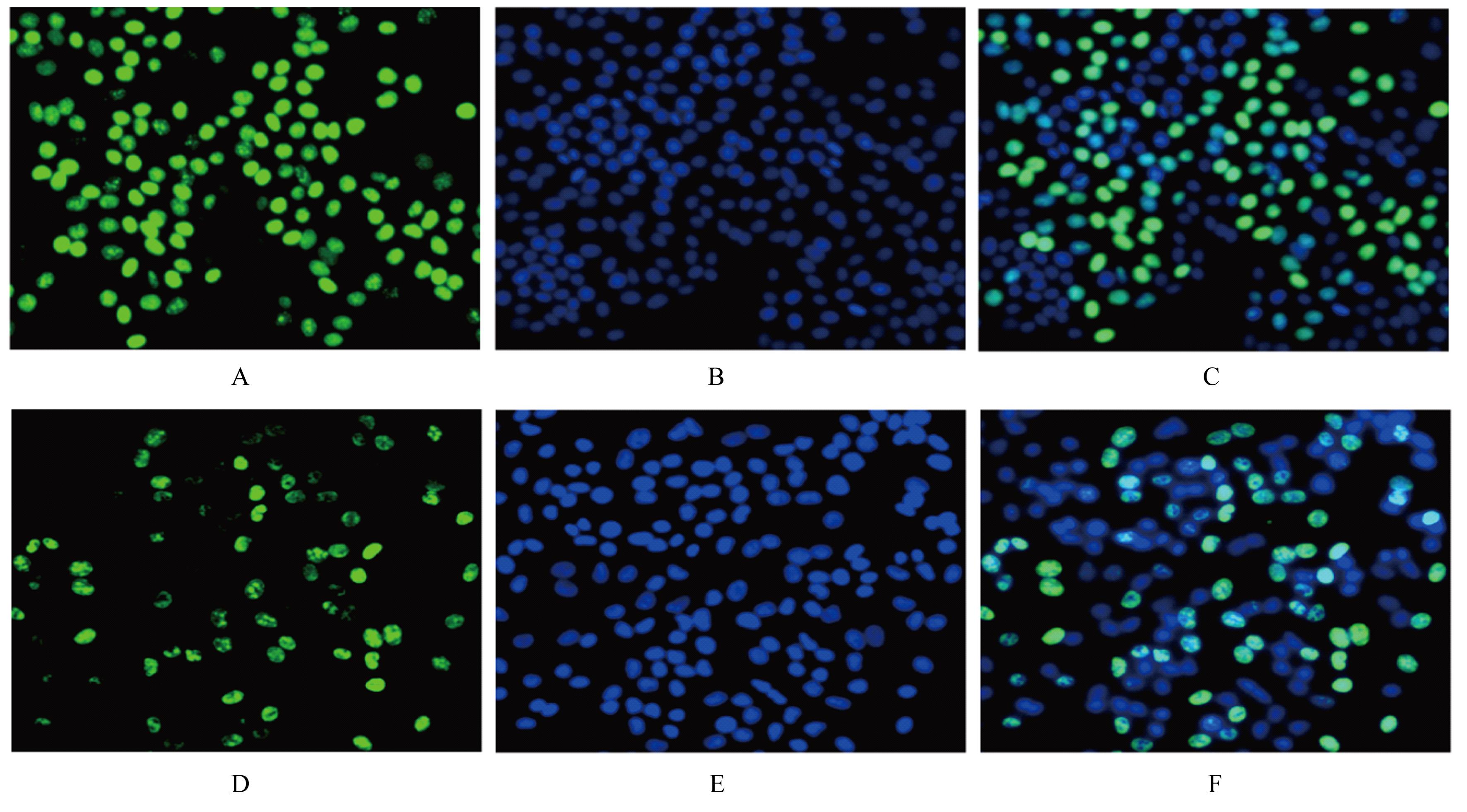
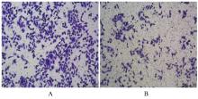
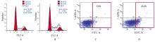
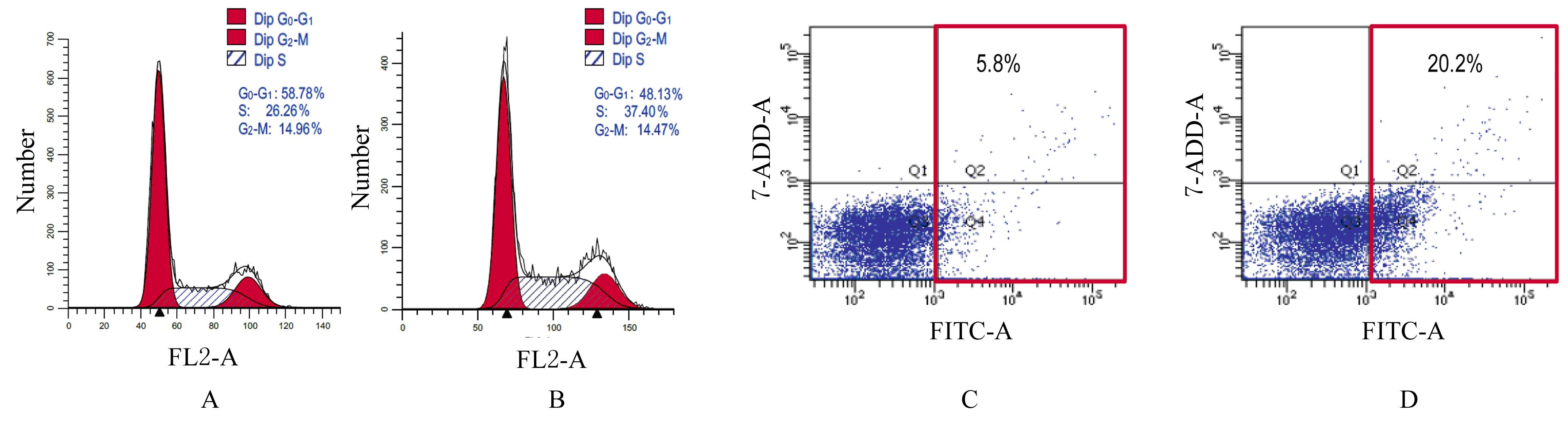
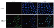
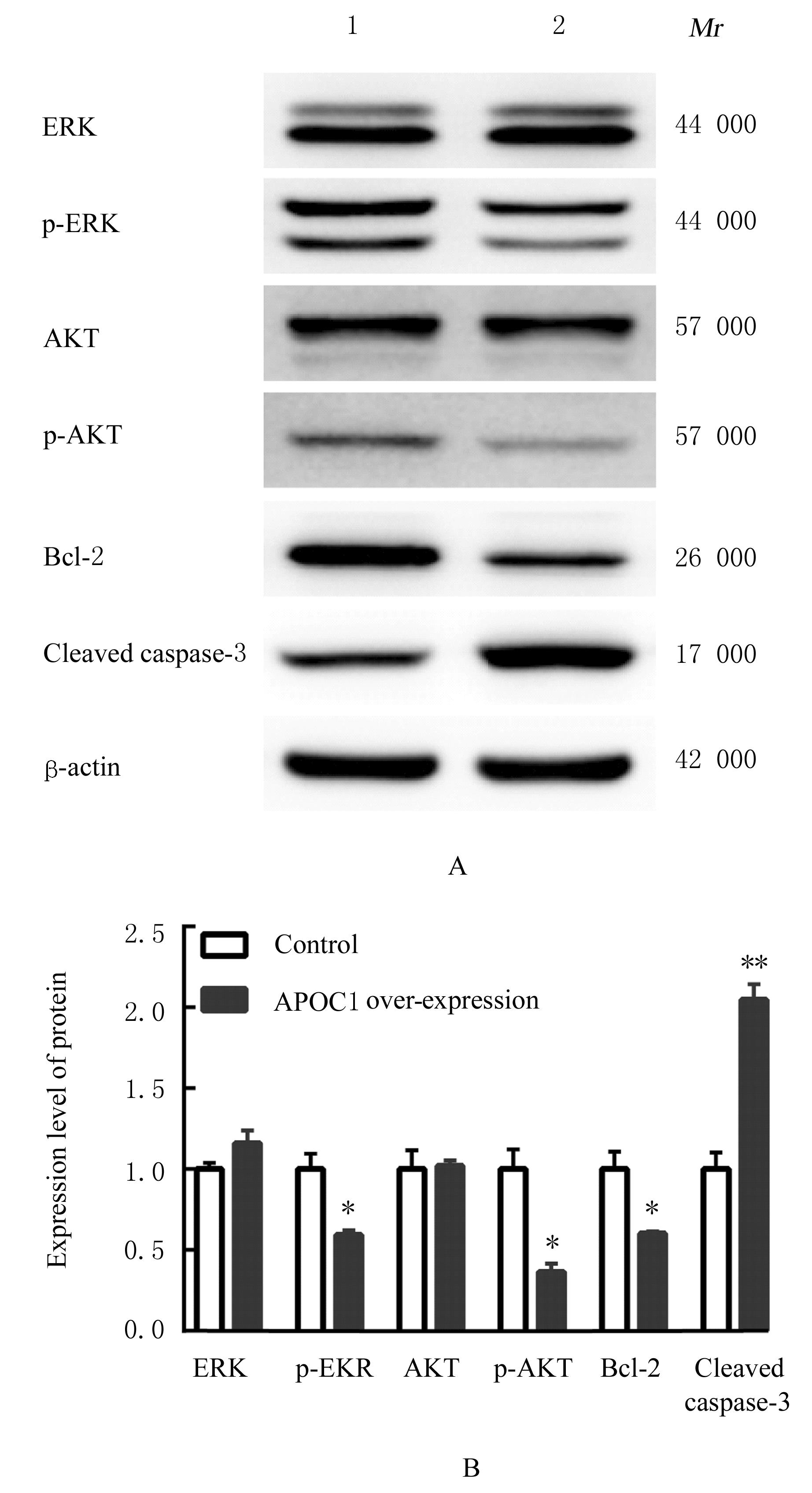
目的 探讨载脂蛋白C1(APOC1)表达对肝癌细胞增殖和凋亡的影响,并初步阐明其相关分子机制。 方法 通过癌症基因组图谱(TCGA)数据库分析肝癌患者癌组织中APOC1 mRNA表达水平及其与患者预后的关系。采用实时荧光定量PCR(RT-qPCR)法检测不同肝癌细胞中APOC1 mRNA表达水平,筛选APOC1低表达的人肝癌HepG2细胞作为研究对象。将pcDNA3.1-APOC1质粒转染至HepG2细胞过表达APOC1(APOC1过表达组),以转染空载体pcDNA3.1的HepG2细胞为对照组,采用MTS法和5-乙炔基-2'-脱氧尿嘧啶核苷(EdU)染色法检测2组细胞增殖活性和增殖率,Transwell小室实验检测2组细胞中迁移细胞数,流式细胞术和TUNEL法检测2组不同细胞周期细胞百分率和细胞凋亡率,Western blotting法检测2组细胞中细胞外调节蛋白激酶(ERK)、磷酸化ERK(p-ERK)、蛋白激酶B(AKT)、磷酸化AKT(p-AKT)、B细胞淋巴瘤2(Bcl-2)和活化型含半胱氨酸的天冬氨酸蛋白水解酶3(cleaved caspase-3)蛋白表达水平。 结果 TCGA数据库分析,肝癌患者癌组织中APOC1 mRNA表达水平低于正常肝组织(P<0.05),并且APOC1 mRNA低表达组肝癌患者预后较差。RT-qPCR法检测,HepG2细胞中APOC1 mRNA表达水平最低,选取该细胞作为后续研究对象。与对照组比较,APOC1过表达组细胞增殖活性和增殖率明显降低(P<0.05或P<0.01),迁移细胞数明显减少(P<0.01), S期细胞百分率和细胞凋亡率明显升高(P<0.01)。与对照组比较,APOC1过表达组细胞中p-ERK、p-AKT和Bcl-2蛋白表达水平明显降低(P<0.05),cleaved caspase-3蛋白表达水平明显升高(P<0.01)。 结论 APOC1高表达能够抑制人肝癌HepG2细胞的增殖,并诱导细胞凋亡,其机制可能与其抑制 p-ERK、p-AKT、Bcl-2蛋白表达和促进cleaved caspase-3蛋白表达有关。

关键词: 载脂蛋白C1, 肝肿瘤, 细胞增殖, 细胞凋亡, 人肝癌HepG2细胞

Abstract:

Objective To discuss the effect of apolipoprotein C1 (APOC1) expression on the proliferation and apoptosis of the hepatocellular carcinoma cells, and to preliminarily clarify the related molecular mechanism. Methods The expression level of APOC1 mRNA in hepatocellular carcinoma tissue and its relationship with the prognosis of the patient were analyzed by The Cancer Genome Atlas (TCGA) Database; real-time fluorescence quantitative PCR (RT-qPCR) method was used to detect the expression levels of APOC1 mRNA in different hepatocellular carcinoma cells; the human liver cancer HepG2 cells with low APOC1 expression were selected as the subjects. The HepG2 cells were transfected with pcDNA3.1-APOC1 plasmid to over-express APOC1 (APOC1 over-expression group),and the HepG2 cells transfected with empty vector pcDNA3.1 were regarded as control group. MTS assay and 5-ethynyl-2'-deoxyuridine (EdU) staining were used to detect the proliferative activities and proliferation rates of the cells in two groups; Transwell chamber assay was used to detect the numbers of migration cells in two groups;flow cytometry and TUNEL assay were used to detect the percentages of the cells at different cell cycles and apoptotic rates in two groups;Western blotting method was used to detect the expression levels of extracellular regulated protein kinase (ERK), phosphorylated ERK (p-ERK), protein kinase B (AKT), phosphorylated AKT (p-AKT), B-cell lymphoma-2 (Bcl-2), and cleaved cysteinyl aspartate specific proteinase-3(cleaved caspase-3) proteins in the cells in two groups. Results The TCGA Database results showed that the expression level of APOC1 mRNA in hepatocellular carcinoma tissue was lower than that in normal liver tissue (P<0.05), and the patients with low expression of APOC1 mRNA had poor prognosis. The RT-qPCR results showed that the expression level of APOC1 mRNA in the HepG2 cells was the lowest, and the HepG2 cells were chosen for the subsequent research. Compared with control group, the proliferative activity and proliferation rate of the cells in APOC1 over-expression group were decreased (P<0.05 or P<0.01),the number of migration cells was decreased (P<0.01),and the percentage of the cells at S phase and the apoptotic rate were significantly increased (P<0.01). Compared with control group, the expression levels of p-ERK, p-AKT, and Bcl-2 proteins in the cells in APOC1 over-expression group were significantly decreased (P<0.05),and the expression level of cleaved caspase-3 protein was increased (P<0.01). Conclusion High expression of APOC1 can inhibit the proliferation of the human liver cancer HepG2 cells and induce the apoptosis,and its mechanism may be related to inhibition of the expressions of p-ERK,p-AKT,Bcl-2 proteins and promotion of the expression of cleaved caspase-3 protein.

Key words: Apolipoprotein C1, Liver neoplasm, Cell proliferation, Apoptosis, Human liver cancer HepG2 cell

中图分类号: 

  • R735.7