吉林大学学报(医学版) ›› 2023, Vol. 49 ›› Issue (6): 1437-1444.doi: 10.13481/j.1671-587X.20230605

• 基础研究 • 上一篇    下一篇

川芎嗪对胶质瘤干细胞裸鼠皮下移植瘤生长、TGF-β信号通路和上皮-间质转化的影响

何涛(),李振江,丁炳谦   

  1. 河南大学淮河医院神经外科, 河南 开封 475000
  • 收稿日期:2022-10-21 出版日期:2023-11-28 发布日期:2023-12-22
  • 通讯作者: 何涛 E-mail:hetaoky@163.com
  • 作者简介:何 涛(1985-),男,河南省开封市人,主治医师,医学硕士,主要从事胶质瘤基础和临床方面的研究。
  • 基金资助:
    河南省卫健委医学科技攻关计划(联合共建)项目(LHGJ20190537)

Influence of ligustrazine on growth of glioma stem cells subcutaneous xenografts in nude mice, TGF-β signaling pathway, and epithelial-mesenchymal transiton

Tao HE(),Zhenjiang LI,Bingqian DING   

  1. Huaihe Hospital of Henan University Neurosurgery Department,Kaifeng 475000,China
  • Received:2022-10-21 Online:2023-11-28 Published:2023-12-22
  • Contact: Tao HE E-mail:hetaoky@163.com

摘要:

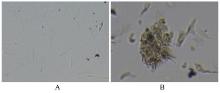
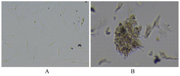
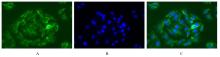
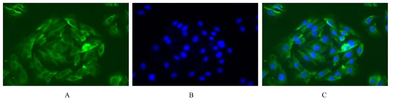
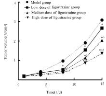
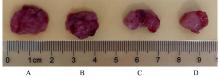
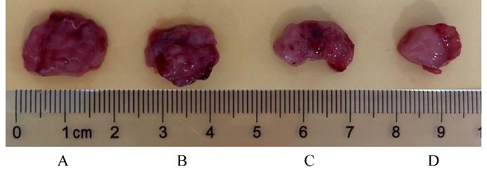
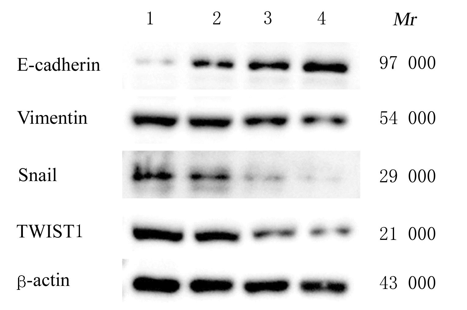
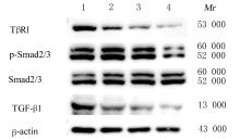
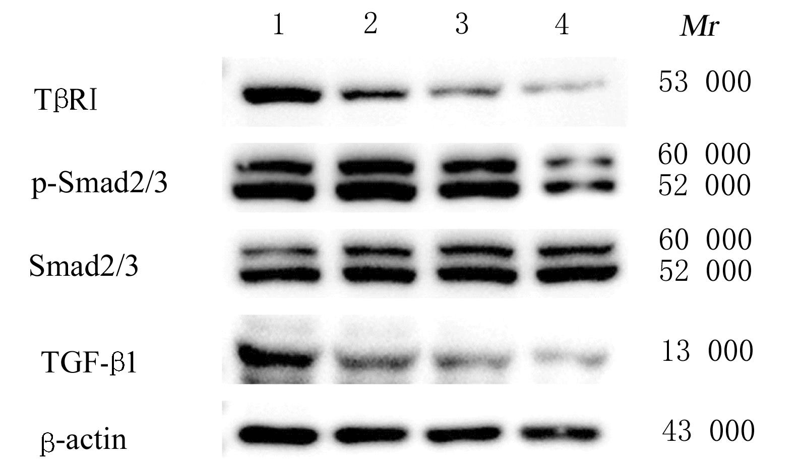
目的 探讨川芎嗪对胶质瘤干细胞(GSCs)裸鼠皮下移植瘤生长、转化生长因子β(TGF-β)信号通路和上皮-间质转化(EMT)的影响,为胶质瘤临床治疗药物的选择提供参考。 方法 收集人脑胶质瘤细胞系U87细胞,培养、分离、富集并鉴定GSCs。40只雌性BALB/c裸鼠,建立GSCs裸鼠皮下移植瘤模型,成模后随机分为模型组和低、中及高剂量川芎嗪组,每组10只。低、中和高剂量川芎嗪组裸鼠每3 d腹腔注射给药1次,给药剂量分别为1.5、3.0和6.0 mg·kg-1,模型组裸鼠仅腹腔注射等量生理盐水,连续15 d。测量各组裸鼠移植瘤体积,称瘤质量,计算瘤质量抑制率,绘制肿瘤生长曲线。HE染色观察各组裸鼠肿瘤组织病理形态表现,实时荧光定量PCR(RT-qPCR)法检测各组裸鼠肿瘤组织中TGF-β1和TGF-β受体Ⅰ(TβRⅠ) mRNA表达水平,Western blotting法检测各组裸鼠肿瘤组织中TGF-β1、TβRⅠ、母亲DPP同源物2/3(Smad2/3)、磷酸化Smad2/3(p-Smad2/3)、E-钙黏蛋白(E-cadherin)、TWIST家族bHLH转录因子1(TWIST1)、波形蛋白(Vimentin)及锌指蛋白(Snail)表达水平。 结果 在U87细胞中成功富集并分离获得GSCs,免疫荧光染色可见干细胞标志物CD133阳性表达。与模型组比较,低、中和高剂量川芎嗪组裸鼠移植瘤体积均减小(P<0.05),且呈剂量依赖性;移植瘤质量均降低(P<0.05),且呈剂量依赖性;低、中和高剂量川芎嗪组瘤质量抑制率逐渐增大,分别为12.72%、39.90%和55.36%。HE染色观察,模型组裸鼠肿瘤组织细胞形态良好,连接紧密,生长密度高,核异型性明显;低、中和高剂量川芎嗪组裸鼠肿瘤组织结构紊乱,细胞密度逐渐降低,核固缩和核裂解逐渐明显,组织坏死区逐渐增大,且随着川芎嗪剂量的增加,上述改善逐渐明显。RT-qRCR法检测,与模型组比较,低、中和高剂量川芎嗪组裸鼠移植瘤组织中TGF-β1和TβRⅠ mRNA表达水平均降低(P<0.05),且呈剂量依赖性。Western blotting法检测,与模型组比较,低、中和高剂量川芎嗪组裸鼠皮下移植瘤组织中E-cadherin蛋白表达水平均升高(P<0.05),TWIST1、Vimentin、Snail、TGF-β1、TβRⅠ和p-Smad2/3蛋白表达水平均降低(P<0.05),且呈剂量依赖性。 结论 川芎嗪可发挥对GSCs裸鼠皮下移植瘤生长的抑制作用,同时可干扰TGF-β1/Smad2/3信号激活和EMT诱导。

关键词: 川芎嗪, 胶质瘤干细胞, 转化生长因子β, 上皮-间质转化, 母亲DPP同源物2/3

Abstract:

Objective To discuss the effect of ligustrazine on growth of the glioma stem cells(GSCs)subcutaneous xenografts in the nude mice, transforming growth factor-β (TGF-β) signaling pathway and epithelial-mesenchymal transition(EMT),and to provide the reference for selecting drugs in the clinical therapy of glioma. Methods The human brain glioma U87 cells were collected and the GSCs were cultured, separated, enriched, and identified. Forty female BALB/c nude mice were used to establish the GSCs subcutaneous xenograft models, which was randomly divided into model group and low, medium, and high doses of ligustrazine groups, and there were 10 mice in each group. The mice in low, medium, and high doses of ligustrazine groups were intraperitoneally injected with ligustrazine once every 3 d, and the dosages were 1.5, 3.0, and 6.0 mg·kg-1, respectively, while the mice in model group were only given equal volume of saline,and this process continued for 15 d. The transplanted tumor volumes and tumor weights were detected, and the inhibitory rates of tumor weights of the mice was caculated. The tumor growth curve was drawn.HE staining was used to observe the pathomorphology of tumor tissue of the mice;real-time fluorescence quantitative PCR (RT-qPCR) method was used to detect the expression levels of TGF-β1 and transforming growth factor β receptor Ⅰ(TβRⅠ) mRNA in tumor tissue of the mice in various groups; Western blotting method was used to detect the expression levels of TGF-β1, TβRⅠ,small mother against decapentaplegic homolog 2/3 (Smad2/3), phosphorylated Smad2/3 (p-Smad2/3),E-cadherin, TWIST family bHLH transcription factor 1 (TWIST1),Vimentin, and Snail proteins in tumor tissue of the mice in various groups. Results The GSCs were successfully enriched and separated from the U87 cells, and the immunofluorescence staining resutls showed the positive expression of stem cell marker CD133. Compared with model group, the transplanted tumor volumes of the nude mice in low, medium, and high doses of ligustrazine groups were significantly decreased (P<0.05), in a dose-dependent manner; the tumor weights were also significantly decreased (P<0.05) in a dose-dependent manner as well. The inhibitory rates of tumor weights in low, medium, and high doses of ligustrazine groups were 12.72%, 39.90%, and 55.36%, respectively.The HE staining results showed that the cells in tumor tissue of the mice in model group were gradually increased and exhibited good morphology and tight connection, the growth density was high, and the atypical nuclei was obviously; the tumor tissue structures of the mice in low, medium, and high doses of ligustrazine groups were disordered, the cell density was gradually decreased, the nuclear condensation and karyorrhexis were increased,the necrotic zones were gradually enlarged, and these improvements became more obvious as the increasing of the dose of ligustrazine. The RT-qPCR results showed that compared with model group, the expression levels of TGF-β1 and TβRⅠ mRNA in tumor tissue of the mice in low, medium, and high doses of ligustrazine groups were decreased (P<0.05) in a dose-dependent manner.The Western blotting results showed that compared with model group, the expression levels of E-cadherin protein in tumor tissue of the mice in low, medium, and high doses of ligustrazine groups were increased (P<0.05),and the expression levels of TWIST1,Vimentin,Snail,TGF-β1,TβRⅠ,and p-Smad2/3 proteins were decreased(P<0.05) in a dose-dependent manner. Conclusion Ligustrazine can inhibit the growth of GSCs subcutaneous xenografts in the nude mice, and can interfere the TGF-β1/Smad2/3 signaling activation and EMT induction.

Key words: Ligustrazine, Glioma stem cell, Transforming growth factor-β, Epithelial-mesenchymal transition, Small mother against decapentraplegic homolog 2/3

中图分类号: 

  • R285.5