吉林大学学报(医学版) ›› 2024, Vol. 50 ›› Issue (3): 638-646.doi: 10.13481/j.1671-587X.20240307

• 基础研究 • 上一篇    

五味子乙素对胰腺癌Pan02细胞增殖的抑制作用及其机制

傅家财1,2,秦玲莎2,杨露2,宋美慧2,张仙映2,刘晓翠2,李凤金2(),齐玲2()   

  1. 1.大理大学药学院,云南 大理 671000
    2.广东医科大学附属清远医院 广东省清远市人民医院,广东 清远 511500
  • 收稿日期:2023-06-13 出版日期:2024-05-28 发布日期:2024-07-01
  • 通讯作者: 李凤金,齐玲 E-mail:wklifengjin@163.com;qiling1718@gzhmu.edu.cn
  • 作者简介:傅家财(1998-),男,辽宁省沈阳市人,在读硕士研究生,主要从事胰腺炎癌转化机制及防治方面的研究。
  • 基金资助:
    国家自然科学基金项目(82203351);广东省中医药局科研项目(20231412);广东省卫健委医学科学技术研究基金项目(A2022163)

Inhibitory effect of Schisandrin B on proliferation of pancreatic cancer Pan02 cells and its mechanism

Jiacai FU1,2,Lingsha QING2,Lu YANG2,Meihui SONG2,Xianying ZHANG2,Xiaocui LIU2,Fengjin LI2(),Ling QI2()   

  1. 1.School of Pharmcy,Dali University,Dali 671000,China
    2.Affiliated Qingyuan Hospital,Guangzhou Medical University,People’s Hospital,Qingyuan City,Guangdong Province,Qingyuan 511500,China
  • Received:2023-06-13 Online:2024-05-28 Published:2024-07-01
  • Contact: Fengjin LI,Ling QI E-mail:wklifengjin@163.com;qiling1718@gzhmu.edu.cn

摘要:

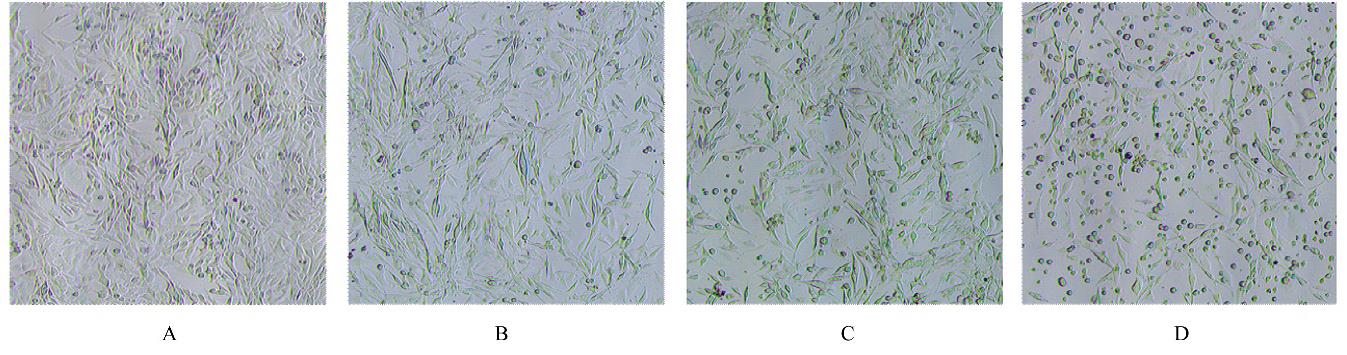
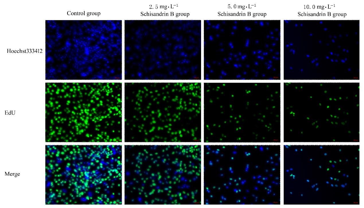
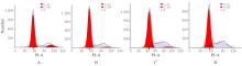
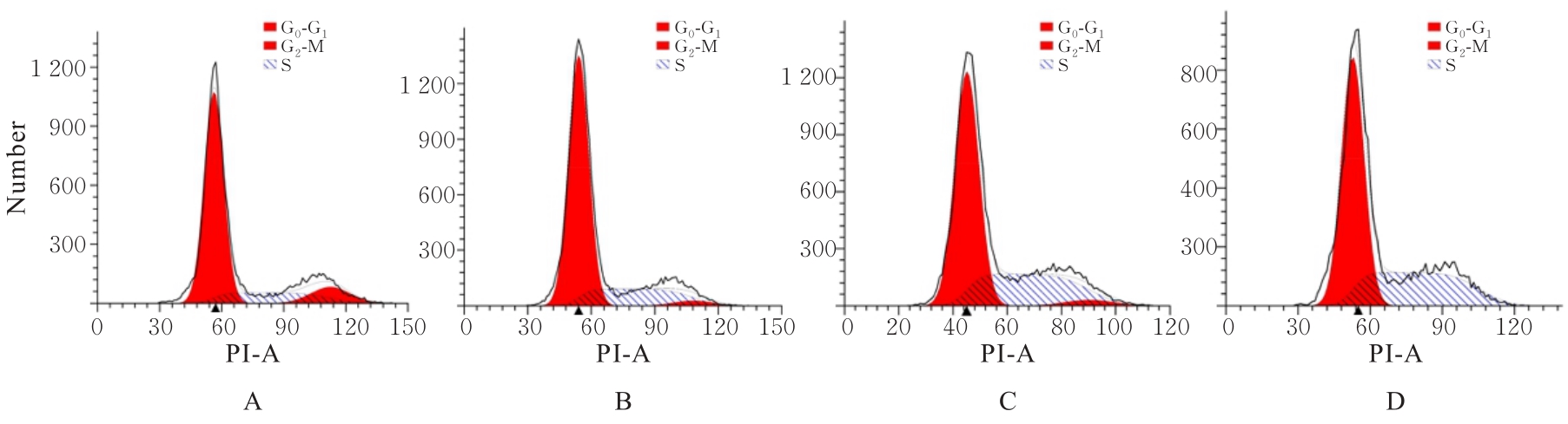
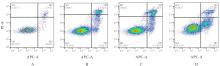
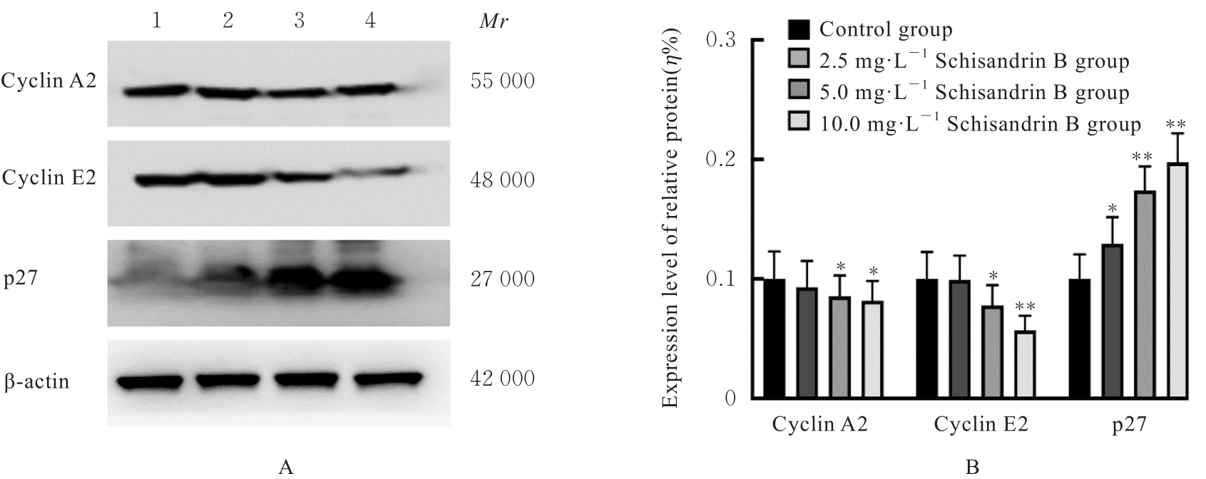
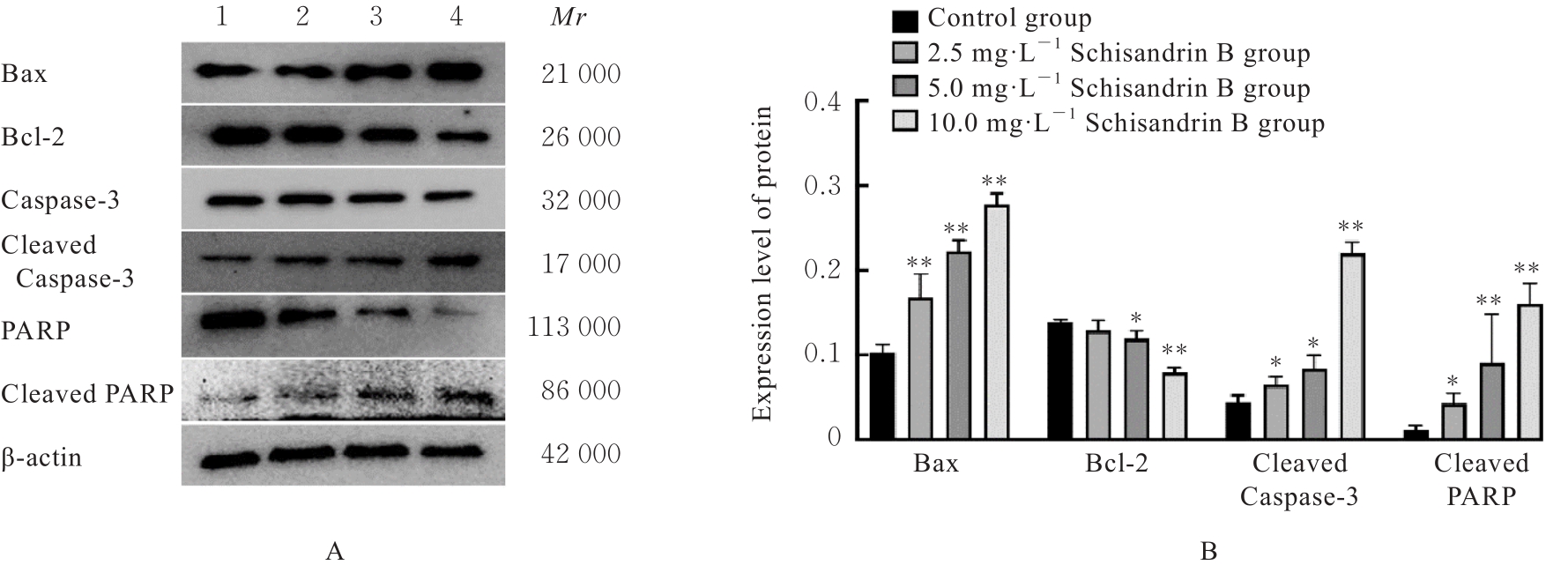
目的 探讨五味子乙素对胰腺癌 Pan02 细胞增殖的抑制作用, 并阐明其作用机制。 方法 采用CCK-8法检测不同浓度(0、0.78、1.56、3.12、6.25、12.50和25.00 mg·L-1 )五味子乙素作用下Pan02细胞增殖率,以选择五味子乙素作用的最适浓度和最佳作用时间。小鼠胰腺癌 Pan02 细胞分为对照组(0 mg·L-1 五味子乙素)、2.5 mg·L-1 五味子乙素组、5.0 mg·L-1五味子乙素组和10.0 mg·L-1 五味子乙素组。光学显微镜观察各组Pan02细胞形态表现,5-乙基-2'-脱氧尿嘧啶核苷(EdU)染色法检测各组Pan02细胞中EdU阳性细胞率,流式细胞术检测各组不同细胞周期Pan02细胞百分率和细胞凋亡率,Western blotting法检测各组Pan02细胞周期和凋亡相关蛋白表达水平。 结果 CCK-8法,五味子乙素作用Pan02细胞48和72 h后,与0 mg·L-1 五味子乙素比较,其他浓度五味子乙素作用下Pan02细胞增殖率明显降低(P<0.01),72 h时细胞抑制作用最明显。选择0、2.5、5.0和10.0 mg·L-1五味子乙素作用Pan02细胞,作用时间为72 h。对照组Pan02细胞呈长梭形,状态良好,紧密且贴壁生长,细胞器和细胞质正常;2.5和5.0 mg·L-1五味子乙素组Pan02细胞体积减小,细胞之间黏连消失,细胞膜虽完整但通透性增强,细胞质皱缩,细胞内部产生空泡结构,部分呈碎片状漂浮于溶液表面;10.0 mg·L-1五味子乙素组Pan02细胞有明显凋亡小体生成,呈现凋亡状态。EdU染色法,与对照组比较,2.5、5.0和10.0 mg·L-1 五味子乙素组 Pan02 细胞中 EdU 阳性细胞率均明显降低(P<0.01)。流式细胞术,与对照组比较,2.5、5.0和10.0 mg·L-1五味子乙素组Pan02细胞S期细胞百分率明显升高(P<0.01),G2/M期细胞百分率明显降低(P<0.01),5.0和10.0 mg·L-1五味子乙素组G0/G1期细胞百分率明显降低(P<0.01);与对照组比较,2.5、5.0和10.0 mg·L-1五味子乙素组Pan02细胞凋亡率明显升高(P<0.01)。Western blotting法,与对照组比较,2.5 mg·L-1五味子乙素组Pan02细胞中p27、B细胞淋巴瘤2(Bcl-2)相关X蛋白(Bax)、裂解的半胱氨酸天冬氨酸蛋白酶3(cleaved Caspase-3)和裂解的多聚二磷酸腺苷(ADP)核糖聚合酶(cleaved PARP)蛋白表达水平明显升高(P<0.05或P<0.01);5.0和10.0 mg·L-1五味子乙素组Pan02细胞中细胞周期蛋白(Cyclin) A2、Cyclin E2 和 Bcl-2 蛋白表达水平均明显降低(P<0.05或P<0.01), p27、 Bax、 cleaved Caspase-3 和cleaved PARP 蛋白表达水平均明显升高(P<0.01)。 结论 五味子乙素具有抑制胰腺癌 Pan02 细胞增殖的作用,其作用机制可能与激活半胱氨酸天冬氨酸蛋白酶3(Caspase-3)通路诱导细胞凋亡和激活p27蛋白并诱导细胞周期S期阻滞有关。

关键词: 五味子乙素, 胰腺肿瘤, Pan02细胞, 细胞凋亡, 细胞周期

Abstract:

Objective To discuss the inhibitory effect of Schisandrin B on the proliferation of pancreatic cancer Pan02 cells, and to clarify the mechanism. Methods CCK-8 method was used to detect the proliferation rates of the Pan02 cells after treated with different concentrations(0, 0.78, 1.56, 3.12, 6.25, 12.50, and 25.00 mg·L-1 ) of Schisandrin B to select the optimal concentration and treatment time of Schisandrin B. The mouse pancreatic cancer Pan02 cells were divided into control group (0 mg·L-1 Schisandrin B), 2.5 mg·L-1 Schisandrin B group, 5.0 mg·L-1 Schisandrin B group, and 10.0 mg·L-1 Schisandrin B group. The morpholoy of Pan02 cells invarious groups was observed with light microscope; 5-ethynyl-2'-deoxyuridine (EdU) staining assay was used to detect the positive expression rates of the Pan02 cells in various groups; flow cytometry was used to detect the percentages of the Pan02 cells at different cell cycles and the apoptotic rates of the cells in various groups; Western blotting method was used to detect the expression levels of cell cycle and apoptosis-related proteins in the cells in various groups. Results The CCK-8 method results showed that after treated with Schisandrin B for 48 and 72 h, compared with 0 mg·L-1 Schisandrin B, the proliferation rates of the Pan02 cells after treated with different concentrations of Schisandrin B were decreased (P<0.01), especially at 72 h. 0.25, 5.0, and 10.0 mg·L-1 Schisandrin B were selected to treat the Pan02 cells, and 72 h was the treatment time. In control group, the Pan02 cells had a spindle shape, with good condition, and grew closely adhered to the wall with normal organelles and cytoplasm, in 2.5 and 5.0 mg·L-1 Schisandrin B groups, the cell volume was decreased, the intercellular adhesion was disappeared, and the cell membrane was intact but more permeable; the cytoplasm shrank and vacuolar structures appeared inside the cells, with some fragmented and floating on the surface of the solution; in 10.0 mg·L-1 Schisandrin B group, the Pan02 cells exhibited notable apoptotic bodies, indicating an apoptotic state. The EdU staining results showed that compared with control group, the rates of EdU positive cells in 2.5, 5.0, and 10.0 mg·L-1 Schisandrin B groups were significantly decreased (P<0.01). The flow cytometry results showed that compared with control group, the percentages of the cells at S phase in 2.5, 5.0, and 10.0 mg·L-1 Schisandrin B groups were significantly increased (P<0.01), while the percentages of the cells at G2/M phase were significantly decreased (P<0.01), and the percentages of the cells at G0/G1 phase in 5.0 amd 1.0 mg·L-1 Schisandrin groups were decreased (P<0.01); compared with control group, the apoptotic rates of the cells in 2.5, 5.0, and 10.0 mg·L-1 Schisandrin B groups were significantly increased (P<0.01). The Western blotting results showed that compared with control group, the expression levels of p27, B-cell lymphoma 2 (Bcl-2) associated X protein (Bax), cleaved cysteine aspartic acid protease-3 (cleaved Caspase-3), and cleaved poly adenosine diphosphate(ADP) ribose polymerase (cleaved PARP) proteins in the cells in 2.5 mg·L-1 Schisandrin B group were significantly increased (P<0.05 or P<0.01), the expression levels of cyclin A2, cyclin E2, and Bcl-2 proteins in the cells in 5.0 and 10.0 mg·L-1 Schisandrin B groups were significantly decreased (P<0.05 or P<0.01), while the expression levels of p27, Bax, cleaved Caspase-3, and cleaved PARP proteins in the cells in 5.0 and 10.0 mg·L-1 Schisandrin B groups were significantly increased (P<0.01). Conclusion Schisandrin B has an inhibitory effect on proliferation of the pancreatic cancer Pan02 cells, and its mechanism may be related to the activation of the cysteine aspartic acid protease-3 (Caspase-3) pathway to induce the apoptosis and activating p27 protein to induce the arrest of cell cycle at S phase.

Key words: Schisandrin B, Pancreas neoplasm, Pan02 cell, Apoptosis, Cell cycle

中图分类号: 

  • R735.9