吉林大学学报(医学版) ›› 2023, Vol. 49 ›› Issue (5): 1125-1133.doi: 10.13481/j.1671-587X.20230503

• 基础研究 • 上一篇    

二氧化硫对大鼠急性心肌缺血损伤后心肌纤维化的改善作用及其机制

刘星,刘佳丽,聂连桂,刘茂军,赵俊雄,汪刘洋,杨军()   

  1. 南华大学衡阳医学院附属第一医院心内科,湖南 衡阳 421001
  • 收稿日期:2022-12-12 出版日期:2023-09-28 发布日期:2023-10-26
  • 通讯作者: 杨军 E-mail:yangjunketizu@163.com
  • 作者简介:刘 星(1997-),男,湖南省常德市人,住院医师,医学硕士,主要从事心血管系统疾病方面的研究。
  • 基金资助:
    国家自然科学基金资助项目(81870230);湖南省科技厅自然科学基金青年基金项目(2021JJ40499)

Improvement effect of SO2 on myocardial fibrosis after acute myocardial ischemic injury in rats and its mechanism

Xing LIU,Jiali LIU,Liangui NIE,Maojun LIU,Junxiong ZHAO,Liuyang WANG,Jun YANG()   

  1. Department of Cardiology,First Affiliated Hospital,Hengyang Medical College,University of South China,Hengyang 421001,China
  • Received:2022-12-12 Online:2023-09-28 Published:2023-10-26
  • Contact: Jun YANG E-mail:yangjunketizu@163.com

摘要:

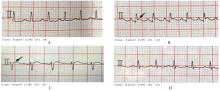
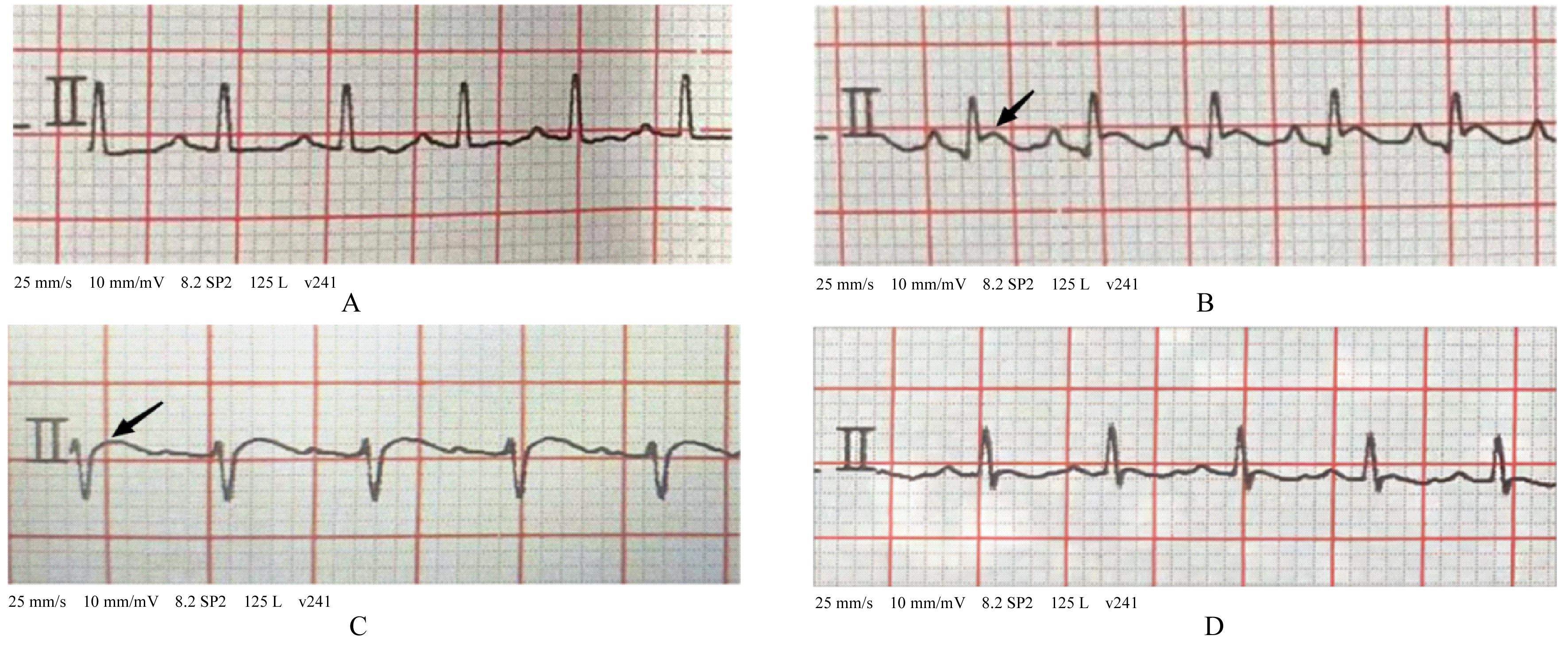
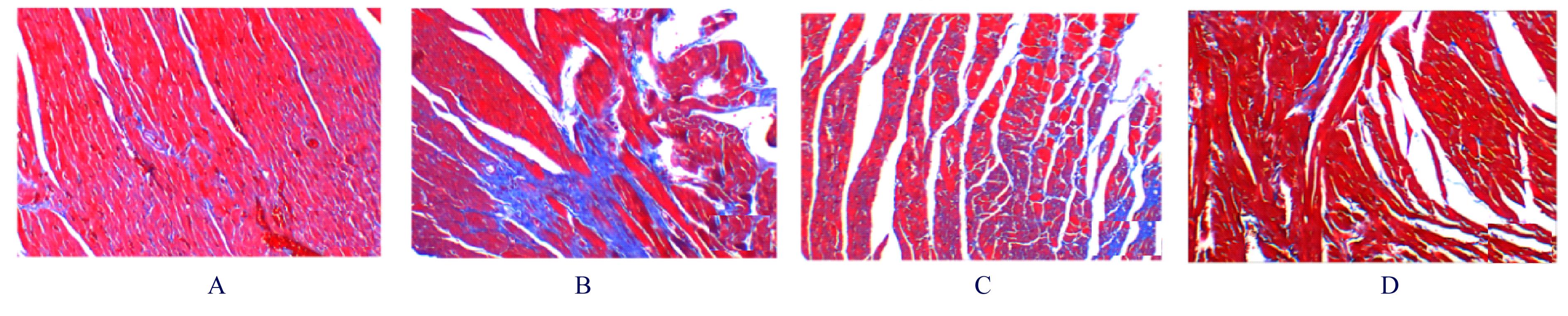
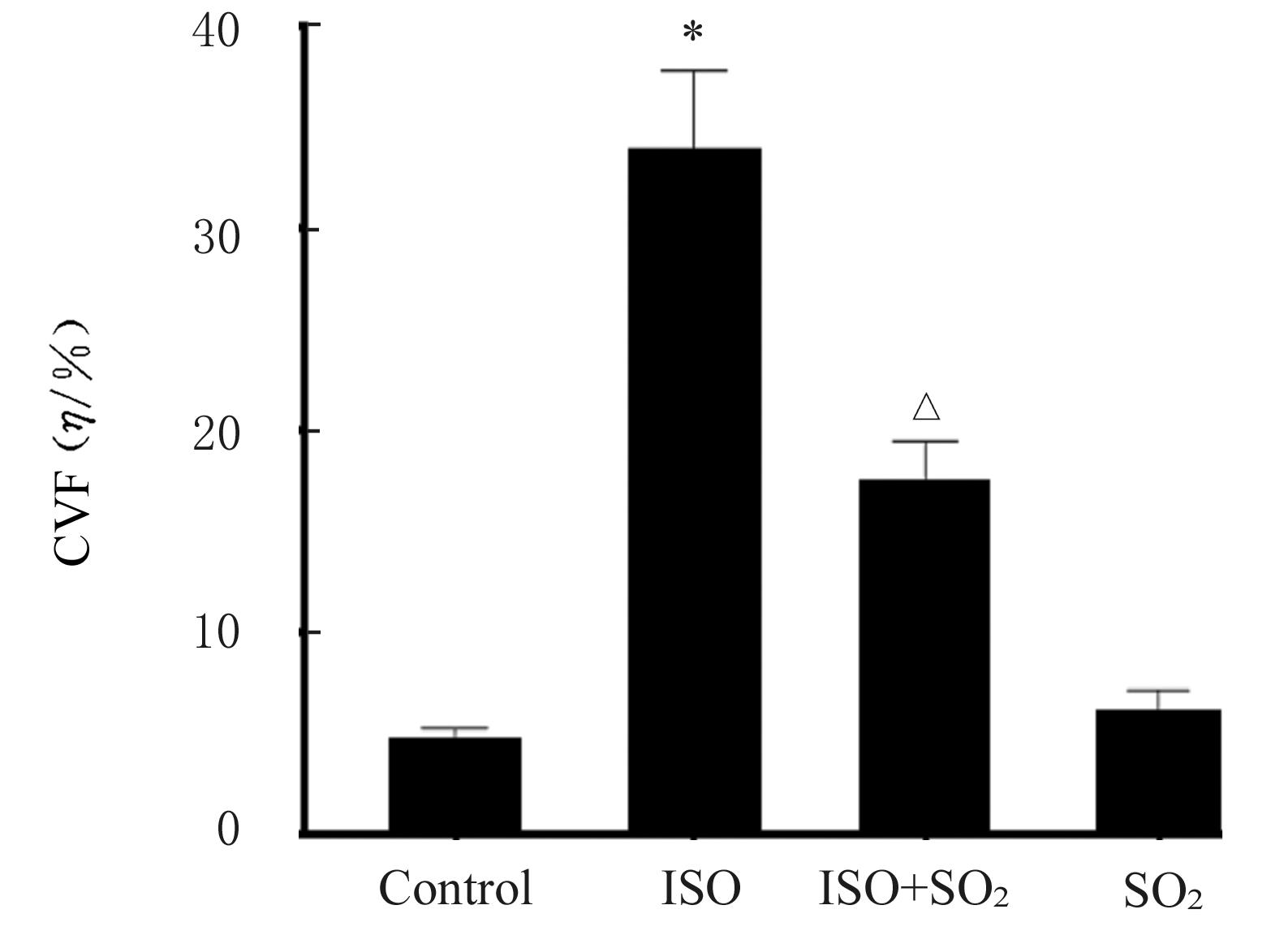
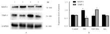
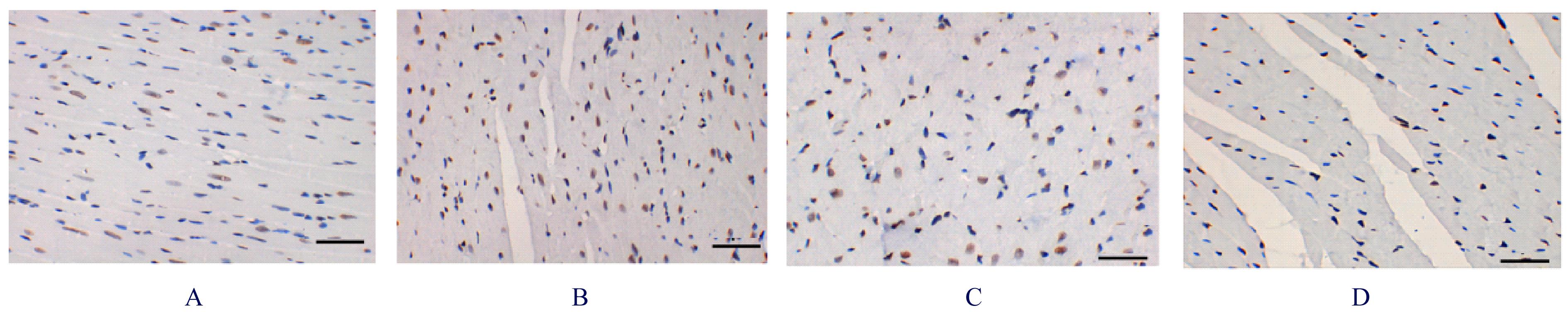
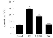
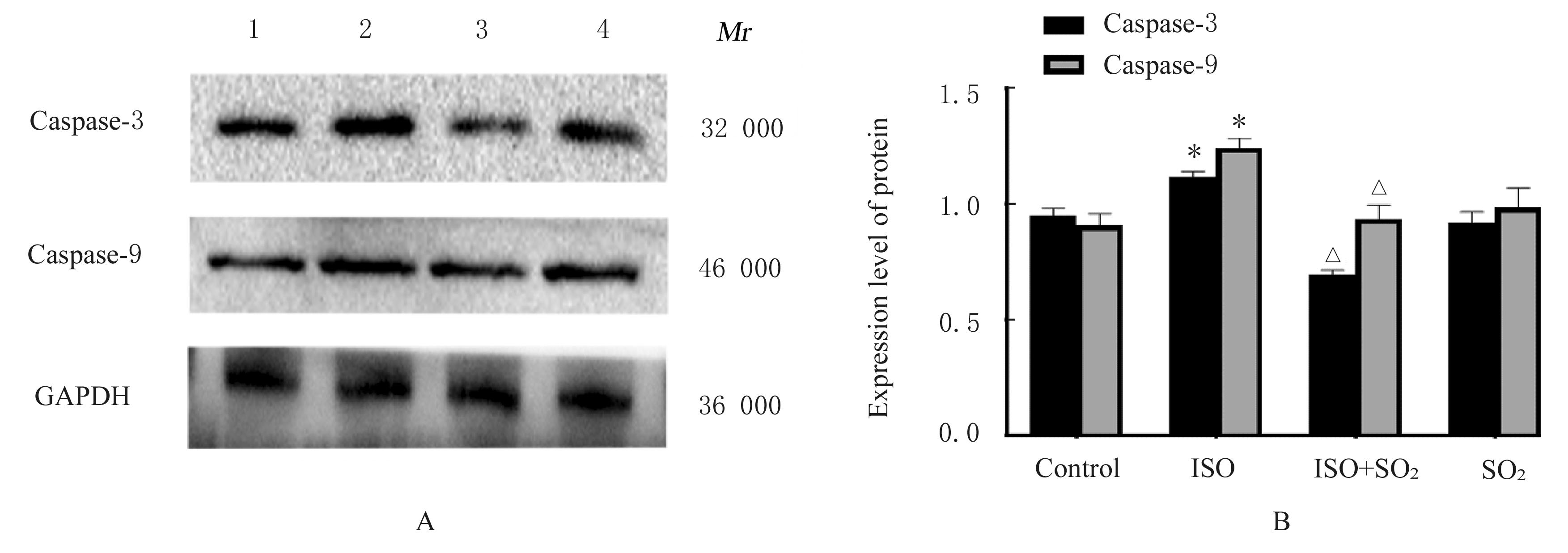
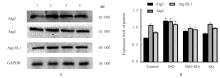
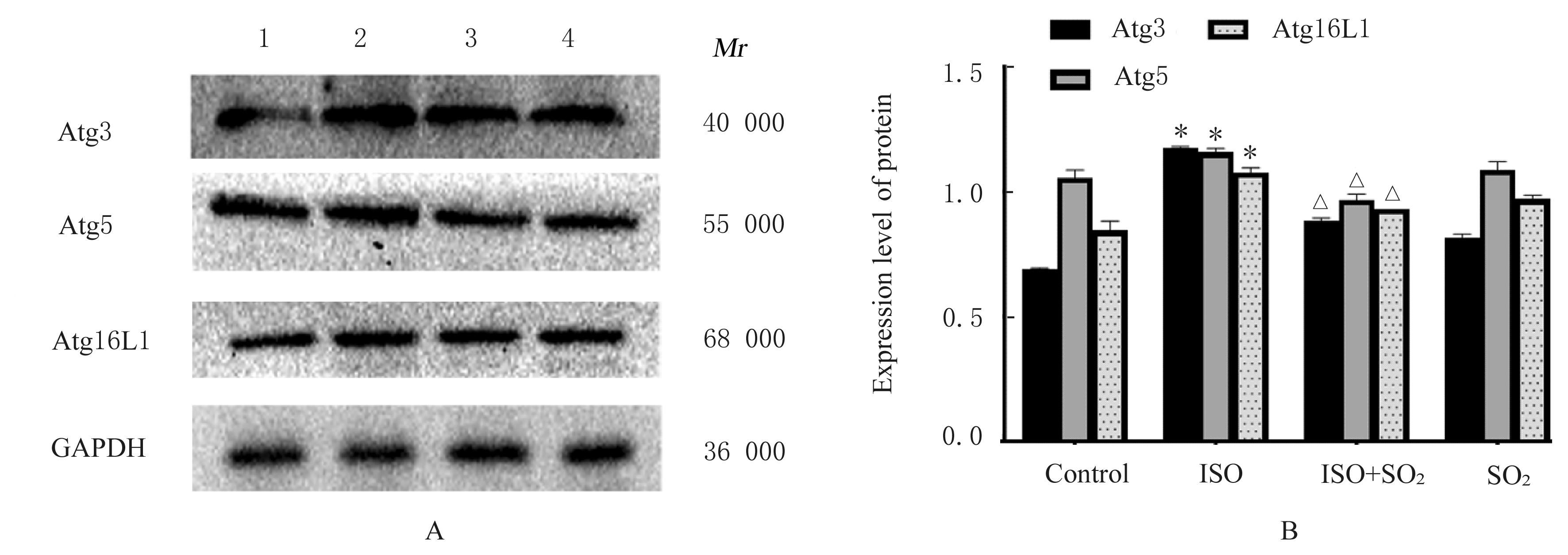
目的 观察二氧化硫(SO2)对大鼠急性心肌缺血损伤后心肌纤维化(MF)的影响,并探讨其作用机制。 方法 24只雄性SD大鼠随机分为对照组(不处理)、异丙肾上腺素(ISO)组(给予ISO)、ISO+SO2组(给予ISO+SO2)和SO2组(给予SO2),每组6只。ISO组和ISO+SO2组大鼠连续2 d腹腔注射大剂量ISO (50 mg·kg-1·d-1)构建急性心肌缺血损伤模型。造模成功后,ISO+SO2组和SO2组大鼠给予Na2SO3溶液(0.54 mmol·kg-1·d-1)和NaHSO3溶液(0.18 mmol·kg-1·d-1),连续4周,对照组和ISO组大鼠给予等量生理盐水。检测4组大鼠血浆肌钙蛋白水平和心电图,超声心动图检测4组大鼠左室短轴缩短率(LVFS)和左室射血分数(LVEF),Masson染色检测4组大鼠心肌组织中胶原沉积情况并计算胶原体积分数(CVF),Tunel染色检测4组大鼠心肌细胞凋亡率,Western blotting法检测4组大鼠心肌组织中自噬相关蛋白3(Atg3)、自噬相关蛋白5(Atg5)、自噬相关蛋白16L1(Atg16L1)、含半胱氨酸的天冬氨酸蛋白水解酶3(Caspase-3)、含半胱氨酸的天冬氨酸蛋白水解酶9(Caspase-9)、基质金属蛋白酶8(MMP-8)和金属蛋白酶组织抑制物2(TIMP-2)蛋白表达水平。 结果 与对照组比较,ISO组和ISO+SO2组大鼠心电图ST段明显抬高,肌钙蛋白水平升高(P<0.05),提示急性心肌缺血损伤大鼠造模成功;与对照组比较,ISO组大鼠LVFS和LVEF降低(P<0.05),心肌组织中CVF升高(P<0.05),心肌细胞凋亡率升高(P<0.05),心肌组织中Atg3、Atg5、Atg16L1、Caspase-3、Caspase-9和MMP-8蛋白表达水平升高(P<0.05),TIMP-2蛋白表达水平降低(P<0.05);与ISO组比较,ISO+SO2组大鼠LVFS和LVEF升高(P<0.05),心肌组织中CVF降低(P<0.05),心肌细胞凋亡率降低(P<0.05),心肌组织中Atg3、Atg5、Atg16L1、Caspase-3、Caspase-9和MMP-8蛋白表达水平降低(P<0.05),TIMP-2蛋白表达水平升高(P<0.05)。 结论 SO2可以改善大鼠急性心肌缺血损伤后MF,其机制可能与抑制心肌细胞过度自噬并减少心肌细胞凋亡有关。

关键词: 二氧化硫, 心肌缺血, 心肌纤维化, 细胞自噬, 细胞凋亡

Abstract:

Objective To observe the effect of sulfur dioxide (SO2) on the myocardial fibrosis(MF) after acute myocardial ischemic injury in the rats, and to explore its mechanism. Methods Twenty-four male SD rats were randomly divided into control group (untreated), isoproterenol (ISO) group (treated with ISO), ISO+ SO2 group (treated with ISO and SO2), and SO2 group (treated with SO2), and there were six rats in each group. The acute myocardial ischemic injury model rats in ISO group and ISO+ SO2 group were induced by intraperitoneal injection with a high dose of ISO (50 mg·kg-1·d-1) for 2 d. After successful modeling, the rats in ISO+ SO2 group and SO2 group were treated with Na2SO3 solution (0.54 mmol·kg-1·d-1) and NaHSO3 solution (0.18 mmol·kg-1·d-1) for four weeks, while the rats in control group and ISO group were treated with equal volume of physiological saline intraperitoneally. The levels of myocardial troponin in plasma and electrocardiogram of the rats in various groups were detected;the left ventricular fractional shortening (LVFS) and left ventricular ejection fraction (LVEF) of the rats in various groups were detected by echocardiography;the collage deposition in myocadium tissue of the rats in various groups was detected by Masson staining and the collagen volume fraction (CVF) was calculated; the apoptotic rates of cardiomyocytes of the rats in various groups were detected by Tunel staining;the expression levels of autophagy-related proteins 3 (Atg3), autophagy-related protein 5 (Atg5), autophagy-related protein 16L1 (Atg16L1),aspartate proteolytic enzyme containing cysteine 3(Caspase-3), aspartate proteolytic enzyme containing cysteine 9(Caspase-9), matrix metalloproteinase 8 (MMP-8), and tissue inhibitor of metalloproteinase 2 (TIMP-2) proteins in myocardium tissue of the rats in various groups were detected by Western blotting method. Results Compared with control group, the ST segment in electrocardiogram of the rats in ISO group and ISO+ SO2 group showed elevation and the levels of myocardial troponin were increased (P<0.05),which indicated the acute myocardial ischemic injury models of the rats were established successfully. Compared with control group, the LVEF and LVFS of the rats in ISO group were decreased (P<0.05), the CVF in myocadium tissue was increased (P<0.05), the apoptotic rate of the cardiomyocytes was increased (P<0.05), and the expression levels of Atg3, Atg5, Atg16L1, Caspase-3, Caspase-9, and MMP-8 proteins in myocardium tissue were increased(P<0.05), and the expression level of TIMP-2 in myocardium tissue was decreased (P<0.05). Compared with ISO group, the LVEF and LVFS of the rats in ISO+ SO2 group was increased (P<0.05), the CVF in myocadium tissue was decreased (P<0.05), the apoptotic rate of the cardiomyocytes was decreased (P<0.05), the expression levels of Atg3, Atg5, Atg16L1, Caspase-3, Caspase-9, and MMP-8 proteins in myocardium tissue were decreased(P<0.05), and the expression level of TIMP-2 in myocardial tissue was increased (P<0.05). Conclusion SO2 can improve the MF after acute myocardial ischemic injury in the rats, and its mechanism may be related to the inhibition of excessive myocardial cell autophagy and reduction of the apoptosis of the cardiomyocytes.

Key words: Sulfur dioxide, Myocardial ischemia, Myocardial fibrosis, Cellular autophagy, Apoptosis

中图分类号: 

  • R542.2