吉林大学学报(医学版) ›› 2024, Vol. 50 ›› Issue (3): 647-657.doi: 10.13481/j.1671-587X.20240308

• 基础研究 • 上一篇    下一篇

D-柠檬烯对胶质母细胞瘤细胞增殖的抑制作用及其机制

王腾飞1,2,陈凤1,2,齐玲2,雷婷1(),宋美慧2()   

  1. 1.大理大学药学院,云南 大理 671000
    2.广州医科大学附属清远医院 广东省清远市人民医院,广东 清远 511500
  • 收稿日期:2023-07-13 出版日期:2024-05-28 发布日期:2024-07-01
  • 通讯作者: 雷婷,宋美慧 E-mail:leiting@dali.edu.cn;songmeihui.gzhmu@foxmail.com
  • 作者简介:王腾飞(1998-),男,山东省烟台市人,在读硕士研究生,主要从事肿瘤与DNA损伤修复方面的研究。
  • 基金资助:
    国家自然科学基金项目(82203351);广东省基础研究委员会基础与应用基础研究基金项目(2021A1515111095);广东省清远市人民医院医学科研基金(20220330)

Inhibitory effect of D-limonene on proliferation of glioblastoma cells and its mechanism

Tengfei WANG1,2,Feng CHEN1,2,Ling QI2,Ting LEI1(),Meihui SONG2()   

  1. 1.School of Pharmaceutical Sciences,Dali University,Dali 671000,China
    2.Institute of Digestive Disease,People’s Hospital,Qingyuan City,Guangdong Province,Qingyuan 511500,China
  • Received:2023-07-13 Online:2024-05-28 Published:2024-07-01
  • Contact: Ting LEI,Meihui SONG E-mail:leiting@dali.edu.cn;songmeihui.gzhmu@foxmail.com

摘要:

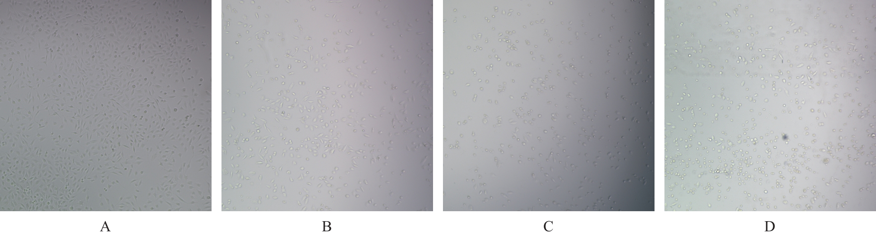
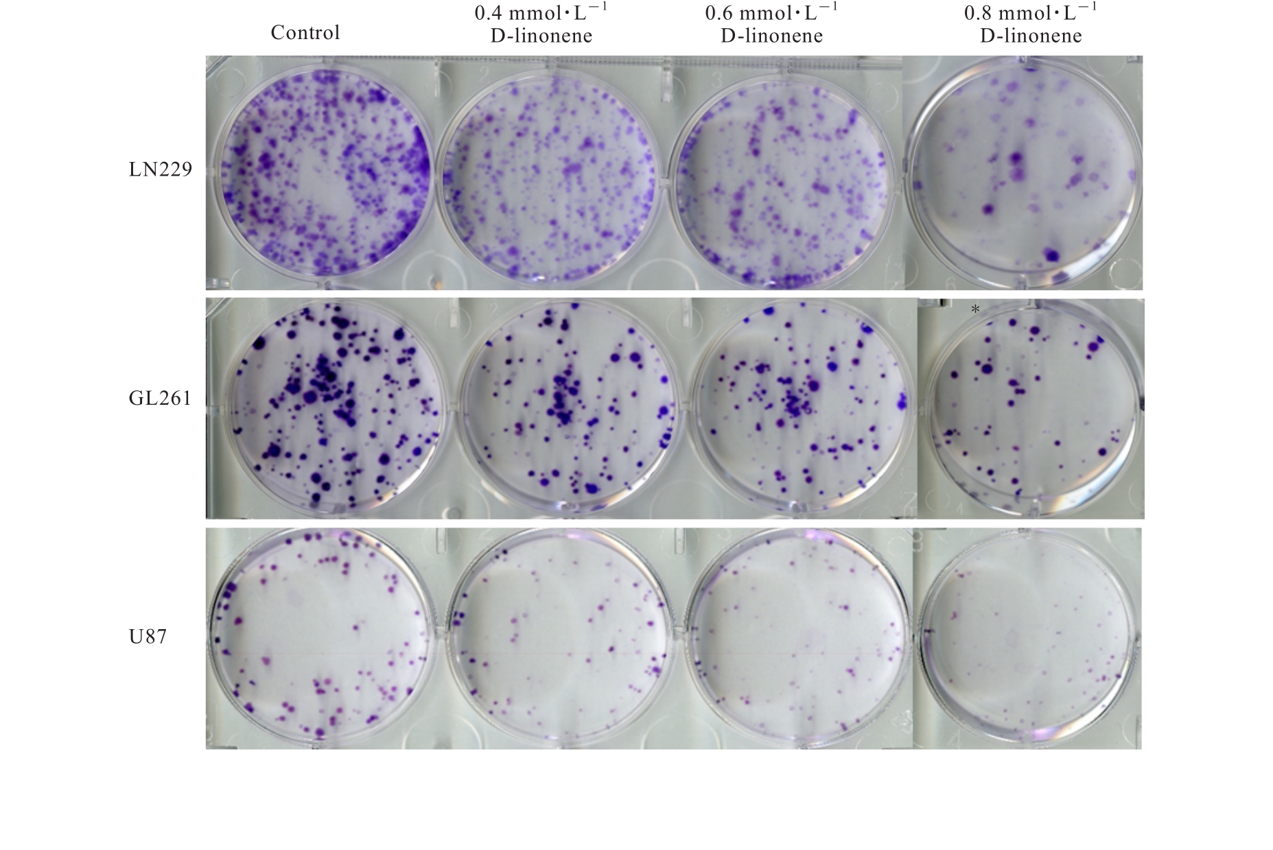
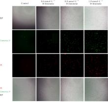
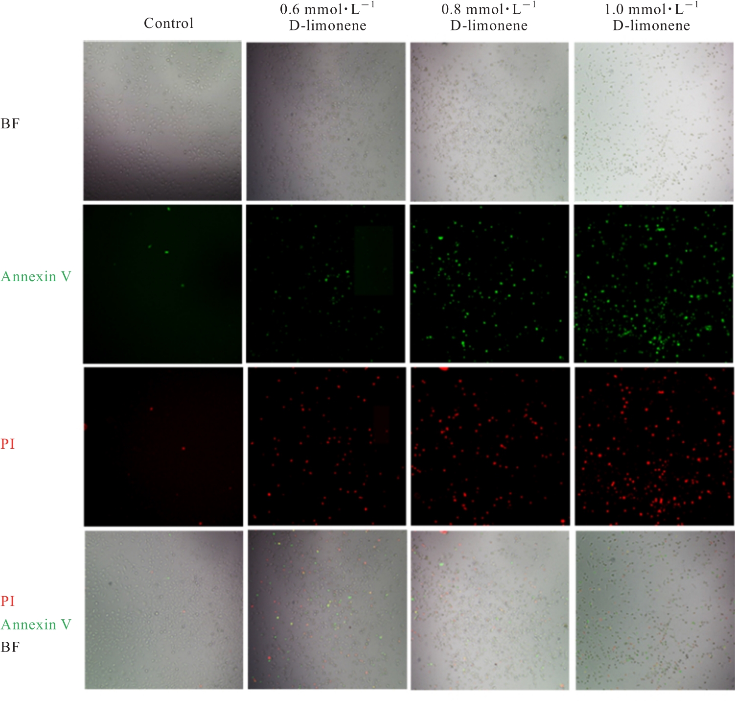
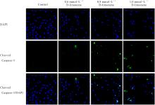
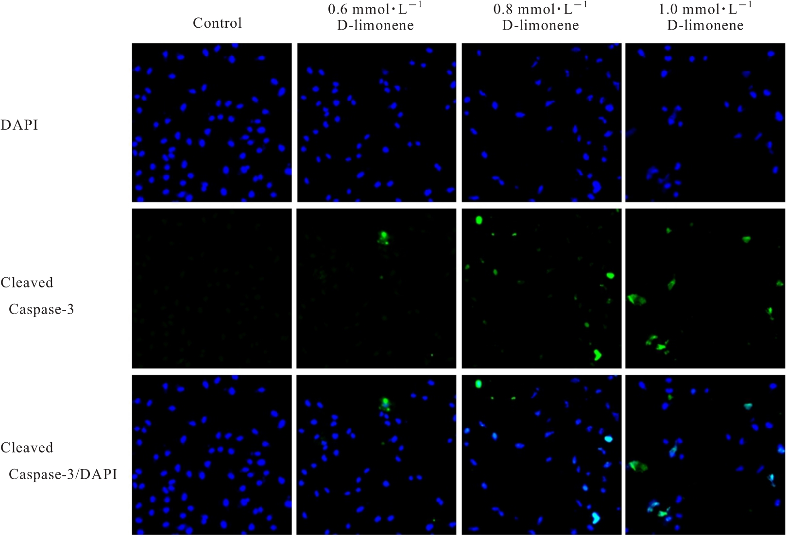
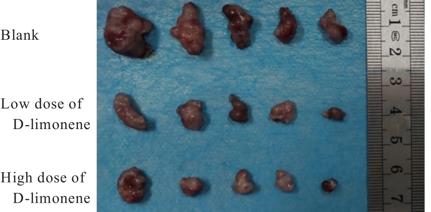
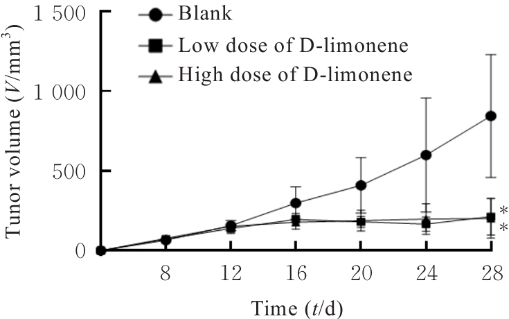
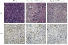
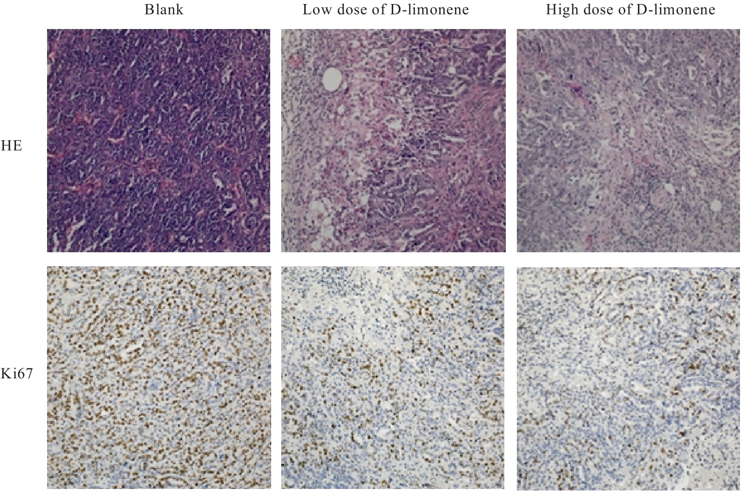
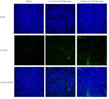
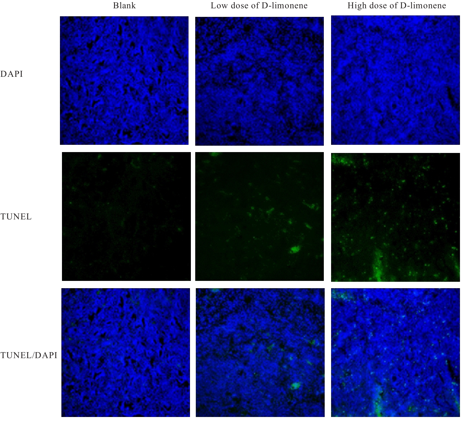
目的 探讨D-柠檬烯对胶质母细胞瘤(GBM)细胞增殖和凋亡的影响,并阐明其可能的作用机制。 方法 将GBM细胞分为对照组(0 mmol·L-1 D-柠檬烯)和0.2、 0.4、 0.6、 0.8及1.0 mmol·L-1 D-柠檬烯组。采用CCK-8法检测各组细胞增殖抑制率,克隆形成法检测各组细胞克隆形成率,Annexin Ⅴ-FITC/PI法检测各组细胞凋亡率,Western blotting法检测各组细胞中蛋白激酶B(AKT)、B细胞淋巴瘤2(Bcl-2)、Bcl-2相关X蛋白(Bax)和多聚二磷酸腺苷(ADP)核糖聚合酶(PARP)蛋白表达水平,免疫荧光法检测各组细胞中裂解的半胱氨酸天冬氨酸蛋白酶(cleaved Caspase)-3 蛋白表达水平。15 只建立皮下移植瘤模型小鼠随机分为空白组(0 mg·kg-1·d-1 D-柠檬烯)、低剂量D-柠檬烯组(200 mg·kg-1·d-1 D-柠檬烯)和高剂量D-柠檬烯组(400 mg·kg-1·d-1 D-柠檬烯),每组5只。计算各组小鼠体内肿瘤抑制率,HE染色和免疫组织化学染色观察各组小鼠皮下肿瘤组织形态表现,并绘制肿瘤生长曲线,免疫组织化学法检测各组小鼠皮下瘤组织中Ki67蛋白阳性表达率,TUNEL染色法检测各组小鼠肿瘤细胞凋亡情况。 结果 对照组细胞呈长梭形,状态良好,紧密且贴壁生长,细胞器和细胞质正常;给药48 h后,0.6 mmol·L-1 D-柠檬烯组细胞体积减小,细胞膜虽完整但通透性增强,细胞质皱缩,细胞内部产生空泡结构,部分呈碎片状漂浮在溶液表面;0.8 和1.0 mmol·L-1 D-柠檬烯组细胞中有明显凋亡小体生成,呈现凋亡状态。CCK-8法,与对照组比较,0.6、0.8和1.0 mmol·L-1 D-柠檬烯组U87、LN229及GL261细胞增殖抑制率均明显升高(P<0.01),0.4 mmol·L-1 D-柠檬烯组U87和GL261细胞增殖抑制率均明显升高(P<0.01)。克隆形成法,与对照组比较,0.4、0.6和0.8 mmol·L-1 D-柠檬烯组U87、LN229及GL261细胞克隆形成率均明显降低(P<0.05或P<0.01)。Annexin Ⅴ-FITC/PI法,与对照组比较,D-柠檬烯处理48 h后,0.6、0.8和1.0 mmol·L-1 D-柠檬烯组LN229细胞凋亡率明显升高(P<0.01)。Western blotting法,与对照组比较,0.6、0.8和1.0 mmol·L-1 D-柠檬烯组LN229细胞中Bax蛋白表达水平均明显升高(P<0.01),AKT和Bcl-2蛋白表达水平均明显降低(P<0.01);0.8和1.0 mmol·L-1 D-柠檬烯组LN229细胞中PARP蛋白表达水平均明显升高(P<0.01)。免疫荧光法,与对照组比较,0.6、0.8和1.0 mmol·L-1 D-柠檬烯组LN229细胞中cleaved Caspase-3蛋白表达水平均明显升高(P<0.01)。与空白组比较,低和高剂量D-柠檬烯组小鼠肿瘤体积均明显减小(P<0.01)。与空白组比较,低和高剂量D-柠檬烯组小鼠肿瘤质量均明显降低(P<0.05),肿瘤抑制率均明显升高(P<0.05)。空白组小鼠肿瘤细胞弥漫分布,细胞核染色加深,核浆比增大;低和高剂量D-柠檬烯组小鼠肿瘤组织出现大量肿瘤细胞变性坏死。与空白组比较,低和高剂量D-柠檬烯组小鼠肿瘤组织中Ki67蛋白阳性表达率均明显降低(P<0.01)。与空白组比较,低和高剂量D-柠檬烯组小鼠肿瘤细胞凋亡率均明显升高(P<0.01)。 结论 D-柠檬烯具有抑制GBM细胞增殖的作用,其作用机制可能与调控AKT蛋白的表达并激活Caspase-3通路诱导凋亡有关。

关键词: D-柠檬烯, 胶质母细胞瘤, 细胞凋亡, 移植瘤, 蛋白激酶B

Abstract:

Objective To discuss the effect of D-limonene on the proliferation and apoptosis of the glioblastoma (GBM) cells,and to clarify its possible mechanism. Methods The GBM cells were divided into control group (0 mmol·L-1 D-limonene) and 0.2, 0.4, 0.6, 0.8, and 1.0 mmol·L-1 D-limonene groups. CCK-8 method was used to detect the inhibitory rates of proliferation of the cells in various groups; clone formation assay was used to detect the clone formation rates of the cells in various groups; Annexin Ⅴ-FITC/PI method was used to detect the apoptotic rates of the cells in various groups; Western blotting method was used to detect the expression levels of protein kinase B (AKT), B-cell lymphoma-2 (Bcl-2), Bcl-2-associated X protein (Bax), and poly adenosine diphosphate(ADP)-ribose polymerase (PARP) proteins in the cells in various groups; imunofluorescence method was used to detect the expression levels of cleaved Caspase-3 protein in the cells in various groups. Fifteen model mice with subcutaneous tumor xenografts were randomly divided into blank group (0 mg·kg-1·d-1 D-limonene), low dose of D-limonene group (200 mg·kg-1·d-1 D-limonene), and high dose of D-limonene group (400 mg·kg-1·d-1 D-limonene), and there were 5 mice in each group.The inhibitory rates of the tumor in vitro in various groups were calculated; HE staining and immunohistochemical staining were used to observe the morphology of subcutaneous tumor tissue of the mice in various groups and the growth curves of the tumor were drawn; immunohistochemical assay was used to detect the positive expression rates of Ki67 protein in subcutaneous tumor tissue of the mice in various groups; TUNEL staining was used to detect the apoptosis of the tumor cells in various groups. Results In control group, the cells were spindle-shaped, in good condition, growing closely and adherently, with normal organelles and cytoplasm. After treated for 48 h, the cells in 0.6 mmol·L-1 D-limonene group showed reduced volume, intact but more permeable cell membranes, shrunken cytoplasm, internal vacuole structures, and some fragments floating in the solution. The cells in 0.8 and 1.0 mmol·L-1 D-limonene groups exhibited significant apoptotic bodies and were in an apoptotic state. The CCK-8 results showed that compared with control group, the inhibitory rates of proliferation of the U87, LN229, and GL261 cells in 0.6, 0.8, and 1.0 mmol·L-1 D-limonene groups were significantly increased (P<0.01), the inhibitory rates of proliferation of the U87 and GL261 cells were significantly increased (P<0.01). The clone formation assay results showed that compared with control group, the clone formation rates of the U87, LN229, and GL261 cells in 0.4, 0.6, and 0.8 mmol·L-1 D-limonene groups were significantly decreased (P<0.05 or P<0.01). The Annexin Ⅴ-FITC/PI results showed that compared with control group, after treated with D-limonene for 48 h, the apoptotic rates of the LN229 cells in 0.6, 0.8, and 1.0 mmol·L-1 D-limonene groups were significantly increased (P<0.01). The Western blotting results showed that compared with control group, the expression levels of Bax proteins in the LN229 cells in 0.6, 0.8, and 1.0 mmol·L-1 D-limonene groups were significantly increased (P<0.01), while the expression levels of AKT and Bcl-2 proteins were significantly decreased (P<0.01), the expression level of PARP protein in the LN229 cells in 0.8 and 1.0 mmol·L-1 D-limonene group was significanthy increased(P<0.01).The immunofluorescence results showed that compared with control group, the expression levels of cleaved Caspase-3 protein in the LN229 cells in 0.6, 0.8, and 1.0 mmol·L-1 D-limonene groups were significantly increased (P<0.01). Compared with blank group, the tumor volumes of the mice in low and high doses of D-limonene groups were significantly decreased (P<0.01). Compared with blank group, the tumor weights of the mice in low and high doses of D-limonene groups were significantly decreased (P<0.05), and the inhitory rates of tumor were significantly increased (P<0.05). The tumor cells in blank group were diffusely distributed, with deepened nuclear staining and increased nucleocytoplasmic ratio; a large number of degenerated and necrotic tumor cells were observed in tumor tissue of the mice in low and high doses of D-limonene groups. Compared with blank group, the positive expression rates of Ki67 protein in tumor tissue of the mice in low and high doses of D-limonene groups were significantly decreased (P<0.01). Compared with blank group, the apoptotic rates of tumor cells of the mice in low and high doses of D-limonene groups were significantly increased (P<0.01). Conclusion D-limonene has the inhibitory effect on the proliferation of the GBM cells; its mechanism may be related to the regulation of AKT protein expression and the activation of the Caspase-3 pathway to induce the apoptosis.

Key words: D-limonene, Glioblastoma, Apoptosis, Transplantation tumor, Protein kinase B

中图分类号: 

  • R739.4