吉林大学学报(医学版) ›› 2024, Vol. 50 ›› Issue (4): 989-999.doi: 10.13481/j.1671-587X.20240413

• 基础研究 • 上一篇    下一篇

Myod1通过调节lncRNA SNHG15和miR-24-3p对氧糖剥夺SH-SY5Y细胞增殖及凋亡的影响

冀方超,张晨昕,任占军,潘云志,逯琦,孙兴元()   

  1. 齐齐哈尔医学院附属第三医院神经内科,黑龙江 齐齐哈尔 161000
  • 收稿日期:2023-07-14 出版日期:2024-07-28 发布日期:2024-08-01
  • 通讯作者: 孙兴元 E-mail:sunxingyuandor@163.com
  • 作者简介:冀方超(1988-),男,黑龙江省齐齐哈尔市人,住院医师,医学硕士,主要从事神经内科基础和临床方面的研究。
  • 基金资助:
    黑龙江省卫健委基金项目(20210303070198)

Effect of Myod1 on proliferation and apoptosis of oxygen-glucose-deprived SHSY5Y cells by regulating lncRNA SNHG15 and miR-24-3p

Fangchao JI,Chenxin ZHANG,Zhanjun REN,Yunzhi PAN,Qi LU,Xingyuan SUN()   

  1. Department of Neurology,Third Affiliated Hospital,Qiqihar Medical University,Qiqihar 161000,China
  • Received:2023-07-14 Online:2024-07-28 Published:2024-08-01
  • Contact: Xingyuan SUN E-mail:sunxingyuandor@163.com

摘要:

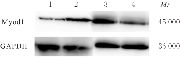
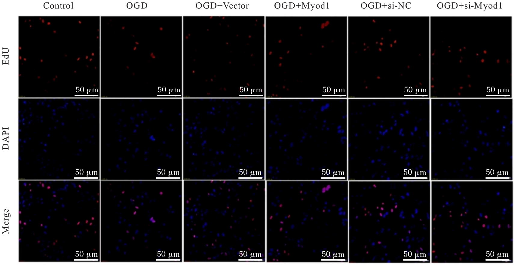
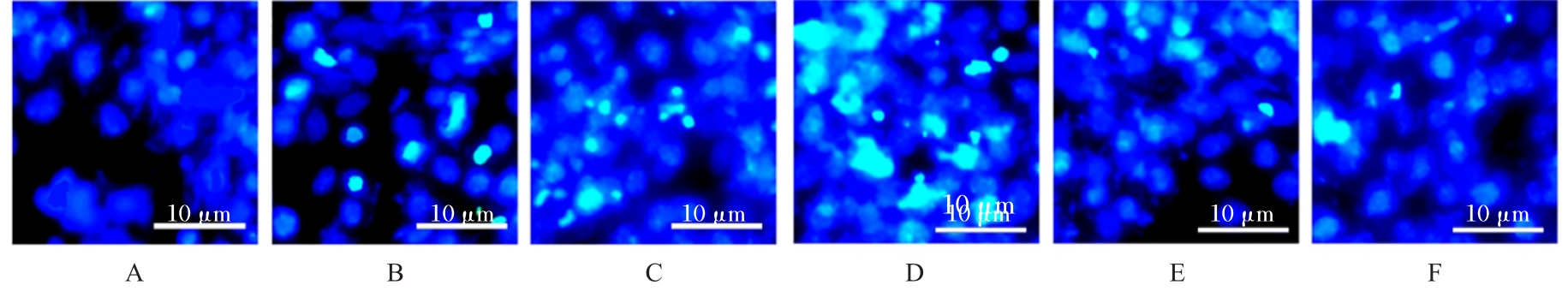
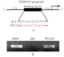
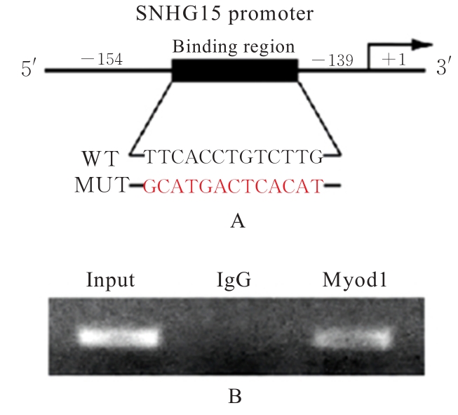
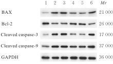
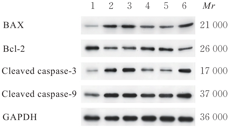
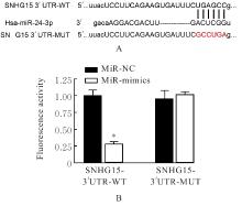
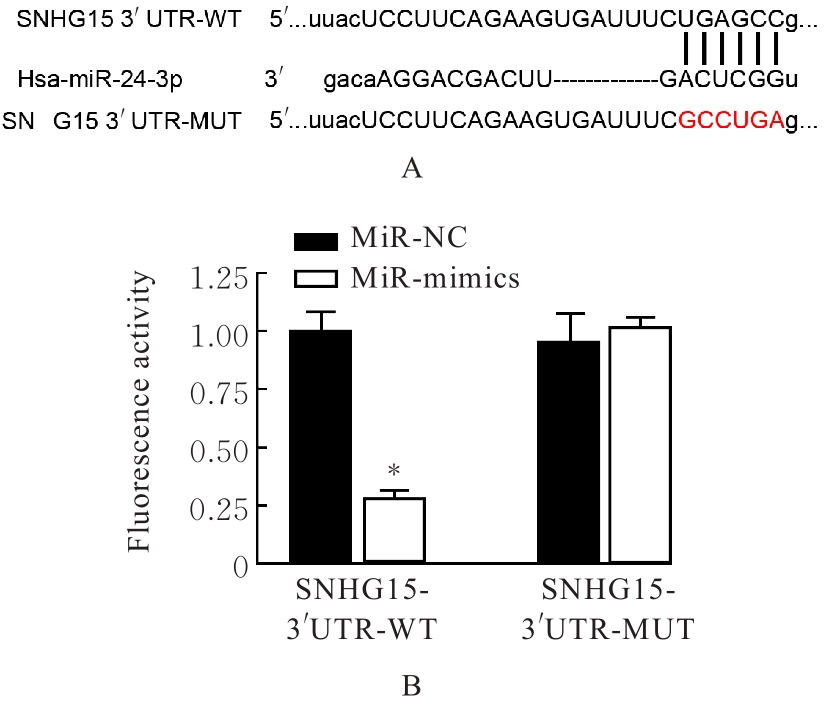
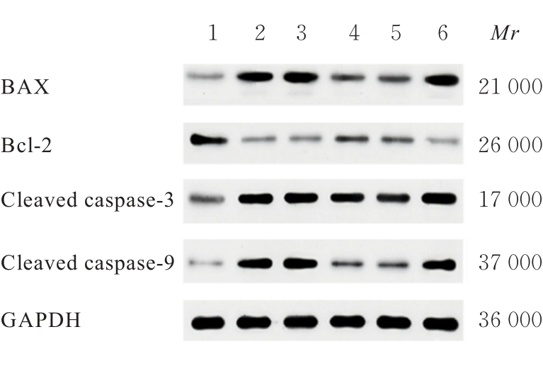
目的 探讨肌源性分化蛋白1(Myod1)对氧糖剥夺(OGD)诱导的SH-SY5Y细胞增殖抑制和凋亡的影响,并阐明其作用机制。 方法 采用实时荧光定量PCR(RT-qPCR)法检测正常对照组研究对象和缺血性脑梗死组患者外周血及正常培养的SH-SY5Y细胞(对照组)和OGD细胞模型(OGD组)细胞中Myod1和长链非编码RNA(lncRNA)小核仁RNA宿主基因15(SNHG15)mRNA表达水平。分别采用si-Myod1、pcDNA3.0-Myod1、si-SNHG15、pcDNA3.0-SNHG15、si-NC、空载质粒(Vector)、miR-NC和miR-24-3p模拟物(miR-mimics)质粒转染SH-SY5Y细胞后,进行OGD处理,将SH-SY5Y细胞分为对照组、OGD组、OGD+Vector组、OGD+Myod1组、OGD+si-NC组、OGD+si-Myod1组、OGD+si-SNHG15组、OGD+si-SNHG15+Vector组、OGD+si-SNHG15+Myod1组、OGD+miR-NC组、OGD+miR-mimics组、OGD+miR-mimics+Vector组和OGD+miR-mimics+SNHG15组。采用CCK-8法检测各组细胞活性,采用5-乙炔基-2'-脱氧尿苷(EdU)染色法检测各组EdU阳性细胞率,采用原位末端转移酶标记(TUNEL)法检测各组TUNEL阳性细胞率,采用Western blotting法检测各组细胞中裂解的含半胱氨酸的天冬氨酸蛋白水解酶3(cleaved caspase-3)、裂解的含半胱氨酸的天冬氨酸蛋白水解酶9(cleaved caspase-9)、B细胞淋巴瘤2(Bcl-2)和Bcl-2相关X蛋白(Bax)蛋白表达水平。染色质免疫共沉淀(CHIP)法评估Myod1和SNHG15之间的关联。双荧光素酶报告基因实验评估Myod1与SNHG15及SNHG15与miR-24-3p的靶向关系。 结果 与正常对照组比较,缺血性脑梗死组患者外周血中Myod1和SNHG15 mRNA表达水平均升高(P<0.05)。与对照组比较,OGD组细胞中Myod1和SNHG15 mRNA表达水平均明显升高(P<0.05)。与OGD组比较,48和72 h时OGD+Myod1组细胞活性和EdU阳性细胞率均降低(P<0.01),TUNEL阳性细胞率升高(P<0.01);OGD+si-Myod1组细胞活性和EdU阳性细胞率均升高(P<0.01),TUNEL阳性细胞率降低(P<0.01)。Myod1可与SNHG15的启动子序列结合。SNHG15可吸附miR-24-3p,Myod1与SNHG15及SNHG15与miR-24-3p存在靶关系。敲低SNHG15后,与OGD组比较,48和72 h时OGD+si-SNHG15组细胞活性和EdU阳性细胞率均升高(P<0.01),TUNEL阳性细胞率降低(P<0.01),细胞中Bax、cleaved caspase-3和cleaved caspase-9蛋白表达水平降低(P<0.01),Bcl-2蛋白表达水平升高(P<0.01);与OGD+si-SNHG15组比较,48和72 h时OGD+si-SNHG15+Myod1组细胞活性和EdU阳性细胞率降低(P<0.05),TUNEL阳性细胞率升高(P<0.05),细胞中Bax、cleaved caspase-3和cleaved caspase-9蛋白表达水平升高(P<0.05),Bcl-2的蛋白表达水平降低(P<0.05)。过表达miR-24-3p和SNHG15后,与OGD组比较,48和72 h时OGD+miR-mimics组细胞活性和EdU阳性细胞率升高(P<0.01),TUNEL阳性细胞率降低(P<0.01),细胞中Bax、cleaved caspase-3和cleaved caspase-9蛋白表达水平降低(P<0.01),Bcl-2蛋白表达水平升高(P<0.01);与OGD+miR-mimics组比较,48和72 h时OGD+miR-mimics+SNHG15组细胞活性和EdU阳性细胞率降低(P<0.05),TUNEL阳性细胞率升高(P<0.05),细胞中Bax、cleaved caspase-3 和 cleaved caspase-9 蛋白表达水平升高(P<0.05),Bcl-2蛋白表达水平降低(P<0.05)。 结论 Myod1可通过与SNHG15启动子区结合进而吸附miRNA-24,促进OGD诱导的SH-SY5Y细胞的增殖抑制和细胞凋亡。

关键词: 肌源性分化蛋白1, 小核仁RNA宿主基因15, 微小RNA-24-3p, SH-SY5Y细胞, 脑梗死

Abstract:

Objective To investigate the effect of myogenic differentiation protein 1 (Myod1) on the proliferation inhibition and apoptosis of the SH-SY5Y cells induced by oxygen-glucose deprivation (OGD), and to elucidate its mechanism. Methods Real-time quantitative fluorescence PCR (RT-qPCR) method was used to detect the mRNA levels of Myod1 and long non-coding RNA (lncRNA) small nucleolar RNA host gene 15 (SNHG15) in peripheral blood of the subjects in normal group and the patients in ischemic cerebral infarction group as well as the normal cultured SH-SY5Y cells(control group) and the cells in OGD model (OGD group). After transfecting SH-SY5Y cells with si-Myod1, pcDNA3.0-Myod1, si-SNHG15, pcDNA3.0-SNHG15、si-NC, Vector, miR-NC, and miR-24-3p mimics, the cells were treated with OGD, and then the SH-SY5Y cells were divided into control group, OGD group, OGD+Vector group, OGD+Myod1 group, OGD+si-NC group, OGD+si-Myod1 group, OGD+si-SNHG15 group, OGD+si-SNHG15+Vector group, OGD+si-SNHG15+Myod1 group, OGD+miR-NC group, OGD+miR-mimics group, OGD+miR-mimics+Vector group, and OGD+miR-mimics+SNHG15 group. CCK-8 method was used to detect the cell activities in various groups; 5-ethynyl-2'-deoxyuridine (EdU) staining was used to detect the rates of EDU positive cells in various groups; the rates of TdT-mediated dUTP nick end labeling (TUNEL) positive cells in various groups were detected by TUNEL staining; Western blotting method was used to detect the expression levels of cleaved caspase-3, cleaved caspase-9, B-cell lymphoma 2 (Bcl-2) and Bcl-2 associated X protein (Bax) proteins in the cells in various groups; the association between Myod1 and SNHG15 was evaluated by chromatin immunoprecipitate (CHIP); dual luciferase reporter gene experiment was used to evaluate the targeting relationships between Myod1 and SNHG15 as well as SNHG15 and miR-24-3p. Results Compared with normal control group, the expression levels of Myod1 and SNHG15 mRNA in peripheral blood of the patients in ischemic cerebral infarction group were significantly increased (P<0.05). Compared with control group, the expression levels of Myod1 and SNHG15 mRNA in the SH-SY5Y cells in OGD group were significantly increased (P<0.05). Compared with OGD group, the cell activities and rates of EdU positive cells in OGD+Myod1 group at 48 and 72 h were decreased (P<0.01), and the rates of TUNEL positive cells were increased (P<0.05); the cell activities and rates of EdU positive cells in OGD+si-Myod1 group were increased (P<0.05), while the rates of TUNEL positive cells were decreased (P<0.01). Myod1 binded to the promoter sequence of SNHG15. SNHG15 could absorb miR-24-3p, and there were target relatronships between Myod1 and SNHG15 as well as SNHG15 and miR-24-3p. After SNHG15 knockdown, compared with OGD group, the cell activities and rates of EdU positive cells in OGD+si-SNHG15 group at 48 and 72 h were increased (P<0.01), and the rates of TUNEL positive cells were decreased (P<0.01), the expression levels of Bax, cleaved caspase-3 and cleaved caspase-9 proteins were decreased (P<0.01), and the expression levels of Bcl-2 protein were increased (P<0.01). Compared with OGD+si-SNHG15 group, the cell activities and rates of EdU positive cells in OGD+si-SNHG15+Myod1 group at 48 and 72 h were decreased (P<0.05), the rates of TUNEL positive cells were (P<0.05), the expression levels of Bax, cleaved caspase-3, and cleaved caspase-9 proteins were increased (P<0.05), and the expression levels of Bcl-2 were decreased (P<0.05). After over-expression of miR-24-3p and SNHG15, compared with OGD group, the cell activities and rates of EdU positive cells in OGD+miR-mimics group at 48 and 72 h were increased (P<0.01), the rates of TUNEL positive cells were significantly decreased (P<0.01), the protein expression levels of Bax, cleaved caspase-3 and cleaved caspase-9 were decreased (P<0.05), and the expression levels of Bcl-2 were increased (P<0.01). Compared with OGD+miR-mimics group, the cell activities and rates of EdU positive cells in OGD+miR-mimics+SNHG15 group at 48 and 72 h were decreased (P<0.05), and the rates of TUNEL positive cells were increased (P<0.05), the expression levels of Bax, cleaved caspase-3 and cleaved caspase-9 proteins were increased (P<0.05), and the expression levels of Bcl-2 protein were decreased (P<0.05). Conclusion Myod1 can promote the proliferation inhibition and apoptosis of OGD-induced SH-SY5Y cells by binding to the SNHG15 promoter region and then absorbing miRNA-24.

Key words: Myogenic differentiation 1, Small nuclear RNA host gene 15, MicroRNA-24-3p, SH-SY5Y cell, Cerebral infarction

中图分类号: 

  • R743