吉林大学学报(医学版) ›› 2024, Vol. 50 ›› Issue (4): 978-988.doi: 10.13481/j.1671-587X.20240412

• 基础研究 • 上一篇    下一篇

miR-761通过调控肿瘤相关巨噬细胞极化对骨肉瘤MG63细胞上皮-间质转化的影响

高世磊,王家强,姚伟涛,田志超,李超,梁潇潇,王鑫()   

  1. 郑州大学附属肿瘤医院 河南省肿瘤医院骨与软组织科,河南 郑州 450008
  • 收稿日期:2023-09-18 出版日期:2024-07-28 发布日期:2024-08-01
  • 通讯作者: 王鑫 E-mail:superwx1984@163.com
  • 作者简介:高世磊(1988-),男,河南省郑州市人,主治医师,医学硕士,主要从事骨肿瘤、软组织肿瘤和骨转移癌诊断及治疗方面的研究。
  • 基金资助:
    河南省科技厅科技发展计划项目(222102310131)

Effect of miR-761 on epithelial-mesenchymal transition in osteosarcoma MG63 cells by regulating tumor-associated macrophage polarization

Shilei GAO,Jiaqiang WANG,Weitao YAO,Zhichao TIAN,Chao LI,Xiaoxiao LIANG,Xin WANG()   

  1. Department of Bone and Soft Tissue,Affiliated Tumor Hospital,Zhengzhou University,Henan Cancer Hospital,Zhengzhou 450008,China
  • Received:2023-09-18 Online:2024-07-28 Published:2024-08-01
  • Contact: Xin WANG E-mail:superwx1984@163.com

摘要:

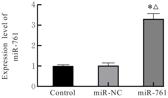
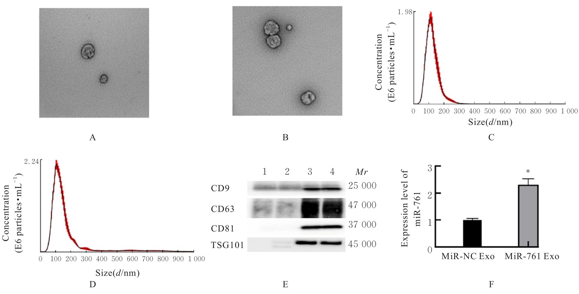
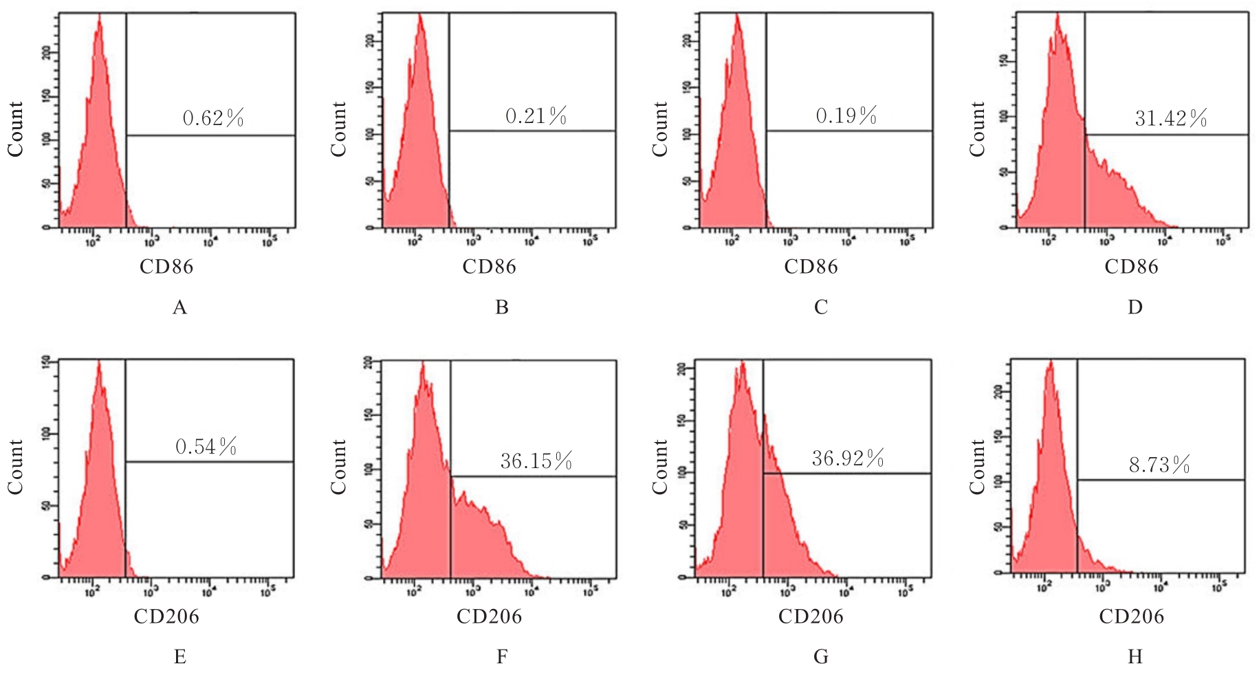
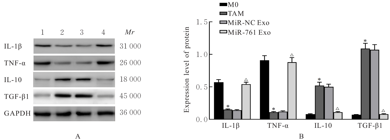
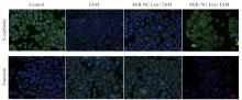
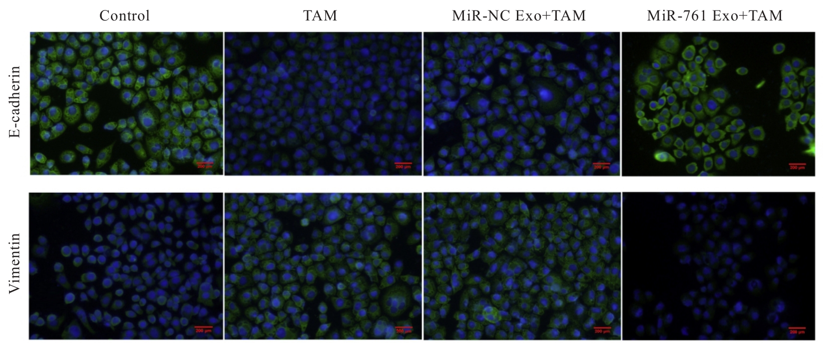
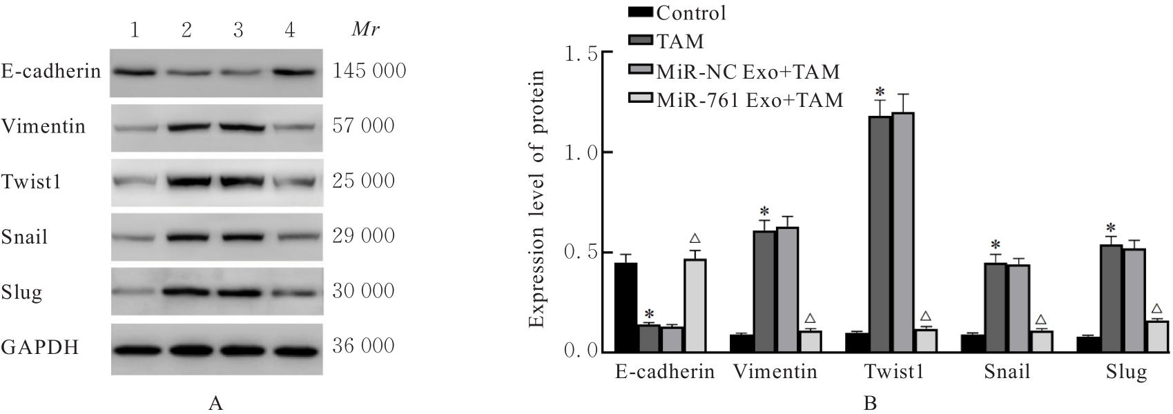
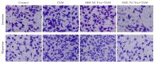
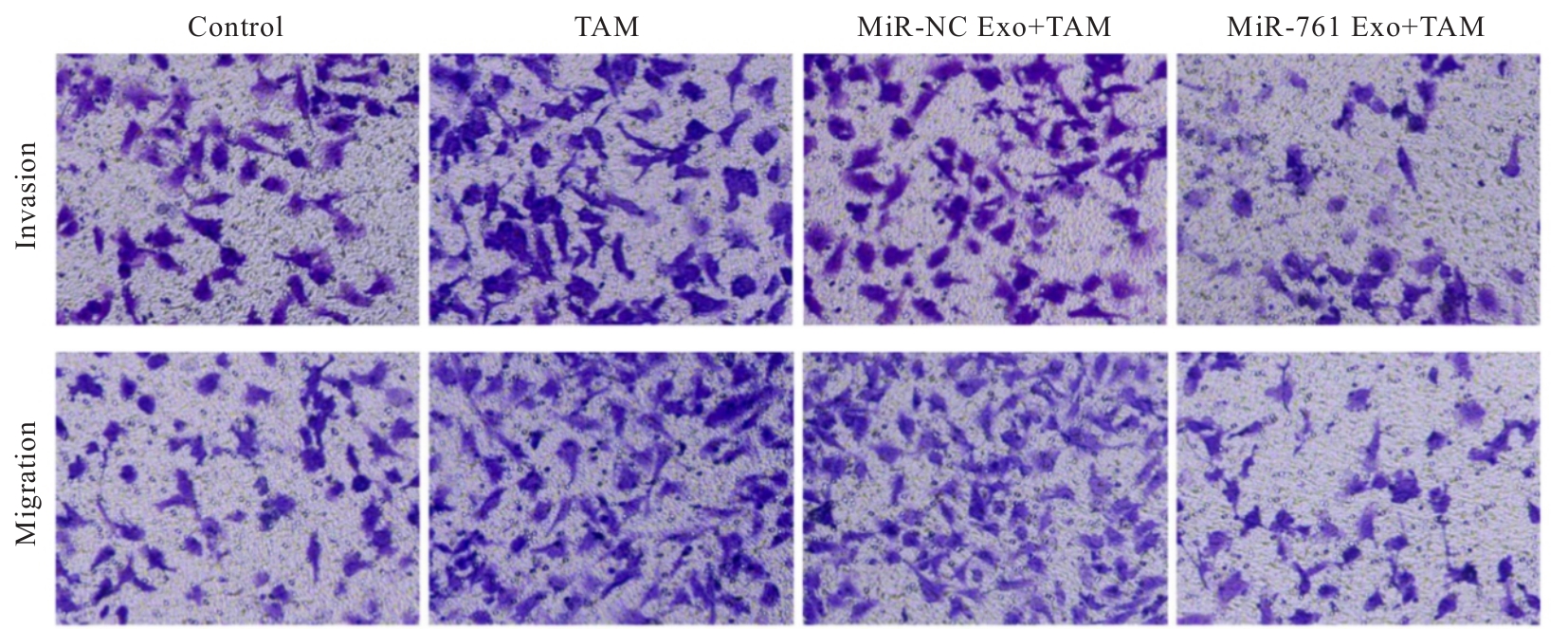
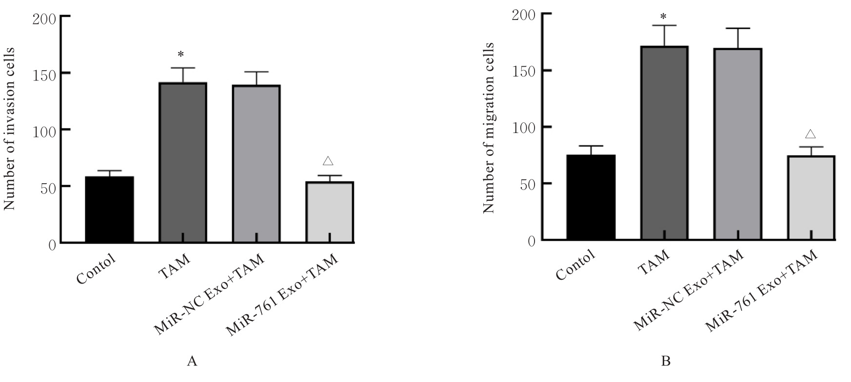
目的 分析外泌体(Exo)微小RNA-761(miR-761)通过调控肿瘤相关巨噬细胞(TAM)极化对骨肉瘤(OS)细胞上皮-间质转化(EMT)进程的影响,阐明其相关的作用机制。 方法 miR-761质粒和阴性对照(miR-NC)质粒分别转染至HEK239细胞中,同时设不转染的细胞为对照组,实时荧光定量PCR(RT-qPCR)法检测转染效果。分离含miR-761的Exo,采用透射电镜观察Exo形态,采用纳米颗粒分析仪检测Exo样品浓度和粒径分布,Western blotting法检测Exo表面标志蛋白表达情况。采用佛波酯(PMA)刺激人单核细胞白血病THP-1细胞成为M0巨噬细胞,采用含miR-761的Exo处理M0巨噬细胞并与OS MG63细胞建立共培养体系,实验分组为M0组、TAM组、miR-761 NC 组和miR-761 Exo 组,收集各组M0巨噬细胞,流式细胞术检测各组细胞中M1巨噬细胞标志物CD86和M2巨噬细胞标志物CD206阳性率,Western blotting法检测各组细胞中M1巨噬细胞分泌因子白细胞介素1β(IL-1β)及肿瘤坏死因子α(TNF-α)和M2巨噬细胞分泌因子白细胞介素10(IL-10)及转化生长因子β1(TGF-β1)蛋白表达水平;采用含miR-761的Exo处理M0巨噬细胞并与MG63细胞建立共培养体系,实验分为对照组、TAM组、miR-NC Exo+TAM组和miR-761 Exo+TAM组,收集各组MG63细胞,免疫荧光染色法观察各组MG63细胞中E-钙黏附蛋白(E-cadherin)和波形蛋白(Vimentin)荧光强度,Western blotting法检测各组细胞中E-cadherin、Vimentin及EMT调控相关转录因子Twist1、Snail和Slug蛋白表达水平,Transwell小室实验检测各组MG63细胞中侵袭和迁移细胞数。 结果 通过转染实验成功获得含miR-761的HEK239细胞,并分离得到Exo。与M0组比较,TAM组巨噬细胞中CD86阳性率降低(P<0.05),CD206阳性率升高(P<0.05),IL-1β和TNF-α蛋白表达水平降低(P<0.05),IL-10和TGF-β1蛋白表达水平升高(P<0.05);与TAM组比较,miR-761 Exo 组巨噬细胞中CD86阳性率升高(P<0.05),CD206阳性率降低(P<0.05),IL-1β和TNF-α蛋白表达水平升高(P<0.05),IL-10和TGF-β1蛋白表达水平降低(P<0.05)。与对照组比较,TAM组MG63细胞中E-cadherin荧光表达强度减弱而Vimentin荧光表达强度增强,E-cadherin蛋白表达水平降低(P<0.05),Vimentin、Twist1、Snail和Slug蛋白表达水平升高(P<0.05),迁移细胞数和侵袭细胞数增加(P<0.05);与 TAM 组比较,miR-761 Exo+TAM 组 MG63 细胞中E-cadherin荧光表达强度增强而Vimentin荧光表达强度减弱,E-cadherin蛋白表达水平升高(P<0.05),Vimentin、Twist1、Snail和Slug蛋白表达水平降低(P<0.05),迁移细胞数和侵袭细胞数减少(P<0.05)。 结论 Exo传递miR-761能够抑制OS细胞EMT进程,进而抑制细胞的迁移和侵袭,其作用机制可能与调控TAM极化作用有关。

关键词: 骨肉瘤, 微小RNA-761, 外泌体, 上皮-间质转化, 肿瘤相关巨噬细胞极化

Abstract:

Objective To discuss the effect of exosome (Exo) microRNA-761 (miR-761) on the epithelial-mesenchymal transition (EMT) process of the osteosarcoma (OS) cells by regulating tumor-associated macrophage (TAM) polarization, and to clarify its related mechanism. Methods The miR-761 plasmid and negative control (miR-NC) plasmid were transfected into the HEK293 cells, and the non-transfected cells were regarded as control group. The transfection efficiency was detected using real-time fluorescence quantitative PCR (RT-qPCR) method.The Exo containing miR-761 was isolated, and the morphology of Exo was observed by transmission electron microscope. The concentration and size distribution of Exo samples were detected by nanoparticle analyzer, and the expression of Exo surface marker protein was detected by Western blotting method.The human monocyte leukemia THP-1 cells were stimulated with phorbol 12-myristate 13-acetate (PMA) to become the M0 macrophages, which were then treated with Exo containing miR-761 and co-cultured with the OS MG63 cells to establish the co-culture system. The experiment was divided into M0 group, TAM group, miR-761 NC group, and miR-761 Exo group. The M0 macrophages were collected from various groups, and the positive rates of M1 macrophage marker CD86 and M2 macrophage marker CD206 in various groups were detected by flow cytometry; the protein expression levels of M1 macrophage secreted factors interleukin-1β (IL-1β) and tumor necrosis factor-α (TNF-α) and M2 macrophage secreted factors interleukin-10 (IL-10) and transforming growth factor-β1 (TGF-β1) in various groups were detected by Western blotting method. The M0 macrophages were treated with Exo containing miR-761 and co-cultured with MG63 cells to establish the co-culture system. The experiment was divided into control group, TAM group, miR-NC Exo+TAM group, and miR-761 Exo+TAM group. The MG63 cells in various groups were collected, and the fluorescence intensities of E-cadherin and Vimentin in the MG63 cells in various groups were observed by immunofluorescence staining; the expression levels of E-cadherin, Vimentin, and EMT regulation-related transcription factors Twist1, Snail, and Slug proteins in the cells in various groups were detected by Western blotting method;the numbers of invasion and migration cells in various groups were detected by Transwell chamber assay. Results The HEK293 cells containing miR-761 were successfully obtained by transfection experiments, and the Exo was isolated. Compared with M0 group, the positive rate of CD86 of the macrophages in TAM group was decreased (P<0.05), while the positive rate of CD206 was increased (P<0.05), the expression levels of IL-1β and TNF-α proteins were decreased (P<0.05), while the expression levels of IL-10 and TGF-β1 proteins were increased (P<0.05). Compared with TAM group, the positive rate of CD86 of the macrophages in miR-761 Exo group was increased (P<0.05), while the positive rate of CD206 was decreased (P<0.05), the expression levels of IL-1β and TNF-α proteins were increased (P<0.05), while the expression levels of IL-10 and TGF-β1 proteins were decreased (P<0.05). Compared with control group, the fluorescence intensity of E-cadherin in the MG63 cells in TAM group was decreased, while the fluorescence intensity of Vimentin was increased, the expression level of E-cadherin protein was decreased (P<0.05), while the expression levels of Vimentin, Twist1, Snail, and Slug proteins were increased (P<0.05), and the numbers of invasion and migration cells were increased (P<0.05). Compared with TAM group, the fluorescence intensity of E-cadherin in the MG63 cells in miR-761 Exo+TAM group was increased, while the fluorescence intensity of Vimentin was decreased, the expression level of E-cadherin protein was increased (P<0.05), while the expression levels of Vimentin, Twist1, Snail, and Slug proteins were decreased (P<0.05), and the numbers of invasion and migration cells were decreased (P<0.05). Conclusion The exo-delivered miR-761 can inhibit the EMT process of the OS cells, thereby inhibiting the cell migration and cell invasion; its mechanism may be related to regulating TAM polarization.

Key words: Osteosarcoma, MicroRNA-761, Exosome, Epithelial-mesenchymal transition, Tumor-associated macrophage polarization

中图分类号: 

  • R738.1