吉林大学学报(医学版) ›› 2024, Vol. 50 ›› Issue (6): 1547-1556.doi: 10.13481/j.1671-587X.20240608

• 基础研究 • 上一篇    

白藜芦醇对颞下颌关节骨关节炎的治疗作用及其机制

孙高1,2,何静1,赵琪1,石剑虹1,廖智羚1,田原野1,吴国民1()   

  1. 1.吉林大学口腔医院口腔颌面外一科与整形美容外科,吉林 长春 130021
    2.吉林大学口腔医院 吉林省牙发育及颌骨重塑与再生重点实验室,吉林 长春 130021
  • 收稿日期:2023-11-14 出版日期:2024-11-28 发布日期:2024-12-10
  • 通讯作者: 吴国民 E-mail:guominwu2006@sina.com
  • 作者简介:孙 高(1994-),男,浙江省宁波市人,在读硕士研究生,主要从事颞下颌关节疾病诊断及治疗等方面的研究。
  • 基金资助:
    吉林省科技厅科技发展计划项目(20230204088YY)

Therapeutic effect of resveratrol on osteoarthritis of temporomandibular joint and its mechanism

Gao SUN1,2,Jing HE1,Qi ZHAO1,Jianhong SHI1,Zhiling LIAO1,Yuanye TIAN1,Guomin WU1()   

  1. 1.Department of Oral and Maxillofacial Surgery Ⅰand Oral Plastic Surgery,Stomatology Hospital,Jilin University,Changchun 130021,China
    2.Jilin Provincal Key Laboratory of Tooth Development and Bone Remodeling and Regeneration,Stomatology Hospital,Jilin University,Changchun 130021,China
  • Received:2023-11-14 Online:2024-11-28 Published:2024-12-10
  • Contact: Guomin WU E-mail:guominwu2006@sina.com

摘要:

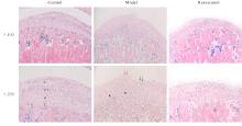
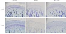
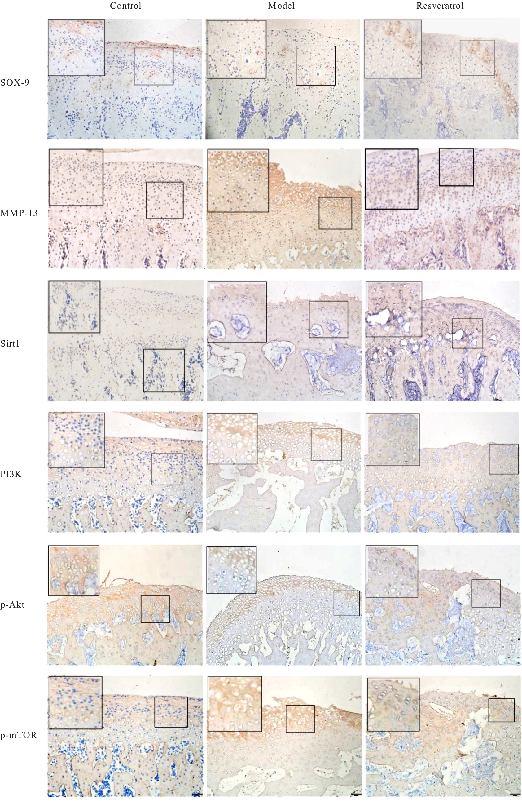
目的 探讨白藜芦醇对颞下颌关节骨关节炎(TMJOA)的治疗作用,并阐明相关作用机制。 方法 45只SD大鼠随机分为对照组、模型组和白藜芦醇组,每组15只。模型组和白藜芦醇组大鼠关节腔内注射20 g·L-1碘乙酸钠(MIA)50 μL,构建TMJOA大鼠模型;对照组大鼠注射等量生理盐水。造模3周后,白藜芦醇组大鼠注射80 μL白藜芦醇溶液,每周1次,连续3周,对照组和模型组大鼠注射等量生理盐水。微型计算机断层扫描技术(Micro-CT)系统检测各组大鼠髁突结构,计算感兴趣区的骨体积分数(BV/TV)、骨小梁厚度(Tb.Th)、骨小梁间距(Tb.p)和骨小梁数(Tb.N)。HE染色和甲苯胺蓝染色观察各组大鼠颞下颌关节(TMJ)组织病理形态表现,免疫组织化学法检测各组大鼠TMJ组织中性别决定区Y框蛋白(SOX)-9、基质金属蛋白酶(MMP)-13、沉默信息调节因子(Sirt)1、磷脂酰肌醇3激酶(PI3K)、磷酸化蛋白激酶B(p-Akt)和磷酸化哺乳动物雷帕霉素靶蛋白(p-mTOR)蛋白表达水平,实时荧光定量PCR(RT-qPCR)法检测各组大鼠TMJ组织中SOX-9、MMP-13、Sirt1、PI3K、哺乳动物雷帕霉素靶蛋白(mTOR)和蛋白激酶B(Akt)mRNA表达水平。 结果 造模3周后,大鼠髁突骨质破坏明显,表面粗糙不平,延续性中断,表明TMJOA大鼠模型造模成功。Micro-CT系统检测,对照组大鼠髁突表面光滑,形态规则,骨质连续完整;模型组大鼠髁突破坏明显,骨质连续性受到不同程度破坏,表面粗糙可伴有不同程度骨质缺损;白藜芦醇组大鼠髁突病变减轻,髁突形态外观得到一定的改善。与对照组比较,模型组大鼠BV/TV和Tb.Th均明显降低(P<0.05),Tb.Sp明显增加(P<0.05);与模型组比较,白藜芦醇组大鼠BV/TV和Tb.Th均明显升高(P<0.05),Tb.Sp明显减少(P<0.05)。HE染色观察,对照组大鼠髁突层次清晰,软骨细胞排列规则,整齐有序;模型组大鼠髁突表面粗糙不平,缺损明显,呈现典型的TMJOA表现;白藜芦醇组大鼠髁突表面略微粗糙,整体分层大致清晰,细胞排列较为有序。甲苯胺蓝染色观察,对照组大鼠髁突肥大层软骨细胞呈蓝紫色,染色明显且均匀;模型组大鼠髁突肥大层软骨细胞淡染,部分区域甚至失染;白藜芦醇组大鼠髁突肥大细胞层染色基本明显且较为均匀。免疫组织化学法检测,与对照组比较,模型组大鼠TMJ组织中MMP-13、PI3K、p-Akt和p-mTOR蛋白表达水平均明显升高(P<0.05),SOX-9和Sirt1蛋白表达水平均明显降低(P<0.05);与模型组比较,白藜芦醇组大鼠TMJ组织中SOX-9和Sirt1蛋白表达水平均明显升高(P<0.05),MMP-13、PI3K、p-Akt和p-mTOR蛋白表达水平均明显降低(P<0.05)。RT-qPCR法检测,与对照组比较,模型组大鼠TMJ组织中MMP-13、PI3K、Akt和mTOR mRNA表达水平均明显升高(P<0.05),SOX-9和Sirt1 mRNA表达水平均明显降低(P<0.05);与模型组比较,白藜芦醇组大鼠TMJ组织中SOX-9和Sirt1 mRNA表达水平均明显升高(P<0.05),MMP-13、PI3K、Akt和mTOR mRNA表达水平均明显降低(P<0.05)。 结论 白藜芦醇对TMJOA有治疗作用,其作用机制可能是通过激活Sirt1、抑制PI3K-Akt-mTOR信号通路实现的。

关键词: 白藜芦醇, 颞下颌关节骨关节炎, 沉默信息调节因子1, 磷脂酰肌醇3激酶, 蛋白激酶B, 哺乳动物雷帕霉素靶蛋白

Abstract:

Objective To discuss the therapeutic effect of resveratrol on the temporomandibular joint osteoarthritis (TMJOA), and to clarify the related mechanism. Methods Forty-five SD rats were randomly divided into control group, model group, and resveratrol group, and there were 15 rats in each group. The rats in model group and resveratrol group were intra-articularly injected with 50 μL of 20 g·L-1 monosodium iodoacetate (MIA) to set TMJOA rat models, while the rats in control group were injected with an equal volume of normal saline. Three weeks after modeling, the rats in resveratrol group received an injection of 80 μL resveratrol solution, once a week for three weeks, while the rats in control and model groups were injected with an equal volume of normal saline. Micro-computed tomography (Micro-CT) system was used to detect the condyle structure and the bone volume fraction (BV/TV), trabecular thickness (Tb.Th), trabecular spacing (Tb.Sp), and trabecular number (Tb.N) of the rats in various groups were calculated; HE staining and toluidine blue staining were used to observe the pathomorphology of temporomandibular joint (TMJ) tissue of the rats in various groups; immunohistochemistry was used to detect the expression levels of SRY-related HMG box (SOX)-9, matrix metalloproteinase (MMP)-13, silent information regulator (Sirt)1, phosphatidylinositol 3-kinase (PI3K), phosphorylated protein kinase B (p-Akt), and phosphorylated mammalian target of rapamycin (p-mTOR) in TMJ tissue of the rats in various groups; real-time quantitative PCR (RT-qPCR) method was used to detect the expression levels of SOX-9, MMP-13, Sirt1, PI3K, mTOR, and Akt mRNA in TMJ tissue of the rats in various groups. Results Three weeks after modeling, condylar bone was destructed, the surface was roughness, and continuity interruption were observed, indicating TMJOA model of the rats was established successfully. The Micro-CT system results showed that the condylar surface of the rats in control group was smooth and regularly shaped, with continuous bone texture; the rats in model group had significant condylar destruction, disrupted continuity, surface roughness, and varying degrees of bone defects; the rats in resveratrol group showed alleviated condylar lesions and improved appearance. Compared with control group, the BV/TV and Tb.Th of the rats in model group were significantly decreased (P<0.05), and Tb.Sp was significantly increased (P<0.05); compared with model group, the BV/TV and Tb.Th of the rats in resveratrol group were significantly increased (P<0.05), and the Tb.Sp was significantly decreased (P<0.05). The HE staining results showed clear layers and orderly chondrocyte arrangement in condyle of the rats in control group; the rats in model group showed rough uneven surface, obvious defects, and typical TMJOA features; the rats in resveratrol group showed slightly rough surface with generally clear layers and orderly arranged cells. The toluidine blue staining results showed distinct blue-purple staining of chondrocytes in hypertrophic layer of the rats in control group; pale staining or even loss of staining in some areas of the rats in model group; and distinct and relatively uniform staining in hypertrophic layer of the rats in resveratrol group. The immunohistochemistry results showed that compared with control group, the expression levels of MMP-13, PI3K, p-Akt, and p-mTOR proteins in TMJ tissue of the rats in model group were significantly increased (P<0.05), while the expression levels of SOX-9 and Sirt1 proteins in TMJ tissue of the rats were significantly decreased (P<0.05); compared with model group, the expression levels of SOX-9 and Sirt1 proteins in TMJ tissue of the rats in resveratrol group were significantly increased (P<0.05), whereas the expression levels of MMP-13, PI3K, p-Akt, and p-mTOR proteins were significantly decreased (P<0.05).The RT-qPCR results showed that compared with control group, the expression levels of MMP-13, PI3K, Akt, and mTOR mRNA in TMJ tissue of the rats in model group were significantly increased (P<0.05), while the expression levels of SOX-9 and Sirt1 mRNA were significantly decreased (P<0.05); compared with model group, the expression levels of SOX-9 and Sirt1 mRNA in TMJ tissue of the rats in resveratrol group were significantly increased (P<0.05), whereas the expression levels of MMP-13, PI3K, Akt, and mTOR mRNA were significantly decreased (P<0.05). Conclusion Resveratrol has therapeutic effect on TMJOA, and its mechanism may be related to the activation of Sirt1 and inhibition of the PI3K-Akt-mTOR signaling pathway.

Key words: Resveratrol, Temporomandibular joint osteoarthritis, Silence information regulation 1, Phosphoinositide 3-kinase, Protein kinase B, Mammalian target of rapamycin

中图分类号: 

  • R684.3