吉林大学学报(医学版) ›› 2025, Vol. 51 ›› Issue (3): 590-598.doi: 10.13481/j.1671-587X.20250304

• 基础研究 • 上一篇    

丝胶通过Akt1调控PI3K/Akt/NF-κB信号通路对链脲佐菌素引起INS-1细胞损伤的保护作用及其机制

陈程1,李警耀2,胡万祥3,刘东慧2(),陈志宏2()   

  1. 1.承德医学院基础医学院生理学教研室,河北 承德 067000
    2.承德医学院基础医学院人体解剖学 教研室,河北 承德 067000
    3.军科正源(天津)生物医药科技有限公司,天津 301700
  • 收稿日期:2024-08-31 接受日期:2024-10-18 出版日期:2025-05-28 发布日期:2025-07-18
  • 通讯作者: 刘东慧,陈志宏 E-mail:313804850@qq.com;czh1971@126.com
  • 作者简介:陈 程(1990-),女,河北省承德市人,讲师,医学硕士,主要从事糖尿病及其并发症中西药预防方面的研究。
  • 基金资助:
    国家自然科学基金项目(81441133);河北省卫健委医学科学研究项目(20210468);承德医学院国家自然科学基金项目培育基金项目(201912)

Protective effect of sericin on streptozotocin-induced INS-1 cell damage by regulating PI3K/Akt/NF-κB signaling pathway through Akt1 and its mechanism

Cheng CHEN1,Jingyao LI2,Wanxiang HU3,Donghui LIU2(),Zhihong CHEN2()   

  1. 1.Department of Physiology,Collegle of Basic Medical Sciences,Chengde Medical College,Chengde 067000,China
    2.Department of Human Anatomy,Collegle of Basic Medical Sciences,Chengde Medical College,Chengde 067000,China
    3.Junkezhengyuan(Tianjin) Biopharmaceutical Technology Co. ,Ltd,Tianjin 301700,China
  • Received:2024-08-31 Accepted:2024-10-18 Online:2025-05-28 Published:2025-07-18
  • Contact: Donghui LIU,Zhihong CHEN E-mail:313804850@qq.com;czh1971@126.com

摘要:

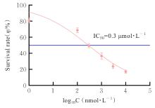
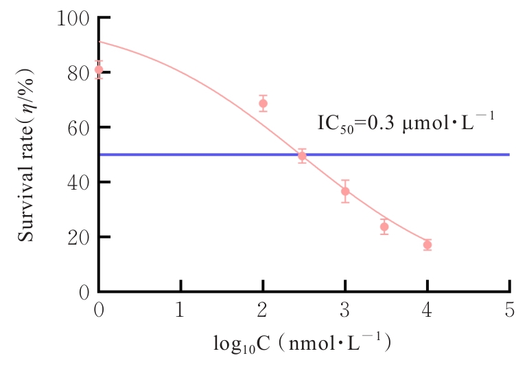
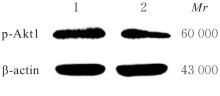
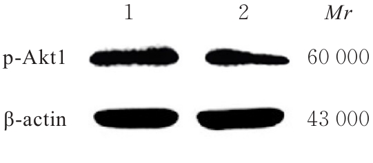
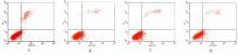
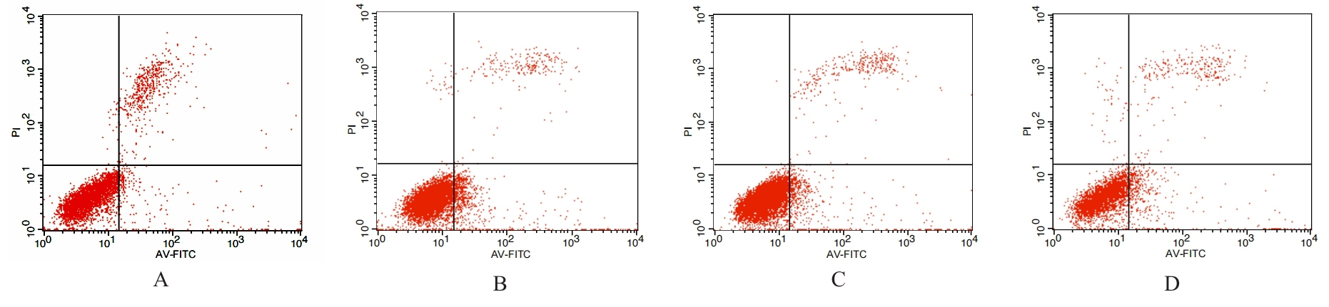
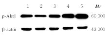
目的 探讨丝胶对链脲佐菌素(STZ)致损伤INS-1细胞磷脂酰肌醇3激酶(PI3K)/蛋白激酶B(Akt)/核因子κB(NF-κB)信号通路和细胞凋亡的影响,并阐明其作用机制。 方法 采用含0、0.1、0.3、1.0、3.0和10.0 μmol·L-1 Akt1抑制剂A-674563及10 mmol·L-1 STZ及600 mg·L-1丝胶的完全培养基培养INS-1细胞,分为0、0.1、0.3、1.0、3.0和10.0 μmol·L-1 A-674563组,另设置对照组(不含药物的完全培养基),采用细胞计数试剂盒8(CCK-8)法检测INS-1细胞存活率,并计算半数抑制浓度(IC50)值,筛选A-674563最佳抑制浓度,并采用Western blotting法验证。将INS-1细胞分为正常对照组(完全培养基)、模型组(10 mmol·L-1 STZ+完全培养基)、低、中和高剂量丝胶组(10 mmol·L-1 STZ+150 mg·L-1丝胶+完全培养基、10 mmol·L-1 STZ+300 mg·L-1丝胶+完全培养基和10 mmol·L-1 STZ+600 mg·L-1丝胶+完全培养基),采用CCK-8法检测各组INS-1细胞存活率,筛选丝胶最佳作用浓度。另将INS-1细胞分为正常对照组(完全培养基)、模型组(10 mmol·L-1 STZ+完全培养基)、丝胶组(10 mmol·L-1 STZ+600 mg·L-1丝胶+完全培养基)和A-674563组(10 mmol·L-1 STZ+600 mg·L-1丝胶+0.3 μmol·L-1 A-674563+完全培养基),采用流式细胞术检测各组INS-1细胞凋亡率,实时荧光定量PCR(RT-qPCR)法检测各组INS-1细胞中Akt1、NF-κB、肿瘤坏死因子α(TNF-α)和白细胞介素6(IL-6)mRNA表达水平,Western blotting法检测各组INS-1细胞磷酸化Akt1(p-Akt1)和NF-κB蛋白表达水平,酶联免疫吸附试验(ELISA)法检测各组INS-1细胞中TNF-α和IL-6水平。 结果 对照组INS-1细胞存活率为100.00%±0.00%,0、0.1、0.3、1.0、3.0和10.0 μmol·L-1 A-674563+10 mmol·L-1 STZ+600 mg·L-1丝胶+完全培养基共同作用后INS-1细胞存活率分别为82.50%±2.28%、69.47%±1.94%、51.51%±1.74%、38.94%± 1.57%、24.79%± 1.14%和19.85%±1.03%。A-674563对INS-1细胞的IC50值为0.3 μmol·L-1,选择0.3 μmol·L-1 A-674563作用INS-1细胞。与0 μmol·L-1 A-674563比较,0.3 μmol·L-1 A-674563+10 mmol·L-1 STZ+600 mg·L-1丝胶+完全培养基共同作用下,INS-1细胞中p-Akt1蛋白表达水平明显降低(P<0.05)。CCK-8法检测,与正常对照组比较,模型组INS-1细胞存活率明显降低(P<0.05);与模型组比较,低、中和高剂量丝胶组INS-1细胞存活率均明显升高(P<0.05);与低和中剂量丝胶组比较,高剂量丝胶组INS-1细胞存活率明显升高(P<0.05),因此选择600 mg·L-1丝胶作用细胞。与正常对照组比较,模型组INS-1细胞存活率明显降低(P<0.05);与模型组比较,丝胶组INS-1细胞存活率明显升高(P<0.05);与丝胶组比较,A-674563组INS-1细胞存活率明显降低(P<0.05)。流式细胞术检测,与正常对照组比较,模型组INS-1细胞凋亡率明显升高(P<0.05);与模型组比较,丝胶组INS-1细胞凋亡率明显降低(P<0.05);与丝胶组比较,A-674563组INS-1细胞凋亡率明显升高(P<0.05)。RT-qPCR法检测,与正常对照组比较,模型组INS-1细胞中Akt1 mRNA表达水平明显降低(P<0.05);与模型组比较,低、中和高剂量丝胶组INS-1细胞中Akt1 mRNA表达水平均明显升高(P<0.05);与低和中剂量丝胶组比较,高剂量丝胶组INS-1细胞中Akt1 mRNA表达水平明显升高(P<0.05)。与正常对照组比较,模型组INS-1细胞中NF-κB、TNF-α和IL-6 mRNA表达水平均明显升高(P<0.05);与模型组比较,丝胶组INS-1细胞中NF-κB、TNF-α和IL-6 mRNA表达水平均明显降低(P<0.05);与丝胶组比较,A-674563组INS-1细胞中NF-κB mRNA表达水平均明显升高(P<0.05)。Western blotting法检测,与正常对照组比较,模型组INS-1细胞中p-Akt1蛋白表达水平明显降低(P<0.05);与模型组比较,低、中和高剂量丝胶组INS-1细胞中p-Akt1蛋白表达水平均明显升高(P<0.05);与低和中剂量丝胶组比较,高剂量丝胶组INS-1细胞中p-Akt1蛋白表达水平明显升高(P<0.05)。与正常对照组比较,模型组INS-1细胞中NF-κB蛋白表达水平明显升高(P<0.05);与模型组比较,丝胶组INS-1细胞中NF-κB蛋白表达水平明显降低(P<0.05);与丝胶组比较,A-674563组INS-1细胞中NF-κB蛋白表达水平明显升高(P<0.05)。ELISA法检测,与正常对照组比较,模型组INS-1细胞中TNF-α和IL-6水平均明显升高(P<0.05);与模型组比较,丝胶组INS-1细胞中TNF-α和IL-6水平均明显降低(P<0.05);与丝胶组比较,A-674563组INS-1细胞中TNF-α和IL-6水平均明显升高(P<0.05)。 结论 丝胶通过靶向Akt1减轻PI3K/Akt/NF-κB信号通路介导的炎症反应和细胞凋亡,对STZ引起的INS-1细胞损伤具有保护作用。

关键词: 丝胶, INS-1细胞, 磷脂酰肌醇3激酶/蛋白激酶B/核因子κB信号通路, 炎症反应, 细胞凋亡

Abstract:

Objective To discuss the effect of sericin on the phosphatidylinositol 3-kinase (PI3K)/protein kinase B (Akt)/nuclear factor-κB (NF-κB) signaling pathway and apoptosis in the streptozotocin (STZ)-damaged INS-1 cells, and to clarify its mechanism. Methods The INS-1 cells were cultured with complete medium containing 0, 0.1, 0.3, 1.0, 3.0, and 10.0 μmol·L-1 Akt1 inhibitor A-674563, 10 mmol·L-1 STZ, and 600 mg·L-1 sericin, and divided into 0, 0.1, 0.3, 1.0, 3.0, and 10.0 μmol·L-1 A-674563 groups, and the control group (complete medium without drugs) was set up. Cell counting kit-8 (CCK-8) method was used to detect the survival rates of the INS-1 cells, and the half-maximal inhibitory concentration (IC50) value was calculated to determine the optimal inhibitory concentration of A-674563, which was further verified by Western blotting method. The INS-1 cells were divided into normal control group (complete medium), model group (10 mmol·L-1 STZ+complete medium), and low, medium, and high doses of sericin groups (10 mmol·L-1 STZ+150 mg·L-1 sericin+complete medium, 10 mmol·L-1 STZ+300 mg·L-1 sericin+complete medium, and 10 mmol·L-1 STZ+600 mg·L-1 sericin+complete medium). CCK-8 method was used to detect the survival rates of the INS-1 cells in various groups to determine the optimal concentration of sericin. Additionally, the INS-1 cells were divided into normal control group (complete medium), model group (10 mmol·L-1 STZ+complete medium), sericin group (10 mmol·L-1 STZ+600 mg·L-1 sericin + complete medium), and A-674563 group (10 mmol·L-1 STZ+600 mg·L-1 sericin+0.3 μmol·L-1 A-674563+complete medium). Flow cytometry was used to detect the apoptotic rates of the INS-1 cells in various groups; real-time fluorescence quantitative PCR (RT-qPCR ) method was used to detect the expression levels of Akt1, NF-κB, tumor necrosis factor-α (TNF-α), and interleukin-6 (IL-6) mRNA in the INS-1 cells in various groups; Western blotting method was used to detect the expression levels of phosphorylated Akt1 (p-Akt1) and NF-κB proteins in the INS-1 cells in various groups; enzyme linked immunosorbent assay(ELISA) method was used to detect the levels of TNF-α and IL-6 in the INS-1 cells in various groups. Results The survival rates of the INS-1 cells in control group was 100.00%±0.00%; in 0, 0.1, 0.3, 1.0, 3.0, and 10.0 μmol·L-1 A-674563+10 mmol·L-1 STZ+600 mg·L-1 sericin+complete medium groups, which were 82.50%±2.28%, 69.47%±1.94%, 51.51%±1.74%, 38.94%±1.57%, 24.79%±1.14%, and 19.85%±1.03%, respectively. The IC?? value of A-674563 for INS-1 cells was 0.3 μmol·L-1, and 0.3 μmol·L-1 A-674563 was selected for subsequent experiments. Compared with 0 μmol·L-1 A-674563, the expression level of p-Akt1 protein in the INS-1 cells after treated with 0.3 μmol·L-1 A-674563+10 mmol·L-1 STZ + 600 mg·L-1 sericin+complete medium was significantly decreased (P<0.05). The CCK-8 results showed that compared with normal control group, the survival rate of the INS-1 cells in model group was significantly decreased (P<0.05); compared with model group, the survival rates of the INS-1 cells in low, medium, and high doses of sericin groups were significantly increased (P<0.05); compared with low and medium doses of sericin groups, the survival rate of the INS-1 cells in high dose of sericin group was significantly increased (P<0.05). Thus, 600 mg·L-1 sericin was selected for subsequent experiments. The CCK-8 results showed that compared with normal control group, the survival rate of the INS-1 cells in model group was significantly decreased (P<0.05); compared with model group, the survival rate of the INS-1 cells in sericin group was significantly increased (P<0.05); compared with sericin group, the survival rate of the INS-1 cells in A-674563 group was significantly decreased (P<0.05). The flow cytometry results showed that compared with normal control group, the apoptotic rate of the INS-1 cells in model group was significantly increased (P<0.05); compared with model group, the apoptotic rate of the INS-1 cells in sericin group was significantly decreased (P<0.05); compared with sericin group, the apoptotic rate of the INS-1 cells in A-674563 group was significantly increased (P<0.05). The RT-qPCR results showed that compared with normal control group, the expression level of Akt1 mRNA in the INS-1 cells in model group was significantly decreased (P<0.05); compared with model group, the expression levels of Akt1 mRNA in the INS-1 cells in low, medium, and high doses of sericin groups were significantly increased (P<0.05); compared with low and medium doses of sericin groups, the expression level of Akt1 mRNA in the INS-1 cells in high dose of sericin group was significantly increased (P<0.05). Compared with normal control group, the expression levels of NF-κB, TNF-α, and IL-6 mRNA in the INS-1 cells in model group were significantly increased (P<0.05); compared with model group, the expression levels of NF-κB, TNF-α, and IL-6 mRNA in the INS-1 cells in sericin group were significantly decreased (P<0.05); compared with sericin group, the expression level of NF-κB mRNA in the INS-1 cells in A-674563 group was significantly increased (P<0.05). The Western blotting results showed that compared with normal control group, the expression level of p-Akt1 protein in the INS-1 cells in model group was significantly decreased (P<0.05); compared with model group, the expression levels of p-Akt1 protein in the INS-1 cells in low, medium, and high doses of sericin groups were significantly increased (P<0.05); compared with low and medium doses of sericin groups, the expression level of p-Akt1 protein in the INS-1 cells in high dose of sericin group was significantly increased (P<0.05). Compared with normal control group, the expression level of NF-κB protein in the INS-1 cells in model group was significantly increased (P<0.05); compared with model group, the expression level of NF-κB protein in the INS-1 cells in sericin group was significantly decreased (P<0.05); compared with sericin group, the expression level of NF-κB protein in the INS-1 cells in A-674563 group was significantly increased (P<0.05). The ELISA results showed that compared with normal control group, the levels of TNF-α and IL-6 in the INS-1 cells in model group were significantly increased (P<0.05); compared with model group, the levels of TNF-α and IL-6 in the INS-1 cells in sericin group were significantly decreased (P<0.05); compared with sericin group, the levels of TNF-α and IL-6 in the INS-1 cells in A-674563 group were significantly increased (P<0.05). Conclusion Sericin alleviates the PI3K/Akt/NF-κB signaling pathway-mediated inflammatory response and apoptosis by targeting Akt1, exerting a protective effect against STZ-induced damage in INS-1 cells.

Key words: Sericin, INS-1 cells, Phosphatidylinositol 3-kinase/protein kinase B/nuclear factor-κB signaling pathway, Inflammatory response, Apoptosis

中图分类号: 

  • R587.1