吉林大学学报(医学版) ›› 2025, Vol. 51 ›› Issue (3): 610-620.doi: 10.13481/j.1671-587X.20250306

• 基础研究 • 上一篇    

骨髓来源的间充质干细胞移植通过ROS/Nrf2信号对血管性痴呆大鼠线粒体自噬的影响及其机制

孙烈乾1,顾梦宇1,杨杰2,王凯漪1,郭高帅1,3,张宏博1,3,张思怡1,3,王堂龙3,杨志伟1,3,贺延妮1,3,杨超1,3()   

  1. 1.湖北中医药大学第一临床学院, 湖北 武汉 430065
    2.湖北省中医院肿瘤科,湖北 武汉 430061
    3.湖北省中西医结合医院老年病科,湖北 武汉 430015
  • 收稿日期:2024-06-20 接受日期:2024-09-14 出版日期:2025-05-28 发布日期:2025-07-18
  • 通讯作者: 杨超 E-mail:yc250820883@163.com
  • 作者简介:孙烈乾(2001-),男,湖北省荆州市人,在读硕士研究生,主要从事中西医结合防治老年病方面的研究。
  • 基金资助:
    湖北省卫健委卫生健康科研项目(WJ2023M097);湖北省科技厅自然科学基金项目(2023AFB914);湖北省中医药管理局青年人才项目(ZY2023Q010);湖北省中西医结合医院“杏林人才工程”青年项目(H2023Q009);湖北省鄂州市科技局基础人才与创新专项(EZ01-007-20230100)

Effect of bone marrow-derived mesenchymal stem cell transplantation on mitochondrial autophagy in rats with vascular dementia through ROS/Nrf2 signaling and its mechanism

Lieqian SUN1,Mengyu GU1,Jie YANG2,Kaiyi WANG1,Gaoshuai GUO1,3,Hongbo ZHANG1,3,Siyi ZHANG1,3,Tanglong WANG3,Zhiwei YANG1,3,Yanni HE1,3,Chao YANG1,3()   

  1. 1.First Clinical Medical College,Hubei University of Chinese Medicine,Wuhan 430065,China
    2.Department of Oncology,Hubei Provincial Hospital of Integrated Chinese and Western Medicine,Wuhan 430061,China
    3.Department of Geriatrics,Hubei Provincial Hospital of Traditional Chinese Medicine,Wuhan 430015,China
  • Received:2024-06-20 Accepted:2024-09-14 Online:2025-05-28 Published:2025-07-18
  • Contact: Chao YANG E-mail:yc250820883@163.com

摘要:

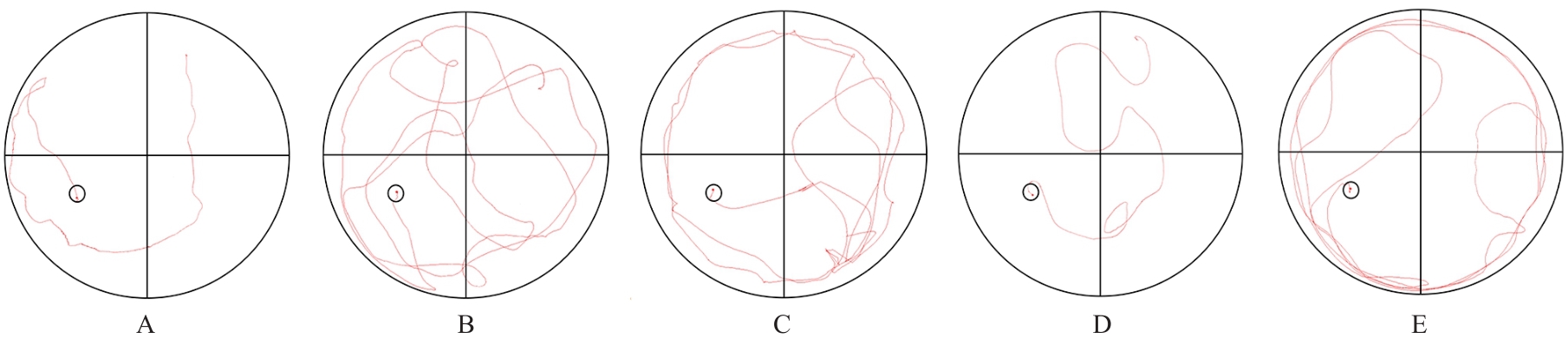
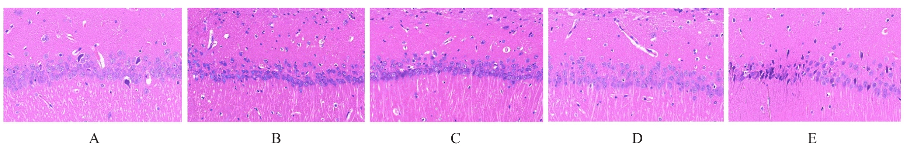
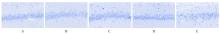
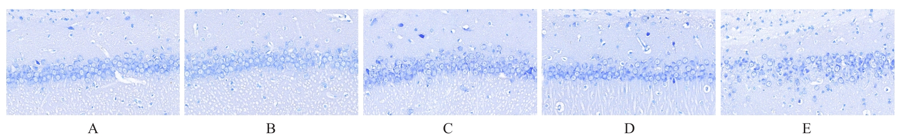
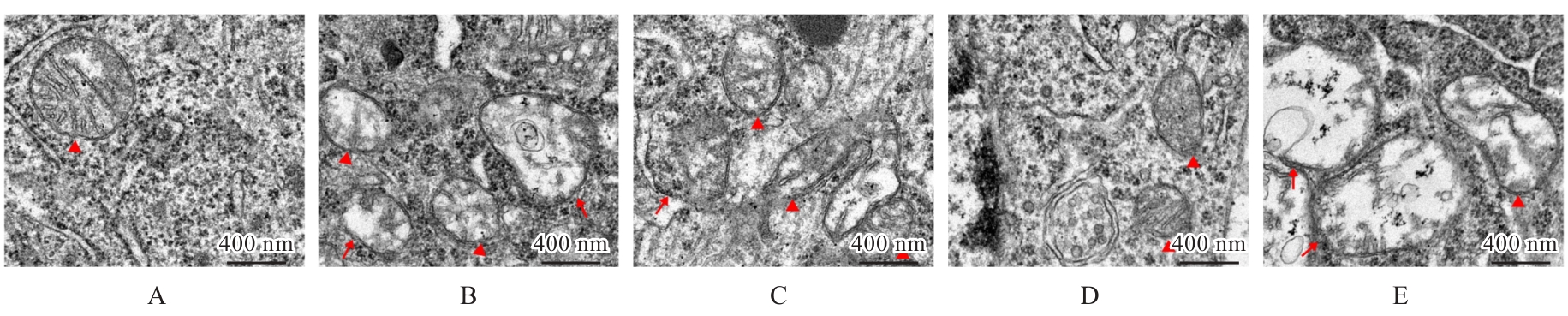
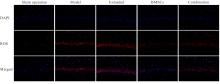
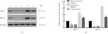
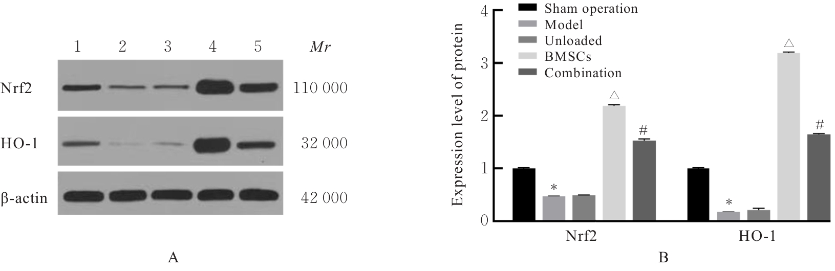
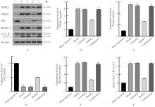
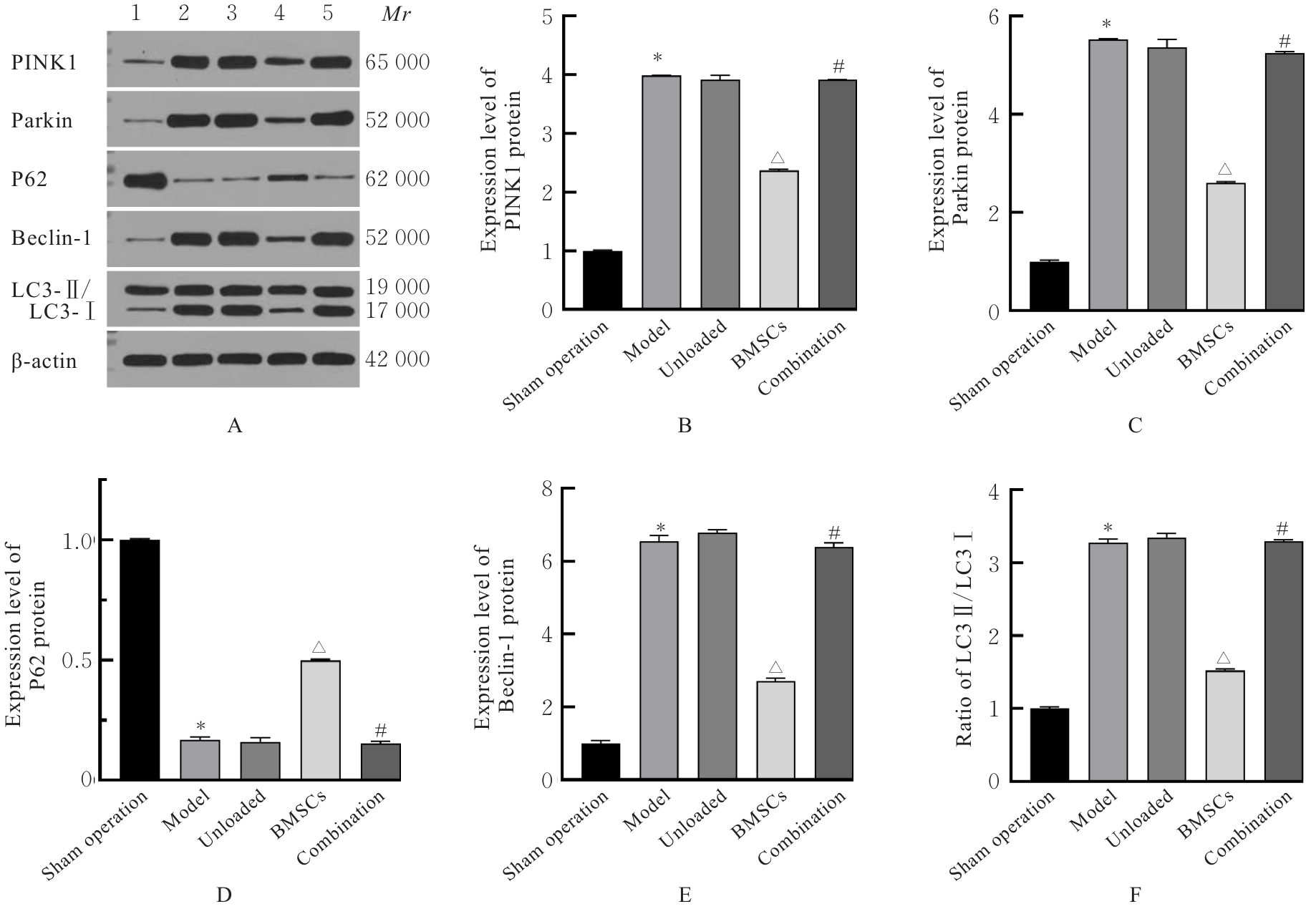
目的 探讨骨髓来源的间充质干细胞(BMSCs)移植通过活性氧(ROS)/核因子E2相关因子2(Nrf2)信号对血管性痴呆(VaD)大鼠线粒体自噬的影响,并阐明其作用机制。 方法 将45只SD雄性成年大鼠随机分为假手术组、模型组、空载组、BMSCs组和BMSCs+ML385(Nrf2抑制剂)组(联合组),每组9只。各组大鼠腹腔注射麻醉后,除假手术组外,其余各组大鼠制备VaD模型。采用Morris水迷宫实验检测各组大鼠的学习记忆能力,HE染色观察各组大鼠脑组织病理形态表现,尼氏染色观察各组大鼠脑组织海马区尼氏体变化情况,透射电镜观察各组大鼠脑组织海马区超微结构,荧光探针法检测各组大鼠脑组织海马区神经元中ROS水平,Western blotting法检测各组大鼠脑组织中Nrf2、血红素加氧酶1(HO-1)、磷酸酶与张力蛋白同源物诱导激酶1(PINK1)、E3泛素蛋白连接酶parkin(Parkin)、苄氯素1(Beclin-1)和泛素结合蛋白P62(P62)蛋白表达水平及微管相关蛋白1A/1B轻链3(LC3-Ⅱ/LC3-Ⅰ)比值。 结果 Morris水迷宫实验,与假手术组比较,模型组大鼠逃避潜伏期明显延长(P<0.01),穿越原平台次数和停留时间均明显减少(P<0.01);与模型组比较,BMSCs组大鼠逃避潜伏期明显缩短(P<0.01),穿越原平台次数和停留时间均明显增加(P<0.01);与BMSCs组比较,联合组大鼠逃避潜伏期明显延长(P<0.01),穿越原平台次数和停留时间均明显减少(P<0.01)。HE染色观察,假手术组大鼠脑组织海马区神经元数量和形态正常,染色均匀,结构清晰,未见明显病变;与假手术组比较,模型组大鼠脑组织海马区组织稀疏,结构紊乱,神经元数量减少且形态不一,染色不均匀,核固缩,可见部分坏死的神经元;与模型组比较,空载组大鼠脑组织海马区可见组织结构紊乱、神经元减少和染色不均等损伤表现,BMSCs组大鼠海马区神经元损伤减轻,形态恢复正常,排列较为整齐,神经元丢失情况明显改善;与BMSCs组比较,联合组大鼠海马区神经元形态不规则,组织结构紊乱,细胞边界不清,染色不均匀,核固缩。尼氏染色观察,假手术组大鼠脑组织海马区神经元排列整齐紧密,形态规则完整,核仁明显,尼氏小体着色深且数量多;与假手术组比较,模型组大鼠脑组织海马区神经元固缩,呈空泡状,尼氏小体着色少且数量稀少;与模型组比较,空载组大鼠脑组织海马区可见尼氏小体着色少且数量稀少等损伤表现,BMSCs组大鼠脑组织海马区神经元固缩减少,细胞形态相对完整,尼氏小体数量相对增多;与BMSCs组比较,联合组大鼠脑组织神经元固缩,形态完整性丧失,尼氏小体破碎且数量减少。透射电镜观察,假手术组大鼠脑组织海马区神经元线粒体呈椭圆形,双层膜结构清晰可见,内部嵴完整;与假手术组比较,模型组大鼠脑组织海马区神经元线粒体肿胀变形,双层膜结构破坏,内部嵴断裂消失,结构模糊,胞质中可见大量自噬小体;与模型组比较,空载组大鼠脑组织海马区线粒体损伤表现仍较明显,胞质中自噬小体数量较多,BMSCs组大鼠脑组织海马区神经元线粒体膜和内部结构有明显改善,损伤程度减轻,胞质中可见少量自噬小体;与BMSCs组比较,联合组大鼠脑组织海马区神经元线粒体肿胀,双层膜结构破坏,内部嵴断裂消失,胞质中可见自噬小体。荧光探针法,与假手术组比较,模型组大鼠脑组织海马区神经元中ROS水平明显升高(P<0.01);与模型组比较,BMSCs组大鼠脑组织海马区神经元中ROS水平明显降低(P<0.01);与BMSCs组比较,联合组大鼠脑组织海马区神经元中ROS水平明显升高(P<0.01)。Western blotting法,与假手术组比较,模型组大鼠脑组织中Nrf2和HO-1蛋白表达水平均明显降低(P<0.01);与模型组比较,BMSCs组大鼠脑组织中Nrf2和HO-1蛋白表达水平均明显升高(P<0.01);与BMSCs组比较,联合组大鼠脑组织中Nrf2和HO-1蛋白表达水平均明显降低(P<0.01)。与假手术组比较,模型组大鼠脑组织中Parkin、PINK1和Beclin-1蛋白表达水平及LC3-Ⅱ/LC3-Ⅰ比值均明显升高(P<0.01),P62蛋白表达水平明显降低(P<0.01);与模型组比较,BMSCs组大鼠脑组织中Parkin、PINK1和Beclin-1蛋白表达水平及LC3-Ⅱ/LC3-Ⅰ比值均明显降低(P<0.01),P62蛋白表达水平明显升高(P<0.01);与BMSCs组比较,联合组大鼠脑组织中Parkin、PINK1和Beclin-1蛋白表达水平及LC3-Ⅱ/LC3-Ⅰ比值均明显升高(P<0.01),P62蛋白表达水平明显降低(P<0.01)。 结论 BMSCs可以减轻大鼠脑组织海马区神经元病理改变,改善VaD大鼠的认知功能,其作用机制可能与调控ROS/Nrf2信号通路抑制线粒体自噬有关。

关键词: 血管性痴呆, 骨髓来源的间充质干细胞, 核因子E2相关因子2, 活性氧, 线粒体自噬

Abstract:

Objective To discuss the effects of bone marrow-derived mesenchymal stem cells (BMSCs) transplantation on mitophagy in the vascular dementia (VaD) rats through reactive oxygen species (ROS)/nuclear factor erythroid 2-related factor 2 (Nrf2) signaling, and to clarify its mechanism. Methods Forty-five male adult SD rats were randomly divided into sham operation group, model group, unloaded group, BMSCs group, and MSCs+ML385 (Nrf2 inhibitor) group (combination group), and there were 9 rats in each group. After intraperitoneal anesthesia, the VaD models were established in all groups except sham operation group. Morris water maze test was used to detect the learning and memory abilities of the rats in various groups; HE staining was used to observe the histopathological morphology of brain tissue of the rats in various groups; Nissl staining was used to observe the changes of Nissl bodies in hippocampus region of brain tissue of the rats in various groups; transmission electron microscope was used to observe the ultrastructure of hippocampus region of the rats in various groups; fluorescence probe method was used to detect the ROS levels in hippocampus neurons in various groups; Western blotting method was used to detect the expression levels of Nrf2, heme oxygenase-1 (HO-1), PTEN-induced putative kinase 1 (PINK1), parkin RBR E3 ubiquitin protein ligase (Parkin), Beclin-1, ubiquitin-binding protein p62 (P62), and microtubule-associated protein 1A/1B-light chain 3 (LC3-Ⅱ/LC3-Ⅰ) ratio in brain tissue of the rats in various groups. Results The Morris water maze results showed that compared with sham operation group, the escape latency of the rats in model group was significantly increased (P<0.01), while the number of crossing time and residence time were significantly decreased (P<0.01). Compared with model group, the escape latency of the rats in BMSCs group was significantly decreased (P<0.01), while the number of crossing time and residence time were significantly increased (P<0.01). Compared with BMSCs group, the escape latency of the rats in combination group was significantly increased (P<0.01), while the number of crossing time and residence time were significantly decreased (P<0.01). The HE staining results showed that hippocampus neurons of the rats in sham operation group were normal in quantity and morphology, with uniform staining and clear structure. Compared with sham operation group, the hippocampus tissue of the rats in model group showed sparse arrangement, disordered structure, reduced neuronal quantity, varied morphology, uneven staining, nuclear pyknosis, and partial neuronal necrosis. Compared with model group, the neuronal damage of the rats in hippocampus regio in BMSCs group was alleviated, with restored morphology and improved neuronal loss. Compared with BMSCs group, the neurons of the rats in hippocampus region in combination group showed irregular morphology, disordered structure, unclear cell boundaries, uneven staining, and nuclear pyknosis. The Nissl staining results showed that the hippocampal neurons in sham operation group were tightly arranged with intact morphology, obvious nucleoli, and abundant darkly stained Nissl bodies. Compared with sham operation group, the neurons in hippocampus region of the rats in model group showed pyknosis, vacuolization, and sparse Nissl bodies. Compared with model group, the BMSCs group showed reduced neuronal pyknosis, relatively intact morphology, and increased Nissl bodies. Compared with BMSCs group, the combination group showed neuronal pyknosis, loss of morphological integrity, and fragmented Nissl bodies. The transmission electron microscope results showed that mitochondria in sham operation group exhibited oval shape with intact double-membrane structure and cristae. Compared with sham operation group, the mitochondria in model group showed swelling, disrupted membranes, broken cristae, and numerous autophagosomes. Compared with model group, the BMSCs group showed improved mitochondrial structure and reduced autophagosomes. Compared with BMSCs group, the combination group showed swollen mitochondria, disrupted membranes, broken cristae, and visible autophagosomes. The fluorescence probe results showed that compared with sham operation group, the ROS levels in the hippocampus neurons in brain tissue of the rats in model group were significantly increased (P<0.01); compared with model group, the ROS levels in hippocampus neurons in brain tissue of the rats in BMSCs group were significantly decreased (P<0.01); compared with BMSCs group, the ROS levels in hippocampus neurons in brain tissue of the rats in combination group were significantly increased (P<0.01). The Western blotting results showed that compared with sham operation group, the expression levels of Nrf2 and HO-1 proteins in brain tissue of the rats in model group were significantly decreased (P<0.01); compared with model group, the expression levels of Nrf2 and HO-1 proteins in brain tissue of the rats in BMSCs group were significantly increased (P<0.01); compared with BMSCs group, the expression levels of Nrf2 and HO-1 proteins in brain tissue of the rats in combination group were significantly decreased (P<0.01); compared with sham operation group, the expression levels of Parkin, PINK1, and Beclin-1 proteins, and LC3-Ⅱ/LC3-Ⅰ ratio of the rats in model group were significantly increased (P<0.01), while the expression level of P62 protein was significantly decreased (P<0.01); compared with model group, the expression levels of Parkin, PINK1, and Beclin-1 proteins, as well as the LC3-Ⅱ/LC3-Ⅰ ratio, of the rats in BMSCs group were significantly decreased (P<0.01), while the expression level of P62 protein was significantly increased (P<0.01); compared with BMSCs group, the expression levels of Parkin, PINK1, and Beclin-1 proteins, as well as the LC3-Ⅱ/LC3-Ⅰ ratio, of the rats in combination group were significantly increased (P<0.01), while the expression level of P62 protein was significantly decreased (P<0.01). Conclusion BMSCs can alleviate the hippocampal neuronal pathological changes and improve cognitive function in the VaD rats, and its mechanism may be related to the regulation of ROS/Nrf2 signaling pathway to inhibit mitophagy.

Key words: Vascular dementia, Bone marrow mesenchymal stem cells, Nuclear factor erythroid 2-related factor 2, Reactive oxygen species, Mitophagy

中图分类号: 

  • R743.9