吉林大学学报(医学版) ›› 2023, Vol. 49 ›› Issue (3): 682-690.doi: 10.13481/j.1671-587X.20230317

• 基础研究 • 上一篇    下一篇

缺氧条件下HIF-1α/ROS对肺癌A549细胞凋亡和侵袭的作用及其机制

黄波1(),丁洁2,郭红荣1,王红娟1,徐建群1,郑泉1   

  1. 1.湖北省武汉市第三医院 武汉大学附属同仁医院呼吸与危重症医学科, 湖北 武汉 430070
    2.湖北省武汉市第三医院 武汉大学附属同仁医院血液透析室, 湖北 武汉 430070
  • 收稿日期:2022-07-15 出版日期:2023-05-28 发布日期:2023-06-20
  • 通讯作者: 黄波 E-mail:doctorhuang82@126.com
  • 作者简介:黄 波(1982-),男,广西壮族自治区贵港市人,副主任医师,医学硕士,主要从事肺癌基础和临床方面的研究。
  • 基金资助:
    湖北省卫健委科研项目(WJ2021F005);湖北省武汉市卫健委医学科研项目(WX21Q43);湖北省武汉市中青年医学骨干人才培养工程项目(武卫通(2019)87号)

Effects of HIF-1α/ROS on apoptosis and invasion of lung cancer A549 cells under hypoxia and its mechanism

Bo HUANG1(),Jie DING2,Hongrong GUO1,Hongjuan WANG1,Jianqun XU1,Quan ZHENG1   

  1. 1.Department of Respiratory and Critical Care Medicine,Wuhan Third Hospital,Hubei Province,Affiliated Tongren Hospital,Wuhan University,Wuhan 430070,China
    2.Hemodialysis Room,Wuhan Third Hospital,Hubei Province,Affiliated Tongren Hospital,Wuhan University,Wuhan 430070,China
  • Received:2022-07-15 Online:2023-05-28 Published:2023-06-20
  • Contact: Bo HUANG E-mail:doctorhuang82@126.com

摘要:

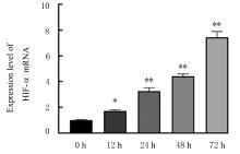
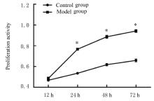
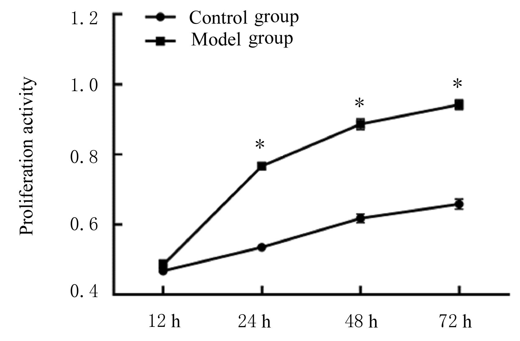
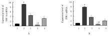
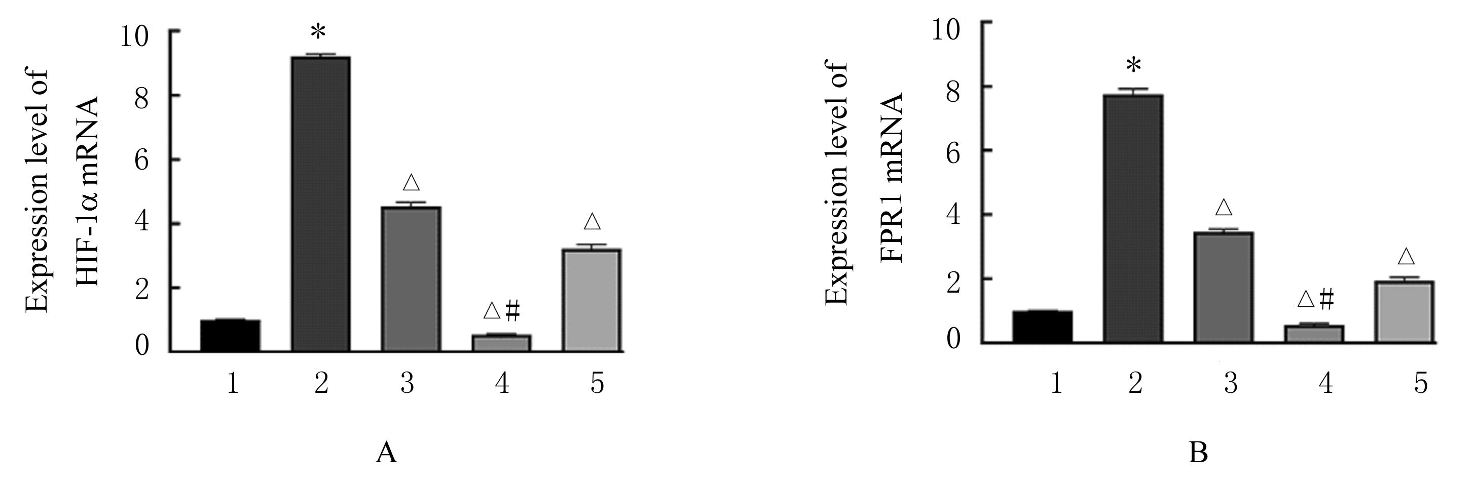
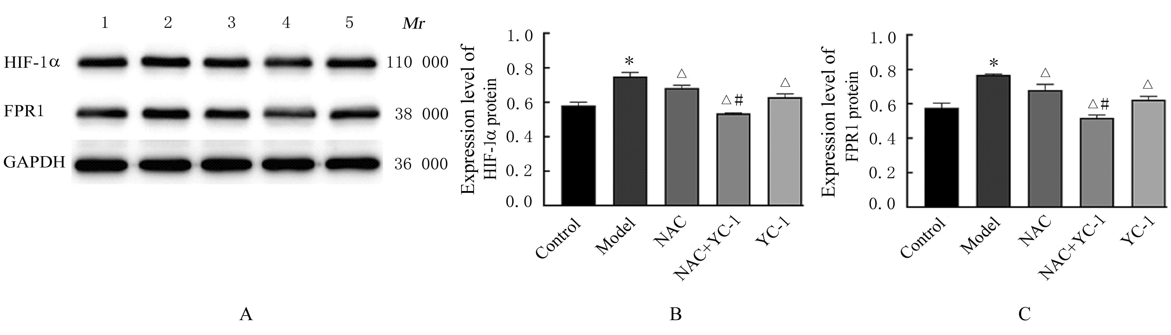
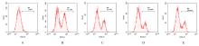
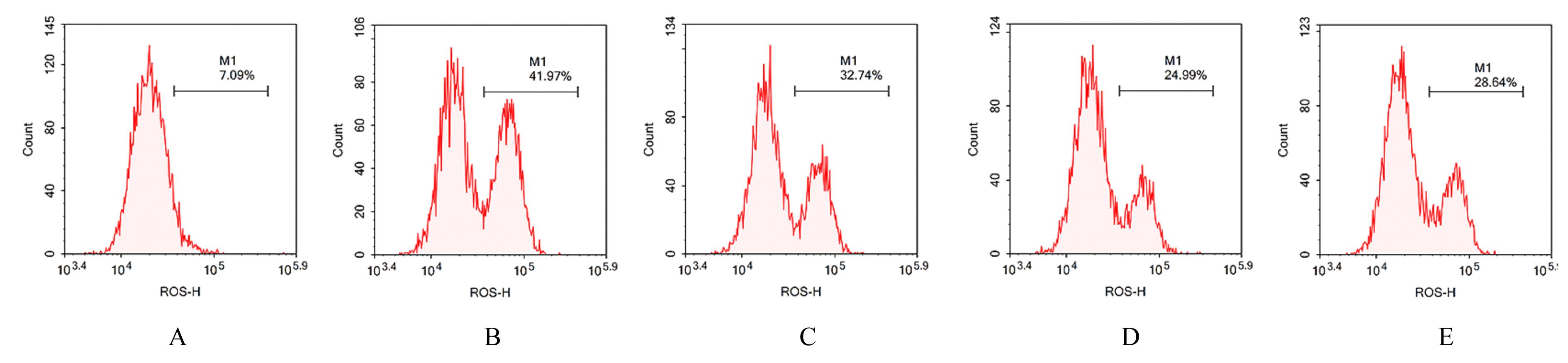
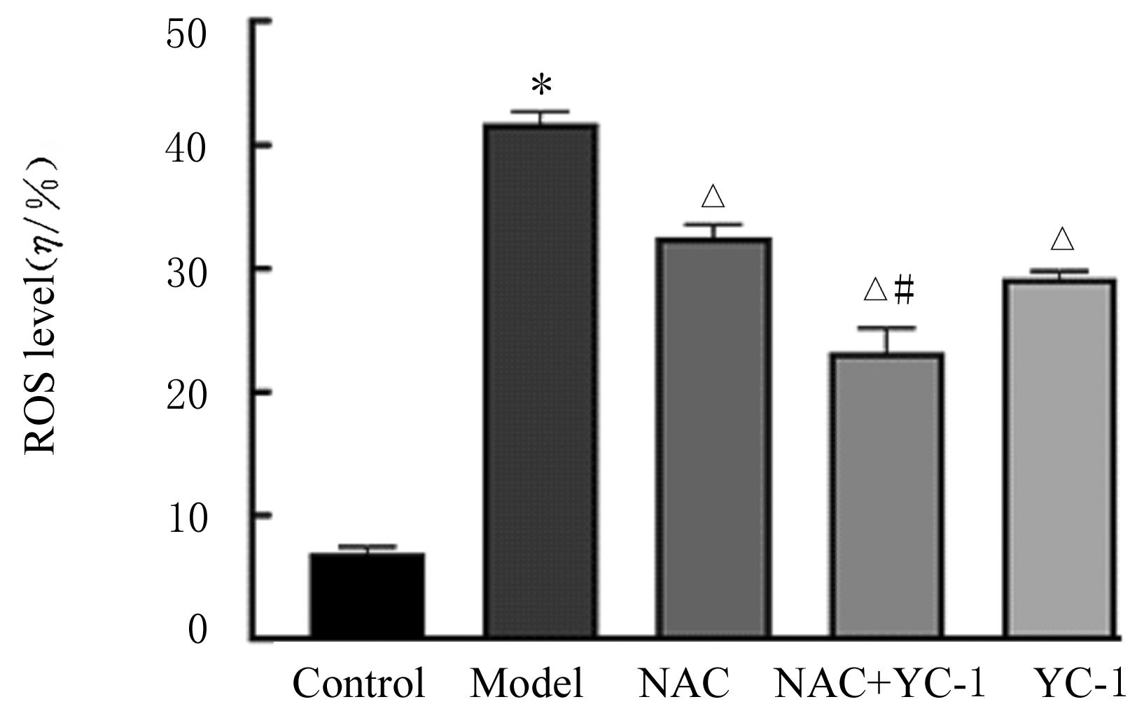
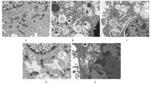
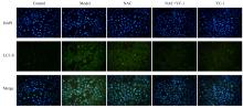
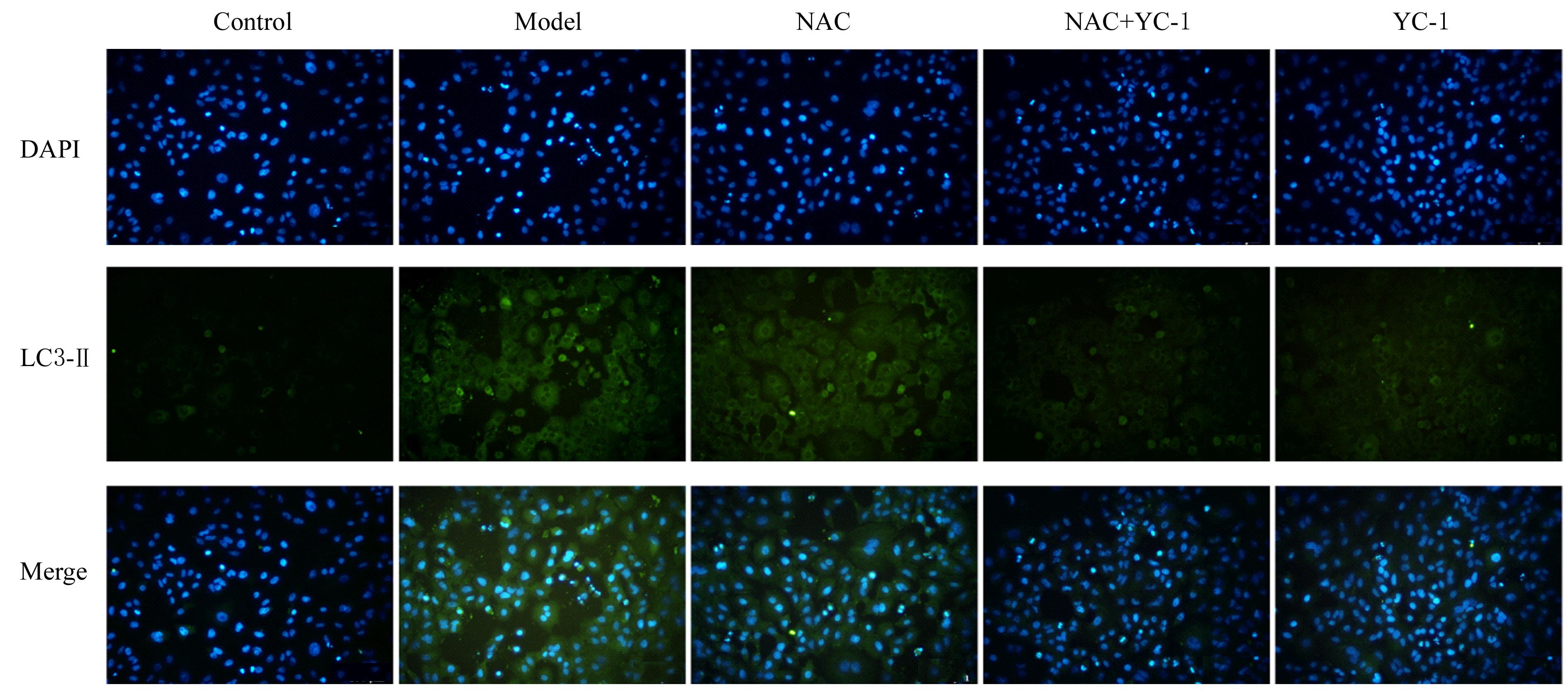
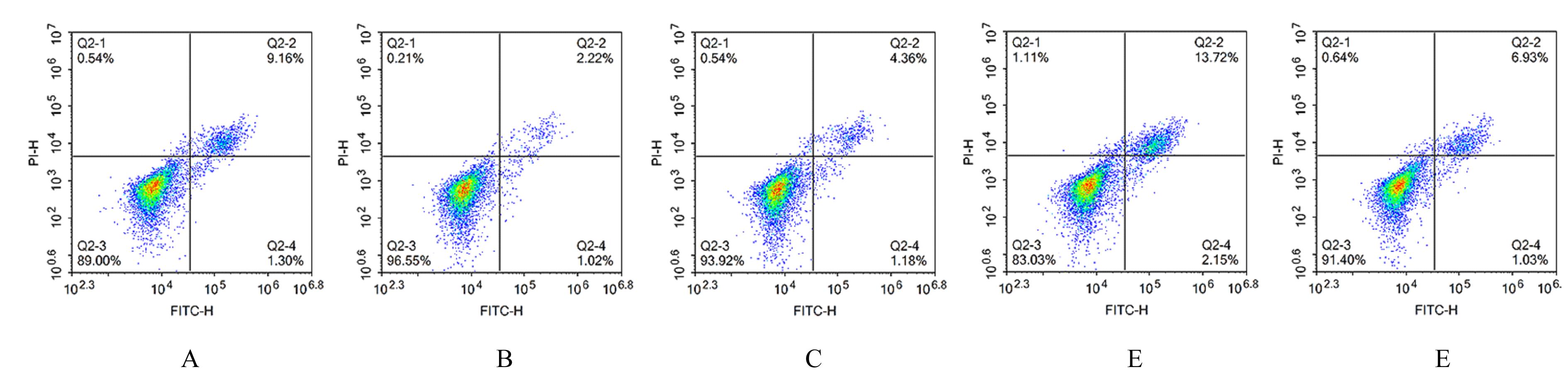
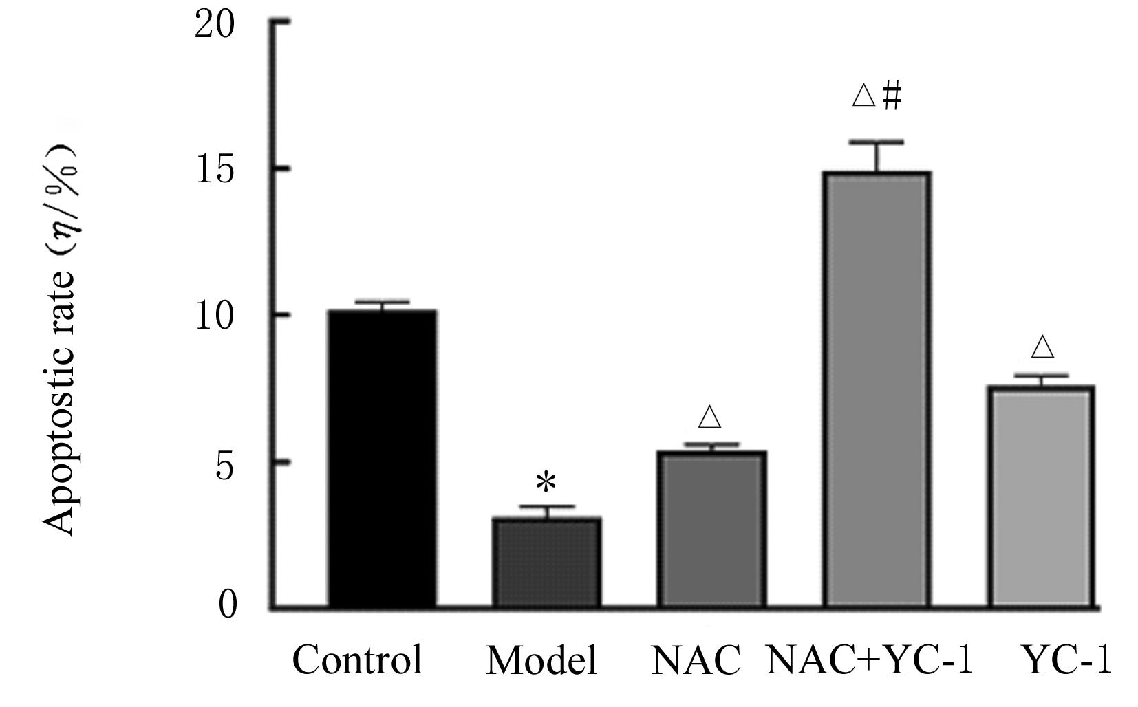
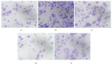
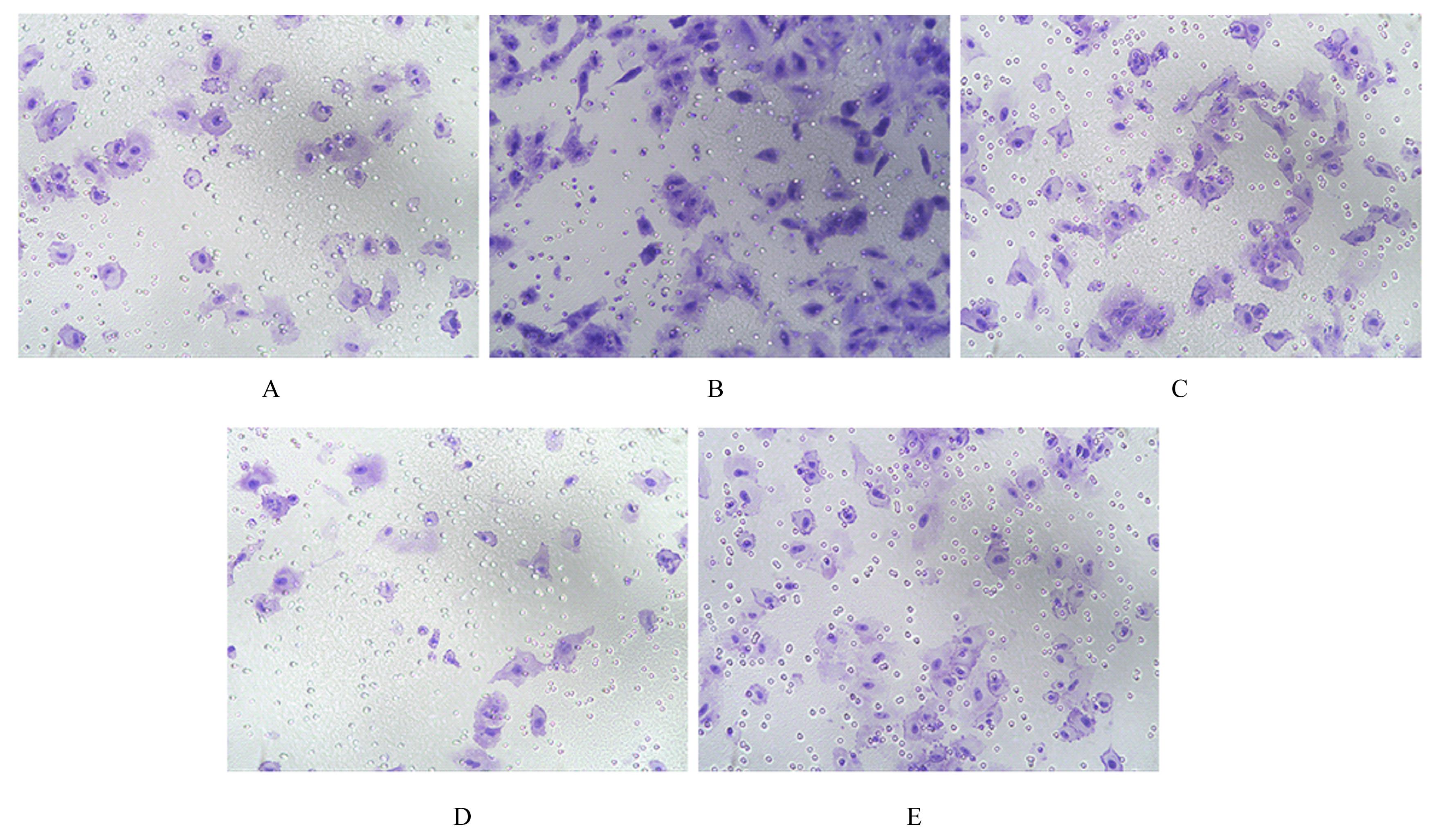
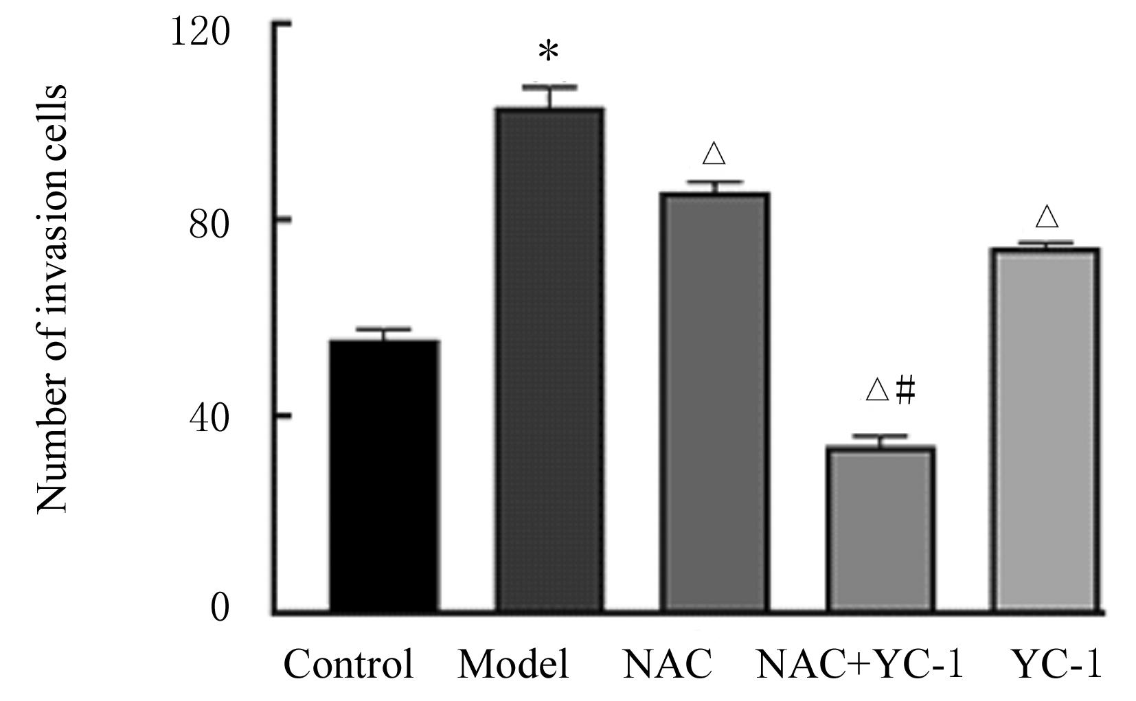
目的 探讨缺氧条件下缺氧诱导因子1α(HIF-1α)/活性氧(ROS)对非小细胞肺癌A549细胞凋亡和侵袭的作用,并阐明其相关机制。 方法 将A549细胞置于缺氧条件下培养12、24、48和72 h,构建缺氧细胞模型,实时荧光定量PCR(RT-qPCR)法检测不同处理时间细胞中HIF-1α mRNA表达水平,CCK-8法检测细胞增殖能力,鉴定缺氧细胞模型并确定最佳诱导时间。将A549细胞分为对照组(21%O2)、模型组(缺氧诱导)、N-乙酰半胱氨酸(NAC)组(20 mmol·L-1 NAC)、NAC+利非西呱(YC-1)组(20 mmol·L-1 NAC+100 μmol·L-1 YC-1)和YC-1组(100 μmol·L-1 YC-1)。RT-qPCR和Western blotting法检测各组细胞中HIF-1α和人甲酰肽受体1(FPR1) mRNA及蛋白表达水平,流式细胞术检测各组细胞中ROS水平,透射电镜观察各组细胞自噬体结构,免疫荧光法检测各组细胞中微管相关轻链3Ⅱ(LC3-Ⅱ)表达情况,流式细胞术检测各组细胞凋亡率,Transwell小室实验检测各组侵袭细胞数。 结果 随着低氧诱导时间的延长,A549细胞中HIF-1α mRNA表达水平和细胞增殖能力均明显升高(P<0.05或P<0.01),最佳低氧诱导时间为24 h。RT-qPCR和Western blotting法检测,与对照组比较,模型组细胞中HIF-1α和FPR1 mRNA及蛋白表达水平明显升高(P<0.01);与模型组比较,NAC组、NAC+YC-1组和YC-1组细胞中HIF-1α和FPR1 mRNA及蛋白表达水平均明显降低(P<0.01);与NAC组比较,NAC+YC-1组细胞中HIF-1α和FPR1 mRNA及蛋白表达水平明显降低(P<0.01)。流式细胞术检测,与对照组比较,模型组细胞中ROS水平明显升高(P<0.01);与模型组比较,NAC组、NAC+YC-1组和YC-1组细胞中ROS水平均明显降低(P<0.01);与NAC组比较,NAC+YC-1组细胞中ROS水平明显降低(P<0.01)。透射电镜观察,对照组细胞中细胞器结构完整,未见自噬体结构;模型组细胞中可见大量自噬体和明显空泡,细胞中细胞器被降解;与模型组比较,NAC组、NAC+YC-1组和YC-1组细胞中自噬体减少,其中NAC+YC-1组细胞中自噬体最少。免疫荧光法检测,与对照组比较,模型组细胞中LC3-Ⅱ表达强度明显增加;与模型组比较,NAC组、NAC+YC-1组和YC-1组细胞中LC3-Ⅱ表达强度明显减少。流式细胞术检测,与对照组比较,模型组细胞凋亡率明显降低(P<0.01);与模型组比较,NAC组、NAC+YC-1组和YC-1组细胞凋亡率明显升高(P<0.01);与NAC组比较,NAC+YC-1细胞凋亡率明显升高(P<0.01)。Transwell小室实验检测,与对照组比较,模型组侵袭细胞数明显增加(P<0.01);与模型组比较,NAC组、NAC+YC-1组和YC-1组侵袭细胞数均明显减少(P<0.01);与NAC组比较,NAC+YC-1组侵袭细胞数明显减少(P<0.01)。 结论 缺氧条件下抑制HIF-1α/ROS可促进肿瘤细胞凋亡,并抑制细胞侵袭,其作用机制与抑制肺癌细胞自噬有关。

关键词: 缺氧, 缺氧诱导因子1α, 活性氧, 自噬, 癌,非小细胞肺

Abstract:

Objective To discuss the effect of hypoxia inducible factor-1α (HIF-1α)/ reactive oxygen species (ROS) on the apoptosis and invasion of the non-small cell lung cancer A549 cells,and to clarify the related mechanism. Methods The A549 cells were cultured under hypoxia condition for 12, 24, 48 and 72 h to construct the hypoxia cell model. The expression levels of HIF-1α mRNA in the cells at different treatment time were detected by real-time fluorescence quantitative PCR(RT-qPCR) method, and the proliferation abilities of the cells were detected by CCK-8 assay;the hypoxia cell model was verified, and the optimal induction time was determined.The A549 cells were divided into control group(21%O2), model group(hypoxia induction), NAC group(20 mmol·L-1 NAC), NAC+YC-1 group(20 mmol·L-1 NAC+100 μmol·L-1 YC-1) and YC-1 (100 μmol·L-1 YC-1)group. The expression levels of HIF-1α and FPR1 mRNA and proteins were detected by RT-qPCR and Western blotting methods; the ROS levels were detected by flow cytometry; the autophagosome structure in the cells was observed by transmission electron microscope; the expressions of microtubule-associated protein light china 3 Ⅱ(LC3-Ⅱ) in the cells in various groups were detected by immunofluorescence; the apoptotic rates of the cells in various groups were detected by flow cytometry; and the number of invasion cells was detected by Transwell chamber assay. Results With the prolongation of hypoxia induction time, the HIF-1α mRNA expression levels in the A549 cells and cell proliferation abilities were increased(P<0.05 or P<0.01), and the optimal hypoxia induction time was 24 h. The results of RT-qPCR and Western blotting methods showed that compared with control group,the expression levels of HIF-1α and FPR1 mRNA and proteins in the cells in model group were significantly increased(P<0.01); compared with model group,the expression levels of HIF-1α and FPR1 mRNA and proteins in the cells in NAC group, NAC+YC-1 group and YC-1 group were significantly decreased(P<0.01).Compared with control group,the level of ROS in the cells in model group was significantly increased (P<0.01); compared with model group,the ROS levels in the cells in NAC group, NAC+YC-1 group, and YC-1 group were significantly decreased(P<0.01);compared with NAC group,the ROS level in the cells in NAC+YC-1 group was significantly decreased(P<0.01).The transmission electron microscope results showed that the organelle structure of the cells in control group was intact and no autophagosome structure was seen; a large number of autophagosomes were seen in the cells in model group, obvious vacuoles were visible, and intracellular organelles were degraded; compared with model group, the number of autophagosomes in the cells in NAC group, NAC+YC-1 group and YC-1 group were decreased,among them the autophagosomes in the cells in NAC+YC-1 group were the least. The immunofluorescence assay results showed that the expression intensity of LC3-Ⅱ in the cells in model group was significantly increased compared with control group, and the expression intensities of LC3-Ⅱ in the cells in NAC group, NAC+YC-1 and YC-1 group were signficantly decreased compared with model group.Compared with control group, the apoptotic rate of the cells in model group was significantly decreased(P<0.01); compared with model group, the apoptotic rates of the cells in NAC group, NAC+YC-1 group and YC-1 group were significantly increased(P<0.01); compared with NAC group,the apoptotic rate of the cells in NAC+YC-1 group was increased(P<0.01).The results of Transwell chamber assay showed that compared with control group the number of the invasion cells in model group was significantly increased(P<0.01); compared with model group, the numbers of invasion cells in NAC group, NAC+YC-1 group and YC-1 group were significantly decreased (P<0.01);compared with NAC group,the number of the in vasion cells in NAC+YC-1 group was decreased(P<0.01). Conclusion Inhibition of HIF-1α/ROS under hypoxia condition can promote the apoptosis and inhibit cell invasion of cancer cells,and its mechanism may be realted to inhibiting the autophagy of lung cancer cells.

Key words: Hypoxia, Hypoxia inducible factor-1α, Reactive oxygen species, Autophagy, Cancer,non-small cell lung

中图分类号: 

  • R734.2