吉林大学学报(医学版) ›› 2025, Vol. 51 ›› Issue (4): 948-957.doi: 10.13481/j.1671-587X.20250411

• 基础研究 • 上一篇    下一篇

藏红花醛对体外高糖诱导的大鼠血管平滑肌细胞增殖、迁移和表型转变的抑制作用

高逸璇1,2,汪鹏1,2,张思龙3,高瑞娟4,马莹芳1,2,张可可1,2,冯丹1,2,黄宗奇1,2,马克涛1,2,李丽1,5,司军强1,2()   

  1. 1.石河子大学医学院新疆地方与民族高发病教育部重点实验室,新疆 石河子 832002
    2.石河子大学医学院生理学教研室,新疆 石河子 832002
    3.新疆维吾尔自治区昌吉州人民医院普外二科,新疆 昌吉 831100
    4.石河子大学第一附属医院放射科,新疆 石河子 832002
    5.嘉兴大学医学院生理学教研室,浙江 嘉兴 314000
  • 收稿日期:2024-06-29 接受日期:2024-11-19 出版日期:2025-07-28 发布日期:2025-08-25
  • 通讯作者: 司军强 E-mail:sijunqiang@shzu.edu.cn
  • 作者简介:高逸璇(1998-),女,甘肃省张掖市人,在读硕士研究生,主要从事心脑血管疾病发病机制方面的研究。
  • 基金资助:
    新疆生产建设兵团科技局指导性科技计划项目(2022DZ004);石河子大学第一附属医院科研项目(QN202208)

Inhibitory effect of safranal on proliferation, migration and phenotypic transformation of vascular smooth muscle cells of rats induced by high glucose in vitro

Yixuan GAO1,2,Peng WANG1,2,Silong ZHANG3,Ruijuan GAO4,Yingfang MA1,2,Keke ZHANG1,2,Dan FENG1,2,Zongqi HUANG1,2,Ketao MA1,2,Li LI1,5,Junqiang SI1,2()   

  1. 1.Key Laboratory of Xinjiang Endemic and Ethnic Diseases,Shihezi University,Shihezi 832002,China
    2.Department of Physiology,School of Medical Sciences,Shihezi University,Shihezi 832002,China
    3.Department of General Surgery,Changji People’s Hospital,Changji Hui Autonomous Prefecture,Xinjiang 831100,China
    4.Department of Radiology,First Affiliated Hospital,Shihezi University,Shihezi 832002,China
    5.Department of Physiology,School of Medical Sciences,Jiaxing University,Jiaxing 314000,China
  • Received:2024-06-29 Accepted:2024-11-19 Online:2025-07-28 Published:2025-08-25
  • Contact: Junqiang SI E-mail:sijunqiang@shzu.edu.cn

摘要:

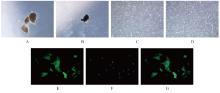
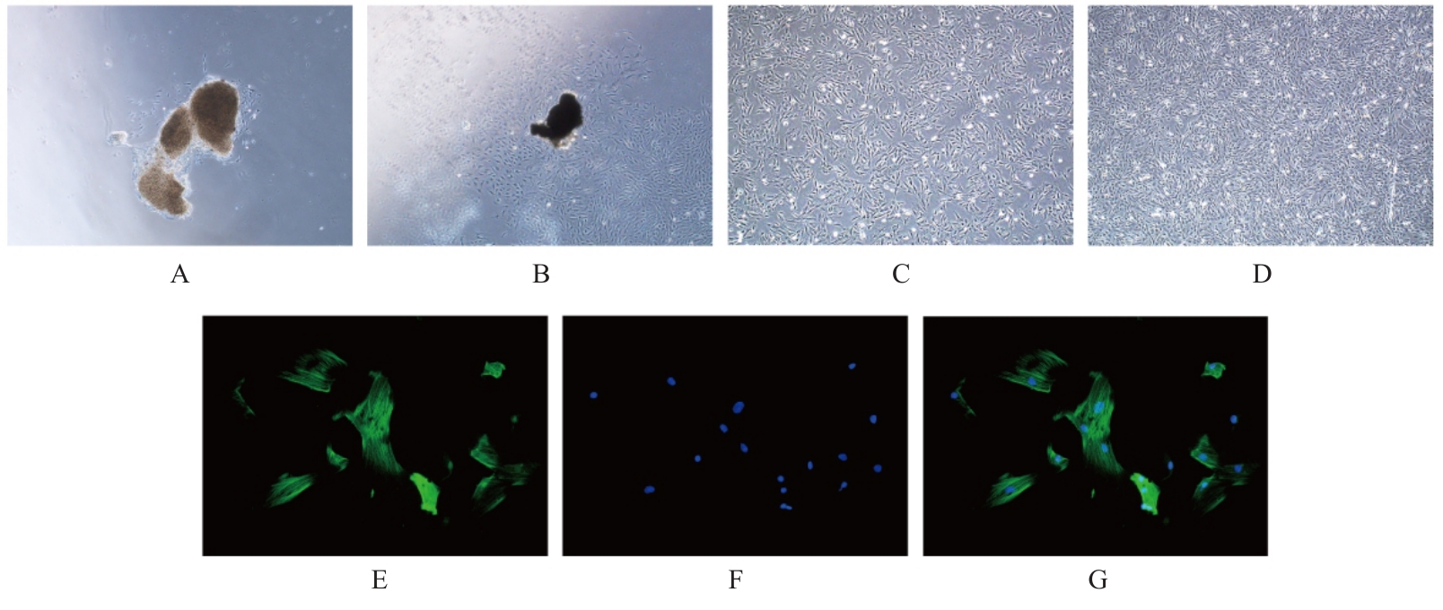
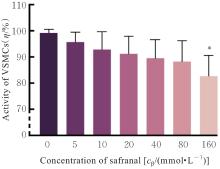
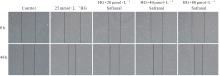
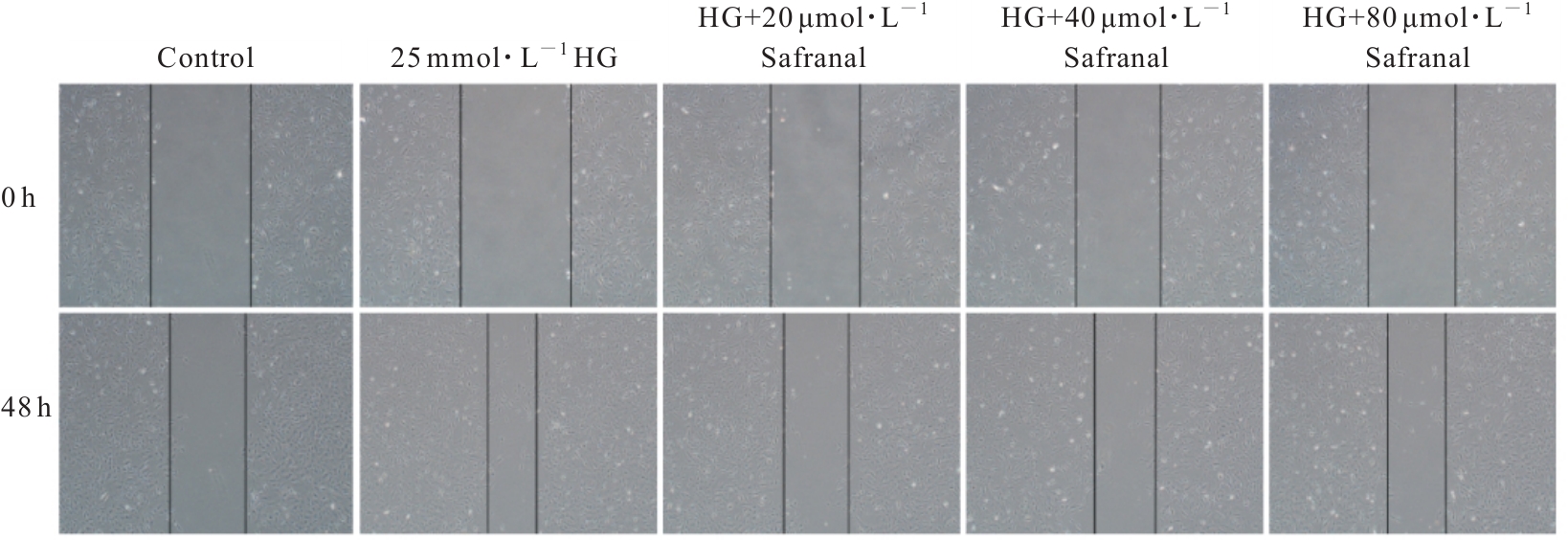
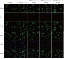
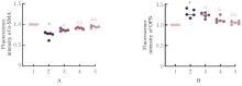
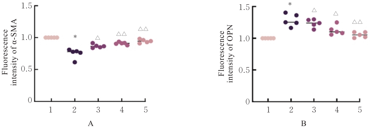
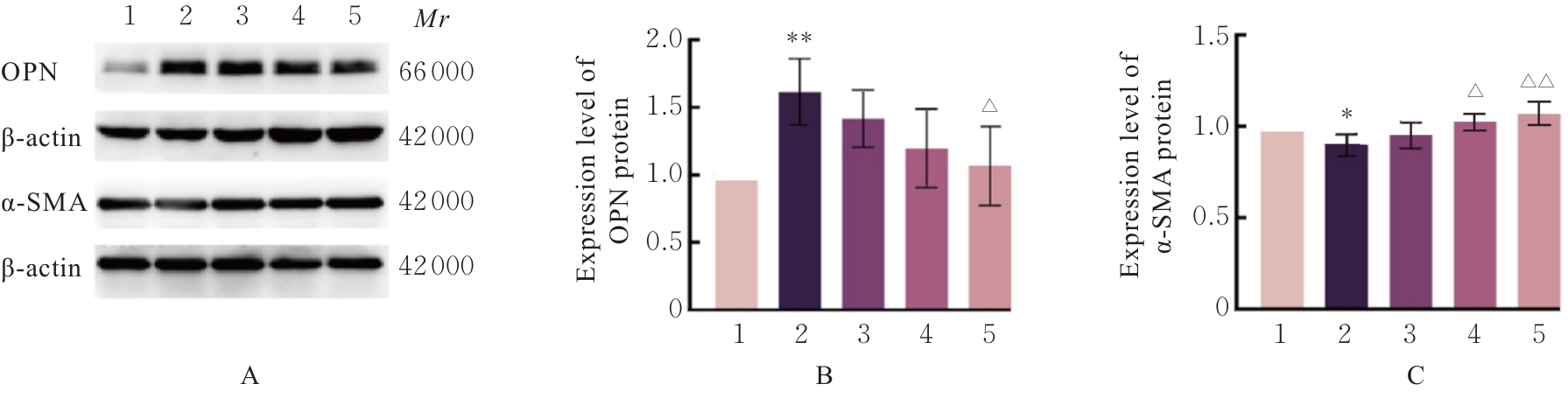
目的 探讨高糖诱导的藏红花醛对大鼠血管平滑肌细胞(VSMCs)增殖、迁移和表型转化的影响,阐明藏红花醛在糖尿病(DM)血管病变防治中的作用。 方法 选择SD大鼠作为实验对象,取大鼠胸主动脉原代培养VSMCs,分为对照组、25 mmol·L-1高糖(HG)组和HG+20、40和80 μmol·L-1藏红花醛组。对照组VSMCs不进行处理,25 mmol·L-1 HG组VSMCs给予25 mmol·L-1 HG预处理,HG+20、40和80 μmol·L-1藏红花醛组VSMCs在25 mmol·L-1 HG组处理的基础上再分别用20、40和80 μmol·L-1藏红花醛进行48 h干预。采用细胞计数试剂盒-8(CCK-8)法确定藏红花醛合适浓度并检测各组VSMCs存活率,细胞划痕愈合实验检测各组VSMCs划痕愈合率,Transwell小室实验检测各组VSMCs迁移细胞数,免疫荧光法检测各组VSMCs中α平滑肌肌动蛋白(α-SMA)和兔抗骨桥蛋白(OPN)荧光强度,Western blotting法检测各组VSMCs中OPN、α-SMA和增殖细胞核抗原(PCNA)蛋白表达水平。 结果 显微镜下可见,体外培养4 d时,胸主动脉组织块边缘有梭形或三角形细胞爬出,其中长梭形为最常见的形态;第14天时细胞逐渐铺满皿底,当细胞密度达到80%~90%时,出现标志性的“谷峰状”生长状态。取第3代细胞进行免疫荧光法鉴定,采用VSMCs特异性标记物α-SMA蛋白进行细胞免疫荧光染色,原代培养VSMCs中α-SMA蛋白均表达为阳性。CCK-8实验,与对照组比较,160 μmol·L-1 藏红花醛组VSMCs活性明显降低(P<0.01),即对VSMCs产生了毒性损伤。20、40和80 μmol·L-1 藏红花醛干预48 h后,VSMCs活性无明显变化,综合考虑藏红花醛的作用效果和毒性后,采用上述3个浓度藏红花醛用于后续的细胞实验。经过48 h的干预,与对照组比较,25 mmol·L-1 HG组VSMCs活性升高(P<0.001);与25 mmol·L-1 HG组比较,HG+20、40和80 μmol·L-1 藏红花醛组VSMCs活性降低(P<0.05)。细胞划痕愈合实验和Transwell小室实验,干预48 h后,25 mmol·L-1 HG组VSMCs划痕愈合率明显高于对照组(P<0.01),穿过Transwell小室的穿膜细胞数明显增多(P<0.05);与25 μmol·L?1 HG组比较,HG+20、40和80 μmol·L-1藏红花醛组VSMCs划痕愈合率降低(P<0.05),穿膜细胞数减少(P<0.05)。免疫荧光染色,与对照组比较,25 mmol·L-1 HG组VSMCs中α-SMA蛋白荧光强度明显减弱(P<0.001),而OPN蛋白荧光强度明显增强(P<0.001);与25 mmol·L-1 HG组比较,HG+20、40和80 μmol·L-1 藏红花醛组VSMCs中α-SMA蛋白荧光强度逐渐增加(P<0.05),OPN蛋白荧光强度逐渐减弱(P<0.05)。Western blotting法,与对照组比较,25 mmol·L-1 HG组VSMCs中α-SMA蛋白表达水平降低(P<0.05),PCNA和OPN蛋白表达水平升高(P<0.01);与25 mmol·L-1 HG组比较,HG+20、40和80 μmol·L-1 藏红花醛组VSMCs中α-SMA蛋白表达水平升高(P<0.05),PCNA和OPN蛋白表达水平降低(P<0.05)。 结论 藏红花醛能够抑制高糖诱导的大鼠VSMCs的增殖、迁移和表型转换。

关键词: 糖尿病, 血管病变, 胸主动脉平滑肌细胞, 细胞迁移, 细胞增殖

Abstract:

Objective To discuss whether safranal affects the proliferation, migration, and phenotypic transformation of the vascular smooth muscle cells (VSMCs) in a high-glucose environment and to clarify the function of safranal in the prevention and treatment of diabetic (DM) vascular complications.? Methods The SD rats were selected as experimental subjects; primary VSMCs were cultured from rat thoracic aortas and divided into control group, 25 mmol·L-1 high glucose (HG) group, HG+ 20 μmol·L-1 safranal group, HG+40 μmol·L-1 safranal group, and HG+80 μmol·L-1 safranal group. The cells in control group received no treatment; the cells in 25 mmol·L-1 HG group were pretreated with 25 mmol·L-1 HG; the cells in HG+20, 40, and 80 μmol·L-1 safranal groups were further treated with 20, 40, and 80 μmol·L-1 safranal respectively for 48 h on the basis of 25 mmol·L-1 HG group. Cell counting kit-8 (CCK-8) method was used to determine the appropriate concentration of safranal and detect the viabilities of the VSMCs in various groups; cell scratch healing assay was used to detect the scratch healing rates of the VSMCs in various groups; Transwell chamber assay was used to detect the numbers of the migration VSMCs in various groups; immunofluorescence method was used to detect the fluorescence intensities of alpha-smooth muscle actin (α-SMA) and rabbit anti-osteopontin (OPN) in the VSMCs in various groups; Western blotting method was used to detect the expression levels of OPN, α-SMA, and proliferating cell nuclear antigen (PCNA) in the VSMCs in various groups. Results Under microscope, on the 4th day of in vitro culture, the spindle-shaped or triangular cells crawled out from the edge of the thoracic aorta tissue blocks, with long spindle being the most common morphology. On the 14th, the cells gradually covered the bottom of the dish; when cell density reached 80%-90%, the characteristic “hills and valleys” growth pattern appeared. Third-generation cells were taken for immunofluorescence identification; immunofluorescence staining with VSMC-specific marker α-SMA showed positive expression of α-SMA protein in the primarily cultured VSMCs. The CCK-8 assay results showed that compared with control group, the cell viability of the cells in 160 μmol·L-1 safranal group was significantly decreased (P<0.01), indicating toxic damage to the cells. Under the conditions of safranal concentrations at 20, 40, and 80 μmol·L-1 respectively, after 48 h intervention on VSMCs, no significant adverse effect on cell viability was observed; considering both the effect and toxicity of safranal, these three concentrations were used in subsequent cell experiments. After 48 h intervention, compared with control group, the activity of the VSMCs in 25 mmol·L-1 HG group was increased (P<0.001); compared with 25 mmol·L-1 HG group, the activities of the VSMCs in HG+20, 40, and 80 μmol·L-1 safranal groups were gradually decreased (P<0.05). The cell scratch healing assay and Transwell assay results showed that after 48 h intervention, the scratch healing rate of the VSMCs in 25 mmol·L-1 HG group was significantly higher than that in control group (P<0.01), and the number of transmembrane cells through the Transwell chamber was significantly increased (P<0.05); compared with 25 mmol·L-1 HG group, the scratch healing rates of the VSMCs in HG+20, 40, and 80 μmol·L-1 safranal groups were gradually decreased (P<0.05), and the number of transmembrane cells was decreased (P<0.05). The immunofluorescence staining results showed that compared with control group, the fluorescence intensity of α-SMA protein in the VSMCs in 25 mmol·L-1 HG group was significantly weakened(P<0.001), while the fluorescence intensity of OPN protein was significantly enhanced (P<0.001); compared with 25 mmol·L-1 HG group, the fluorescence intensities of α-SMA protein in the VSMCs in HG+20, 40, and 80 μmol·L-1 safranal groups were gradually increased (P<0.05), and the fluorescence intensities of OPN were gradually weakened (P<0.05). The Western blotting method results showed that compared with control group, the expression level of α-SMA protein in the VSMCs in 25 mmol·L-1 HG group was decreased (P<0.05), and the expression levels of PCNA and OPN proteins were increased (P<0.01); compared with 25 mmol·L-1 HG group, the expression level of α-SMA protein in the VSMCs in HG+20, 40, and 80 μmol·L-1 safranal groups were increased (P<0.05), and the expression levels of PCNA and OPN proteins were decreased (P<0.05). Conclusion Safranal can inhibit the proliferation, migration, and phenotypic transformation of the VSMCs induced by high glucose.

Key words: Diabetes mellitus, Vascular diseases, Thoracic aorta smooth muscle cells, Cell migration, Cell proliferation

中图分类号: 

  • R363.1