吉林大学学报(医学版) ›› 2026, Vol. 52 ›› Issue (2): 451-459.doi: 10.13481/j.1671-587X.20260217

• 基础研究 • 上一篇    

右美托咪定对心肌缺血/再灌注损伤模型大鼠心肌损伤的保护作用及其机制

李爱梅,韩静霏,邓莉,陈思宇()   

  1. 新疆医科大学第一附属医院麻醉科,新疆 乌鲁木齐 830054
  • 收稿日期:2025-05-20 接受日期:2025-08-06 出版日期:2026-03-28 发布日期:2026-04-15
  • 通讯作者: 陈思宇 E-mail:53615367@qq.com
  • 作者简介:李爱梅(1987-),女,江苏省江阴市人,副主任医师,医学博士,主要从事围术期心肌保护方面的研究。
  • 基金资助:
    新疆维吾尔自治区科技厅自然科学基金项目(2022D01C229)

Protective effect of dexmedetomidine on myocardial injury in myocardial ischemia/reperfusion injury model rats

Aimei LI,Jingfei HAN,Li DENG,Siyu CHEN()   

  1. Department of Anesthesiology,First Affiliated Hospital,Xinjiang Medical University,Urumqi 830054,China
  • Received:2025-05-20 Accepted:2025-08-06 Online:2026-03-28 Published:2026-04-15
  • Contact: Siyu CHEN E-mail:53615367@qq.com

摘要:

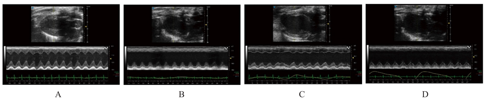
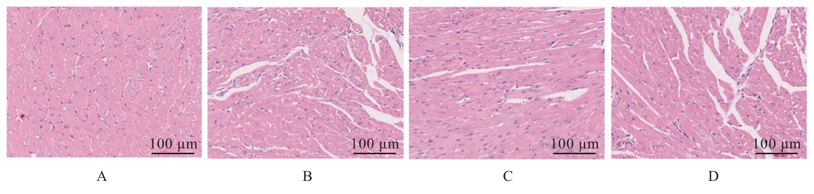
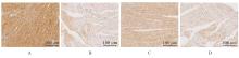
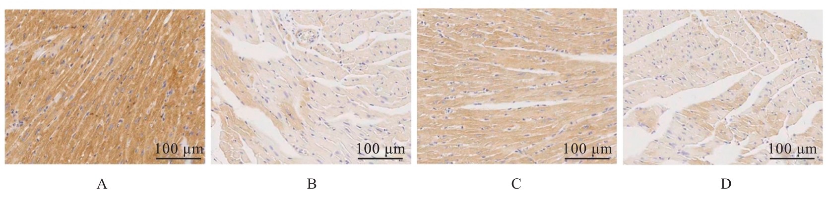
目的 探讨右美托咪定(Dex)对大鼠心肌缺血/再灌注损伤(MIRI)的保护作用,并阐明其作用机制。 方法 取48只SD大鼠,随机分为对照组、模型组、Dex组和Dex+Compound C组,每组12只。采用冠状动脉左前降支结扎法构建MIRI模型,利用超声心动图评估各组大鼠心功能,HE染色观察各组大鼠心肌组织病理形态表现,试剂盒检测各组大鼠血清中心肌肌钙蛋白I(cTnI)水平和己糖激酶(HK)、 磷酸果糖激酶(PFK)及丙酮酸激酶(PKM)活性, 高效液相色谱(HPLC)法检测各组大鼠心肌能量代谢物三磷酸腺苷(ATP)、二磷酸腺苷(ADP)和一磷酸腺苷(AMP)水平并计算能荷(EC),免疫组织化学法和Western blotting法检测各组大鼠心肌组织中Toll样受体4(TLR4)、巨噬细胞迁移抑制因子(MIF)、腺苷酸活化蛋白激酶(AMPK)及磷酸化AMPK(p-AMPK)蛋白表达情况。 结果 与对照组比较,模型组大鼠左心室射血分数(EF)、缩短分数(FS)、心输出量(CO)和每搏输出量(SV)均明显降低(P<0.05);血清中cTnI水平明显升高(P<0.05),HK、PFK和PKM活性明显降低(P<0.05);心肌组织中ATP和ADP水平及EC均明显降低(P<0.05),AMP水平明显升高(P<0.05);心肌组织中TLR4和MIF蛋白表达水平升高(P<0.05),p-AMPK/AMPK比值降低(P<0.05)。与模型组比较,Dex组大鼠EF、FS、CO和SV均明显升高(P<0.05);血清中cTnI水平明显降低(P<0.05),HK和PKM活性明显升高(P<0.05);心肌组织中ATP和ADP水平及EC明显升高(P<0.05);心肌组织中TLR4和MIF蛋白表达水平降低(P<0.05),p-AMPK/AMPK比值升高(P<0.05)。与Dex组比较,Dex+Compound C组大鼠EF、FS、CO和SV明显降低(P<0.05),血清中cTnI水平升高(P<0.05),PKM活性降低(P<0.05);心肌组织中ATP和ADP水平及EC明显降低(P<0.05);心肌组织中TLR4和MIF蛋白表达水平升高(P<0.05),p-AMPK/AMPK比值降低(P<0.05)。 结论 Dex通过抑制TLR4/MIF信号通路并激活AMPK磷酸化,进而改善MIRI后心肌能量代谢失衡及心肌功能障碍,其疗效可被AMPK抑制剂Compound C拮抗,Dex通过调控TLR4/MIF/AMPK信号通路发挥心肌保护作用。

关键词: 右美托咪定, 心肌缺血再灌注损伤, Toll样受体4, 能量代谢, 一磷酸腺苷活化蛋白激酶

Abstract:

Objective To discuss the protective effect of dexmedetomidine (Dex) on myocardial ischemia/reperfusion injury (MIRI) in the rats, and to clarify its mechanism. Methods Forty-eight SD rats were randomly divided into control group, model group, Dex group, and Dex+Compound C group, with 12 rats in each group. The MIRI model was established by ligation of left anterior descending coronary artery. Echocardiography was used to evaluate the cardiac function of the rats in various groups; HE staining was used to observe the pathomorphology of myocardium tissue of the rats in various groups; kits were used to detect the serum level of cardiac troponin I (cTnI) and the activities of hexokinase (HK), phosphofructokinase (PFK), and pyruvate kinase (PKM) of the rats in various groups; high performance liquid chromatography (HPLC) was used to detect the levels of myocardial energy metabolites adenosine triphosphate (ATP), adenosine diphosphate (ADP), and adenosine monophosphate (AMP) in myocardium tissue of the rats in various groups, and the energy charge (EC) was calculated; immunohistochemistry and Western blotting method were used to detect the expressions of Toll-like receptor 4 (TLR4), macrophage migration inhibitory factor (MIF), AMP-activated protein kinase(AMPK) and phosphorylated AMPK (p-AMPK) proteins in myocardium tissue of the rats in various groups. Results Compared with control group, the left ventricular ejection fraction (EF), fractional shortening (FS), cardiac output (CO), and stroke volume (SV) of the rats in model group were significantly decreased (P<0.05); the serum level of cTnI was significantly increased (P<0.05), and the activities of HK, PFK, and PKM were significantly decreased (P<0.05); the levels of ATP and ADP and EC in myocardium tissue were significantly decreased (P<0.05), while the level of AMP was significantly increased (P<0.05); the expression levels of TLR4 and MIF proteins in myocardium tissue were increased (P<0.05), and the p-AMPK/AMPK ratio was decreased (P<0.05). Compared with model group, the EF, FS, CO, and SV of the rats in Dex group were significantly increased (P<0.05); the serum level of cTnI was significantly decreased (P<0.05), and the activities of HK and PKM were significantly increased (P<0.05); the levels of ATP and ADP and EC in myocardium tissue were significantly increased (P<0.05); the expression levels of TLR4 and MIF proteins in myocardium tissue were decreased (P<0.05), and the p-AMPK/AMPK ratio was increased (P<0.05). Compared with Dex group, the EF, FS, CO, and SV of the rats in Dex+Compound C group were significantly decreased (P<0.05), the serum level of cTn I was increased (P<0.05), and the PKM activity was decreased (P<0.05); the levels of ATP and ADP and EC in myocardium tissue were significantly decreased (P<0.05); the expression levels of TLR4 and MIF proteins in myocardium tissue were increased (P<0.05), and the p-AMPK/AMPK ratio was decreased (P<0.05). Conclusion Dex improves the imbalance of myocardial energy metabolism and myocardial dysfunction after MIRI by inhibiting TLR4/MIF signaling and activating AMPK phosphorylation. Its therapeutic effect can be antagonized by the AMPK inhibitor Compound C; Dex plays the cardioprotective effect by regulating the TLR4/MIF/AMPK signaling pathway.

Key words: Dexmedetomidine, Myocardial ischemia-reperfusion injury, Toll-like receptor 4, Energy metabolism, Adenosine monophosphate-activated protein kinase

中图分类号: 

  • R542.2