吉林大学学报(医学版) ›› 2021, Vol. 47 ›› Issue (3): 652-659.doi: 10.13481/j.1671-587X.20210315

• 基础研究 • 上一篇    下一篇

MCM10沉默对乳腺癌MDA-MB-231细胞的增殖抑制作用及其机制

巩培1,刘竟然1,赵世敏1,王玉珍1,谢基明2()   

  1. 1.内蒙古农业大学生命科学学院生物制药工程系,内蒙古 呼和浩特 010108
    2.内蒙古自治区人民 医院检验科,内蒙古 呼和浩特 010020
  • 收稿日期:2020-10-06 出版日期:2021-05-28 发布日期:2021-05-28
  • 通讯作者: 谢基明 E-mail:xclinton@sina.com
  • 作者简介:巩 培(1982-),男,内蒙古自治区呼和浩特市人,讲师,在读医学博士,主要从事小分子药物开发方面的研究。
  • 基金资助:
    国家自然科学基金项目(81360394);内蒙古自治区高等学校科学技术研究项目(NJZY21472)

Inhibitory effect of MCM10 silencing on proliferation of breast cancer MDA-MB-231 cells and its mechanism

Pei GONG1,Jingran LIU1,Shimin ZHAO1,Yuzhen WANG1,Jiming XIE2()   

  1. 1.Department of Biopharmaceutical Engingeering,College of Life Science,Inner Mongolia Agricultural University,Hohhot 010018,China
    2.Clinical Laboratory,Inner Mongolia People’s Hospital,Hohhot 010020,China
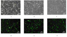
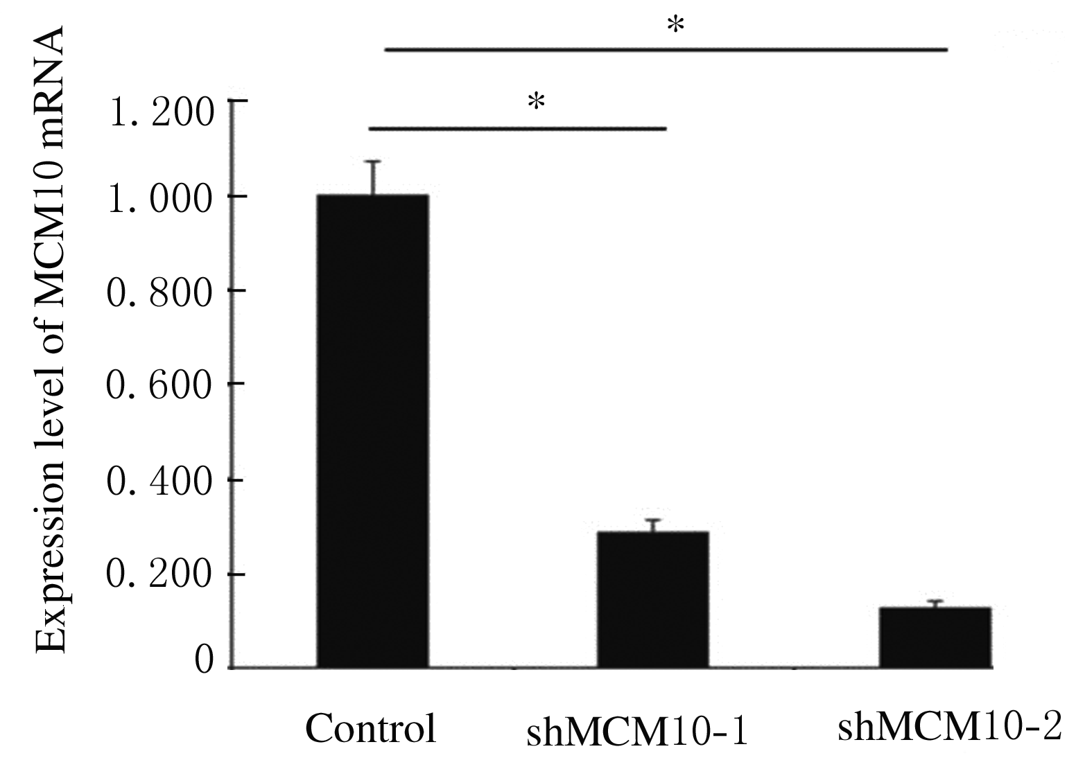
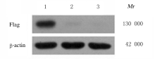
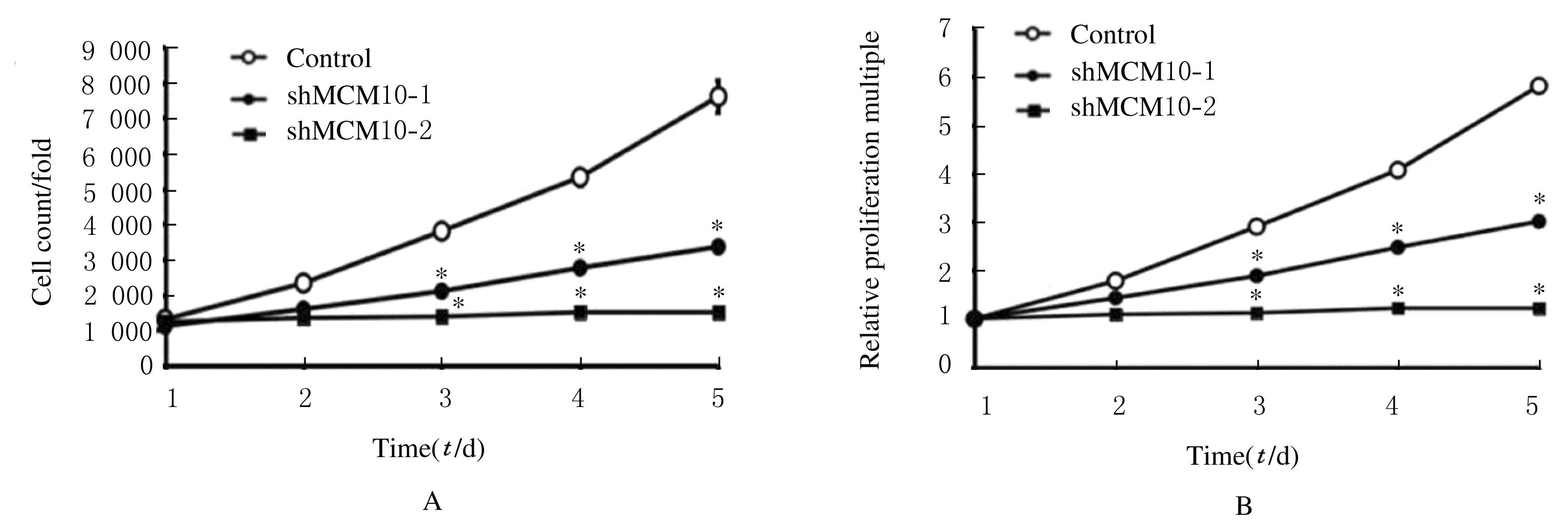
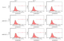
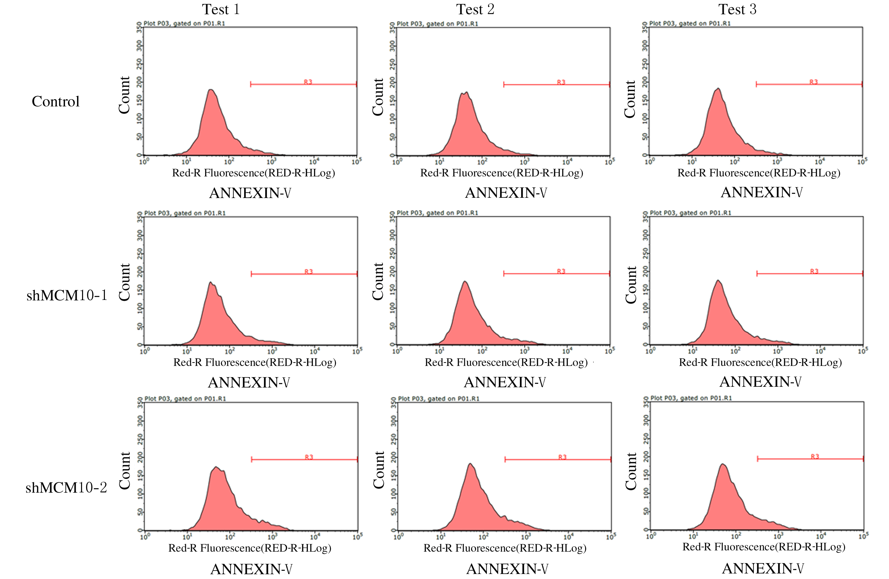
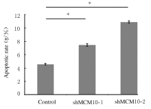
  • Received:2020-10-06 Online:2021-05-28 Published:2021-05-28
  • Contact: Jiming XIE E-mail:xclinton@sina.com

摘要: 目的

探讨微小染色体维持蛋白10 (MCM10)基因沉默对乳腺癌细胞的增殖抑制作用,并阐明其作用机制。

方法

将人三阴性乳腺癌(TNBC)MDA-MB-231细胞分为对照组、MCM10干扰组1(shMCM10-1组)和MCM10干扰组2(shMCM10-2组)。构建MCM10慢病毒干扰载体MCM10-1和MCM10-2,分别感染shMCM10-1组和shMCM10-2组MDA-MB-231细胞,对照组MDA-MB-231细胞感染阴性对照病毒。采用实时荧光定量PCR(RT-qPCR)法和Western blotting法检测各组MDA-MB-231细胞中MCM10 mRNA和蛋白表达水平,Cellomics计数法检测各组MDA-MB-231细胞的增殖率,流式细胞术检测各组MDA-MB-231细胞凋亡率,采用Caspase3/7活性检测试剂盒检测各组MDA-MB-231细胞中Caspase3/7活性。

结果

与对照组比较,shMCM10-1组和shMCM10-2组MDA-MB-231细胞中MCM10 mRNA表达率明显降低(P<0.01),未检测到MCM10蛋白表达;与对照组比较,shMCM10-1组和shMCM10-2组MDA-MB-231细胞相对增殖倍数明显降低(P<0.01),MDA-MB-231细胞凋亡率明显升高(P<0.01),MDA-MB-231细胞中Caspase3/7 活性明显升高(P<0.01)。

结论

MCM10基因沉默可以抑制MDA-MB-231细胞增殖,促进细胞凋亡,增加细胞中Caspase3/7 活性。

关键词: 微小染色体维持蛋白10, 慢病毒载体, 乳腺肿瘤, MDA-MB-23细胞, 细胞凋亡

Abstract: Objective

To investigate the inhibitory effect of mini-chromosome maintenance protein 10(MCM10) silencing on the proliferation of breast cancer cells,and to elucidate its mechanism.

Methods

The human triple negative breast cancer (TMBC)MDA-MB-231 cells were divided into control group,MCM10 interference group 1 (shMCM10-1 group)and MCM10 interference group 2(shMCM10-2 group). The lentivirus interference vectors of MCM10-1 group and MCM10-2 were constructed and infected into the MDA-MB231 cells in shMCM10-1 group and shMCM10-2 group,respectively. The MDA-MB-231 cells in control group were infected with the negative control virus. The expression levels of MCM10 in the MDA-MB-231 cells in various groups were measured by Real-time fluorescence quantitative PCR(RT-qPCR) and Western blotting methods; the proliferation of the MDA-MB-231 cells in various groups was monitored by Cellomics Count assay;the apoptotic rates of the MDA-MB-231 cells in various groups were detected by Flow cytometry, and the activities of Caspase3/7 in the MDA-MB-231 cells in various groups were measured by Caspase 3/7 activity detection kit.

Results

The expression levels of MCM10 mRNA in the MDA-MB-231 cells in shMCM10-1 group and shMCM10-2 group were significantly lower than that in control group (P<0.01), and the expression of MCM10 protein was not found.Compared with control group, the relative proliferation multiples of the MDA-MB-231 cells in shMCM10-1 group and shMCM10-2 group were significantly decreased (P<0.01),and the apoptotic rates of the MDA-MB-231 cells in shMCM10-1 group and shMCM10-2 group were significantly increased (P<0.01),and the activities of Caspase 3/7 in the MDA-MB-231 cells in shMCM10-1 group and shMCM10-2 group were significantly increased (P<0.01).

Conclusion

MCM10 gene silencing can inhibit the proliferation of the MDA-MB-231 cells, and promote the apoptosis of the MDA-MB-231 cells and increase the activities of Caspase3/7 in the MDA-MB-231 cells.

Key words: mini-chromosome maintenance protein 10, lentivirus vector, breast neoplasm, MDA-MB-231 cells, apoptosis

中图分类号: 

  • R730.2