吉林大学学报(医学版) ›› 2024, Vol. 50 ›› Issue (6): 1519-1525.doi: 10.13481/j.1671-587X.20240605

• 基础研究 • 上一篇    

3D打印PLA/PTMC-Ca₃(PO₄)₂复合支架制备及其对兔骨髓间充质干细胞成骨分化的影响

刘鑫钢,陈旭,刘亚东,苗锦虎,邵国喜()   

  1. 吉林大学第二医院创伤外科,吉林 长春 130022
  • 收稿日期:2023-12-26 出版日期:2024-11-28 发布日期:2024-12-10
  • 通讯作者: 邵国喜 E-mail:sgx@jlu.edu.cn
  • 作者简介:刘鑫钢(1998-),男,山东省临沂市人,在读硕士研究生,主要从事骨缺损修复材料方面的研究。
  • 基金资助:
    吉林省科技厅科技发展计划项目(20210204113YY)

Preparation of 3D-printed PLA/PTMC-Ca₃(PO₄)₂ composite scaffolds and their effects on osteogenic differentiation of bone marrow mesenchymal stem cells of rabbits

Xingang LIU,Xu CHEN,Yadong LIU,Jinhu MIAO,Guoxi SHAO()   

  1. Department of Trauma Surgery,Second Hospital,Jilin University,Changchun 130022,China
  • Received:2023-12-26 Online:2024-11-28 Published:2024-12-10
  • Contact: Guoxi SHAO E-mail:sgx@jlu.edu.cn

摘要:

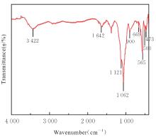
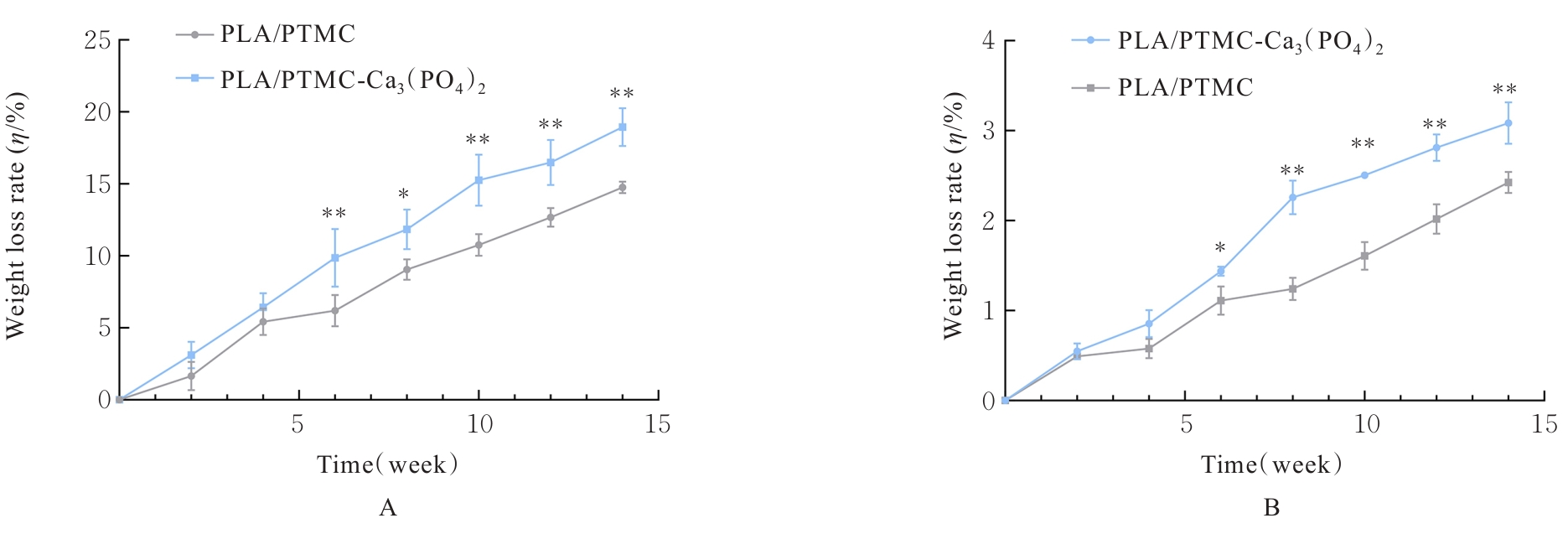
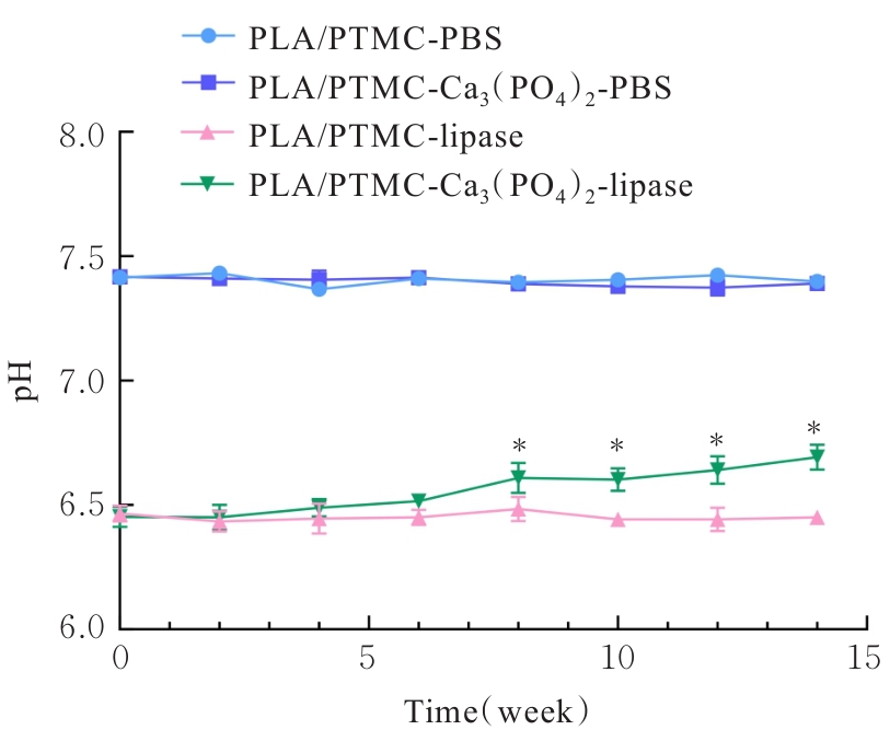
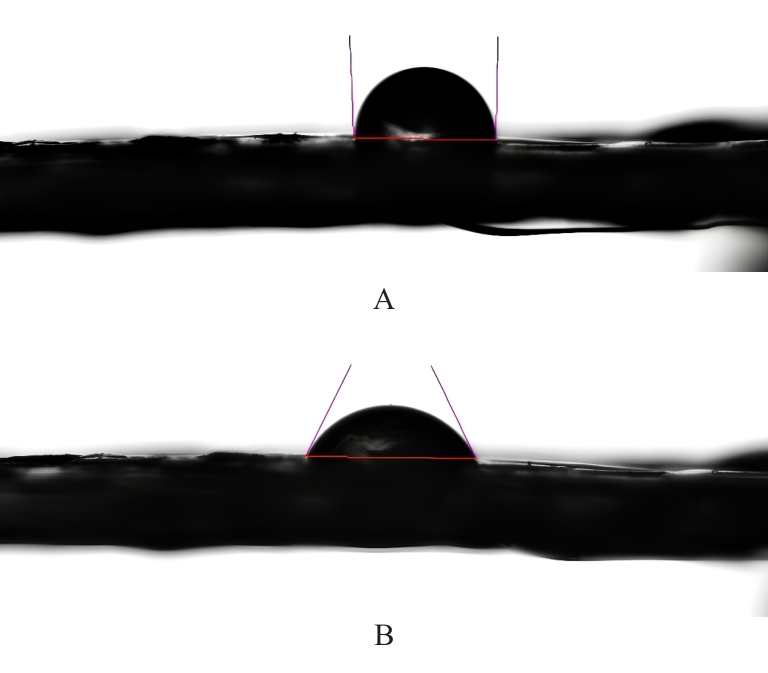
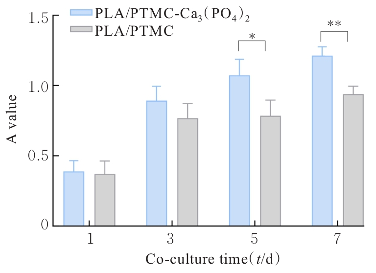
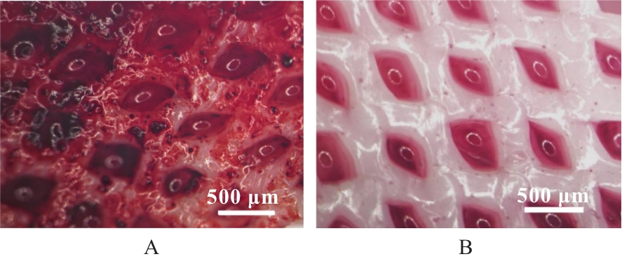
目的 探讨通过3D打印技术制备的聚乳酸(PLA)/聚三亚甲基碳酸酯(PTMC)和PLA/PTMC-磷酸三钙[Ca?(PO?)?]复合多孔支架对兔骨髓间充质干细胞(BMSCs)的影响,阐明其在骨缺损修复中的应用价值。 方法 混合材料后使用桌面挤丝机制备PLA/PTMC和PLA/PTMC-Ca?(PO?)? 线材,使用CATIA V5-6R2019建模软件设计支架,并使用CreatBot F430 3D打印机进行支架制作。红外光谱检测PLA/PTMC-Ca?(PO?)? 支架化学结构,体外降解实验检测2种支架降解失重率和pH值,角接触测量仪检测2种支架亲水性。取3只出生2~5 d的白色新西兰乳兔,提取BMSCs,CCK-8法检测2种支架共培养细胞增殖活性,茜素红染色观察2种支架共培养细胞成骨分化情况。 结果 红外光谱检测证实成功制备出含有PLA、PTMC和β-Ca?(PO?)? 3种物质的复合支架。降解6~14周,与PLA/PTMC支架比较,PLA/PTMC-Ca?(PO?)? 支架在脂肪酶溶液和磷酸盐缓冲液(PBS)中的降解速率均明显升高(P<0.05或P<0.01);降解8~14周,与PLA/PTMC支架比较,PLA/PTMC-Ca?(PO?)? 支架在脂肪酶溶液中的pH值明显升高(P<0.01)。与PLA/PTMC支架比较,PLA/PTMC-Ca?(PO?)? 支架接触角明显减小(P<0.01)。细胞共培养第5和7天时,与PLA/PTMC支架比较,PLA/PTMC-Ca?(PO?)?支架共培养细胞增殖活性均明显升高(P<0.05或P<0.01)。共培养21 d后,2种支架与BMSCs重叠生长,局部均形成钙化结节,茜素红将其局部染成橘红色;与PLA/PTMC支架比较,PLA/PTMC-Ca?(PO?)? 支架共培养细胞矿化钙结节数量增多,密度增大,颜色加深。 结论 通过3D打印技术成功制备出含有PLA、PTMC和β-Ca?(PO?)? 的PLA/PTMC-Ca?(PO?)? 复合多孔支架,其降解性能良好,在生物相容性、亲水性和成骨诱导性等方面均表现出优势,是一种性能优良的骨缺损修复材料。

关键词: 骨缺损, 骨再生, 3D打印, 聚乳酸, 聚三亚甲基碳酸酯

Abstract:

Objective To discuss their effects of PLA (polylactic acid)/PTMC (polytrimethylene carbonate) and PLA/PTMC-calcium phosphate [Ca?(PO?)?] composite porous scaffolds prepared by 3D printing technology on bone marrow mesenchymal stem cells (BMSCs) of the rabbits, and to clarify their application values in bone defect repairment. Methods After mixing the materials, PLA/PTMC and PLA/PTMC-Ca3(PO42 filaments were prepared by desktop filament extruder. The scaffolds were designed by CATIA V5-6R2019 modeling software and fabricated using CreatBot F430 3D printer. The chemical structure of the PLA/PTMC-Ca3(PO42 scaffold was detected by infrared spectroscopy. In vitro degradation experiments were used to detect the degradation weight loss rates and pH values of the two scaffolds. A contact angle measuring instrument was used to detect the hydrophilicities of the two scaffolds. The BMSCs were extracted from three newborn New Zealand white rabbits (2-5-day-old); CCK-8 method was used to detect the proliferation activities of the cells co-cultured with two scaffolds, and Alizarin red staining was used to observe the osteogenic differentiation of the cells co-cultured with two scaffolds. Results Infrared spectroscopy confirmed the successful preparation of composite scaffolds containing PLA, PTMC, and β-Ca?(PO?)?. During degradation for 6-14 weeks, compared with PLA/PTMC scaffold, the degradation rates of the PLA/PTMC-Ca3(PO42 scaffold in lipase solution and phosphate-buffered saline (PBS) were significantly increased (P<0.05 or P<0.01). During degradation for 8-14 weeks, compared with PLA/PTMC scaffold, the pH value of the PLA/PTMC-Ca?(PO?)? scaffold in lipase solution was significantly increased (P<0.01). Compared with PLA/PTMC scaffold, the contact angle of the PLA/PTMC-Ca?(PO?)? scaffold was significantly decreased (P<0.01). On days 5 and 7 of cell co-culture, compared with PLA/PTMC scaffold, the proliferation activity of the cells co-cultured with PLA/PTMC-Ca3(PO42 scaffold was significantly increased (P<0.05 or P<0.01). After 21 d of co-culture, both scaffolds overlapped with BMSCs and locally formed calcified nodules, which were stained orange by Alizarin red. Compared with PLA/PTMC scaffold, the number of mineralized calcium nodules in the cells co-cultured with PLA/PTMC-Ca?(PO?)? scaffold was increased, with greater density and deeper color. Conclusion The PLA/PTMC-Ca3(PO42 composite porous scaffolds containing PLA, PTMC, and β-Ca3(PO42 are successfully prepared by 3D printing technology. These scaffolds exhibit good degradation properties and show advantages in biocompatibility, hydrophilicity, and osteogenic induction; they are excellent materials for the bone defect repairment.

Key words: Bone defect, Bone regeneration, 3D printing, Polylactic acid, Polytrimethylene carbonate

中图分类号: 

  • R331