吉林大学学报(医学版) ›› 2025, Vol. 51 ›› Issue (1): 191-201.doi: 10.13481/j.1671-587X.20250123

• 临床研究 • 上一篇    

自分泌运动因子和溶血磷脂酸受体3在慢性阻塞性肺疾病患者血清及肺组织中的表达及其意义

蒋佩芩1,张政1,黄钟2(),卢献灵1()   

  1. 1.石河子大学第一附属医院呼吸与危重症医学科,新疆 石河子 832000
    2.石河子大学第一附属 医院急诊医学中心,新疆 石河子 832000
  • 收稿日期:2024-02-10 接受日期:2024-04-07 出版日期:2025-01-28 发布日期:2025-03-06
  • 通讯作者: 黄钟,卢献灵 E-mail:10082404@qq.com;luxianlingmary@163.com
  • 作者简介:蒋佩芩(1996-),女,湖南省怀化市人,在读硕士研究生,主要从事呼吸内科学基础和临床方面的研究。
  • 基金资助:
    新疆生产建设兵团科技局兵团指导性科技计划项目(2022ZD039);石河子大学自主资助支持校级科研立项项目(ZZZC201715A)

Expressions of autotaxin and lysophosphatidic acid receptor 3 in serum and lung tissue of patients with chronic obstructive pulmonary disease and their significances

Peiqin JIANG1,Zheng ZHANG1,Zhong HUANG2(),Xianling LU1()   

  1. 1.Department of Pulmonary and Critical Care Medicine,First Affiliated Hospital,Shihezi University,Shihezi 832000,China
    2.Emergency Medical Center,First Affiliated Hospital,Shihezi University,Shihezi 832000,China
  • Received:2024-02-10 Accepted:2024-04-07 Online:2025-01-28 Published:2025-03-06
  • Contact: Zhong HUANG,Xianling LU E-mail:10082404@qq.com;luxianlingmary@163.com

摘要:

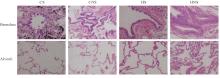
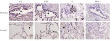
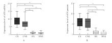
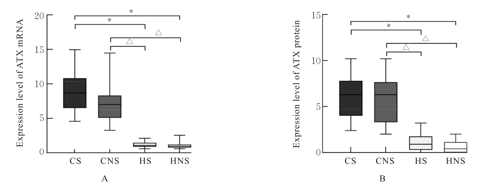
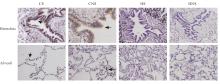
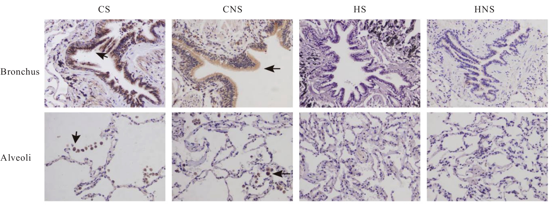
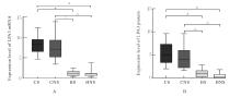
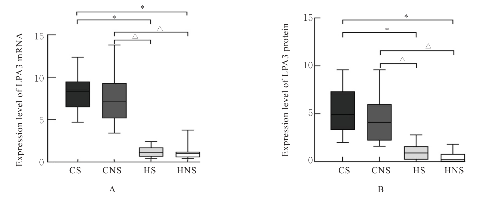
目的 探讨自分泌运动因子(ATX)和溶血磷脂酸受体3(LPA3)在慢性阻塞性肺疾病(COPD)患者血清及肺组织中的表达情况,阐明ATX和LPA3在COPD发生发展过程中的作用。 方法 收集40例COPD患者纳入急性加重期组(AECOPD组),经治疗后处于稳定期者纳入COPD稳定期组,共计40例,并收集40名健康体检者纳入对照组,采用酶联免疫吸附试验(ELISA)检测各组研究对象血清中ATX水平。另收集80例行肺叶切除术的患者,分为COPD吸烟组(CS组,n=20)、COPD不吸烟组(CNS组,n=20)、非COPD吸烟组(HS组,n=20)和非COPD不吸烟组(HNS组,n=20),收集各组研究对象的一般资料,HE染色观察各组患者肺组织的病理形态表现,免疫组织化学染色法检测各组患者肺组织中ATX和LPA3蛋白表达水平,实时荧光定量PCR(RT-qPCR)法检测各组患者肺组织中ATX和LPA3 mRNA表达水平,Pearson相关分析符合正态分布的连续变量的相关性,Spearman相关分析评估其他变量间的相关性。 结果 与对照组比较,COPD稳定期组研究对象第1秒用力呼气容积占预计值百分比(FEV1%pred)和第1秒用力呼气容积占用力肺活量百分比(FEV1/FVC)均明显降低(P<0.05)。与COPD稳定期组和对照组比较,AECOPD组患者血清中ATX水平升高(P<0.05);与对照组比较,COPD稳定期组患者血清中ATX水平升高(P<0.05)。AECOPD组患者血清中ATX水平与COPD评估测试量表(CAT)评分呈正相关关系(r=0.581,P<0.001),与吸烟史、白细胞计数(WBC)、中性粒细胞百分比(NEUT%)、中性粒细胞与淋巴细胞比值(NLR)和体质量指数(BMI)无相关性(P>0.05);COPD稳定期组患者血清中ATX水平与WBC和CAT评分均呈正相关关系(r=0.384,P=0.014;r=0.463,P=0.003),与FEV1%pred和FEV1/FVC均呈负相关关系(r=-0.393,P=0.012;r=-0.353,P=0.025);与CS组和CNS组比较,HS组和HNS组患者FEV1%pred及FEV1/FVC均升高(P<0.05);免疫组织化学染色,与CS组比较,HS组和HNS组患者肺组织中ATX及LPA3蛋白表达水平降低(P<0.05);与CNS组比较,HS组和HNS组患者肺组织中ATX及LPA3蛋白表达水平降低(P<0.05)。RT-qPCR法,与CS组比较,HS组和HNS组患者肺组织中ATX及LPA3 mRNA表达水平降低(P<0.05);与CNS组比较,HS组和HNS组患者肺组织中ATX及LPA3 mRNA表达水平降低(P<0.05)。相关性分析,COPD患者肺组织中ATX蛋白表达水平与LPA3蛋白表达水平呈正相关关系(r=0.723,P<0.001)。 结论 COPD患者肺组织中ATX和LPA3 mRNA及蛋白表达水平均升高,AECOPD组和COPD稳定期组患者血清ATX水平均升高,二者可能参与COPD的炎症反应,促进COPD的发生发展。

关键词: 慢性阻塞性肺疾病, 自分泌运动因子, 溶血磷脂酸, 受体, 吸烟

Abstract:

Objective To discuss the expressions of autotaxin (ATX) and lysophosphatidic acid receptor 3 (LPA3) in serum and lung tissue of the chronic obstructive pulmonary disease (COPD) patients, and to clarify the role of ATX and LPA3 in the occurrence and development of COPD. Methods A total of 40 COPD patients were collected and brought into acute exacerbation of COPD group (AECOPD group); after treatment, those stabilized patients were included in COPD stable group (n=40); additionally, 40 healthy individuals were recruited as control group. Enzyme-linked immunosorbent assay (ELISA) was used to detect the ATX levels in the serum of the subjects in various groups. Another 80 patients who underwent lobectomy were divided into COPD smoking group (CS group, n=20), COPD non-smoking group (CNS group, n=20), non-COPD smoking group (HS group, n=20), and non-COPD non-smoking group (HNS group, n=20). The general informations of the subjects in various groups were collected. HE staining was used to observe the pathomorphology of lung tissue of the patients in various groups;immunohistochemical staining was used to detect the expression levels of ATX and LPA3 proteins in lung tissue of the patients in various groups;real-time fluorescence quantitative PCR (RT-qPCR) method was used to detect the expression levels of ATX and LPA3 mRNA in lung tissue of the patients in various groups;Pearson correlation analysis was used to evaluate the correlations of continuous variables with normal distribution, and Spearman correlation analysis was used to evaluate the correlations of other variables. Results Compared with control group, the percentage of forced expiratory volume in the first second to predicted value (FEV1%pred) and the percentage of forced expiratory volume in the first second to forced vital capacity (FEV1/FVC) of the patients in COPD stable group were significantly decreased (P<0.05). Compared with COPD stable group and control group, the level of ATX in serum of the patients in AECOPD group was increased (P<0.05); compared with control group, the level of ATX in serum of the patients in COPD stable group was increased (P<0.05). The serum ATX level of the patients in AECOPD group was positively correlated with COPD Assessment Test (CAT) scores (r=0.581, P<0.001) and showed no correlations with smoking history, white blood cells (WBC), percentage of neutrophils (NEUT%), neutrophil to lymphocyte ratio (NLR), and body mass index (BMI) (P>0.05). The serum ATX level of the patients in COPD stable group was positively correlated with WBC and CAT score (r=0.384, P=0.014; r=0.463, P=0.003) and negatively correlated with FEV1%pred and FEV1/FVC (r=-0.393, P=0.012; r=-0.353, P=0.025). Compared with CS group and CNS group, the FEV1%pred and FEV1/FVC of the patients in HS group and HNS group were increased (P<0.05). The immunohistochemical staining results showed that compared with CS group, the expression levels of ATX and LPA3 proteins in lung tissue of the patients in HS group and HNS group were decreased (P<0.05). Compared with CNS group, the expression levels of ATX and LPA3 proteins in lung tissue of the patients in HS group and HNS group were decreased (P<0.05). The RT-qPCR results showed that compared with CS group, the expression levels of ATX and LPA3 mRNAs in lung tissue of the patients in HS group and HNS group were decreased (P<0.05); compared with CNS group, the expression levels of ATX and LPA3 mRNAs in lung tissue of the patients in HS group and HNS group were decreased (P<0.05). The correlation analysis results showed that the expression level of ATX protein in lung tissue of the COPD patients was positively correlated with the expression level of LPA3 protein (r=0.723, P<0.001). Conclusion The expression levels of ATX and LPA3 mRNA and proteins in lung tissue of the COPD patients are increased. The serum ATX levels in both AECOPD group and COPD stable group are increased, suggesting that these factors may be involved in the inflammatory response of COPD, promoting its occurrence and development.

Key words: Chronic obstructive pulmonary disease, Autotaxin motility factor, Lysophosphatidic acid, Receptor, Smoking

中图分类号: 

  • R563.9