吉林大学学报(医学版) ›› 2024, Vol. 50 ›› Issue (3): 587-595.doi: 10.13481/j.1671-587X.20240301

• 基础研究 •    下一篇

NLRP3炎症小体在大鼠单侧输尿管梗阻引起肾间质纤维化中的作用及其机制

阮颖新1,贾俊亚1,武占飞1,商文雅1,张鹏宇2()   

  1. 1.天津医科大学总医院肾内科,天津 300052
    2.国家恶性肿瘤临床医学研究中心 天津医科大学 肿瘤医院输血科,天津 300060
  • 收稿日期:2023-07-14 出版日期:2024-05-28 发布日期:2024-07-01
  • 通讯作者: 张鹏宇 E-mail:pzhang@tmu.edu.cn
  • 作者简介:阮颖新(1977-),女,黑龙江省哈尔滨市人,主治医师,医学博士,主要从事肾脏病诊治方面的研究。
  • 基金资助:
    国家重点研发计划项目(2019YFF0216502);天津市卫健委医学重点学科(专科)建设项目(TJYXZDXK-071C)

Effect of NLRP3 inflammatome in renal interstitial fibrosis induced by unilateral ureteral obstruction in rats and its mechanism

Yingxin RUAN1,Junya JIA1,Zhanfei WU1,Wenya SHANG1,Pengyu ZHANG2()   

  1. 1.Department of Nephrology, General Hospital, Tianjin Medical University, Tianjin 300052, China
    2.Department of Blood Transfusion, Tumor Hospital, Tianjin Medical University, National Clinical Research Center for Cancer, Tianjin 300060, China
  • Received:2023-07-14 Online:2024-05-28 Published:2024-07-01
  • Contact: Pengyu ZHANG E-mail:pzhang@tmu.edu.cn

摘要:

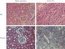
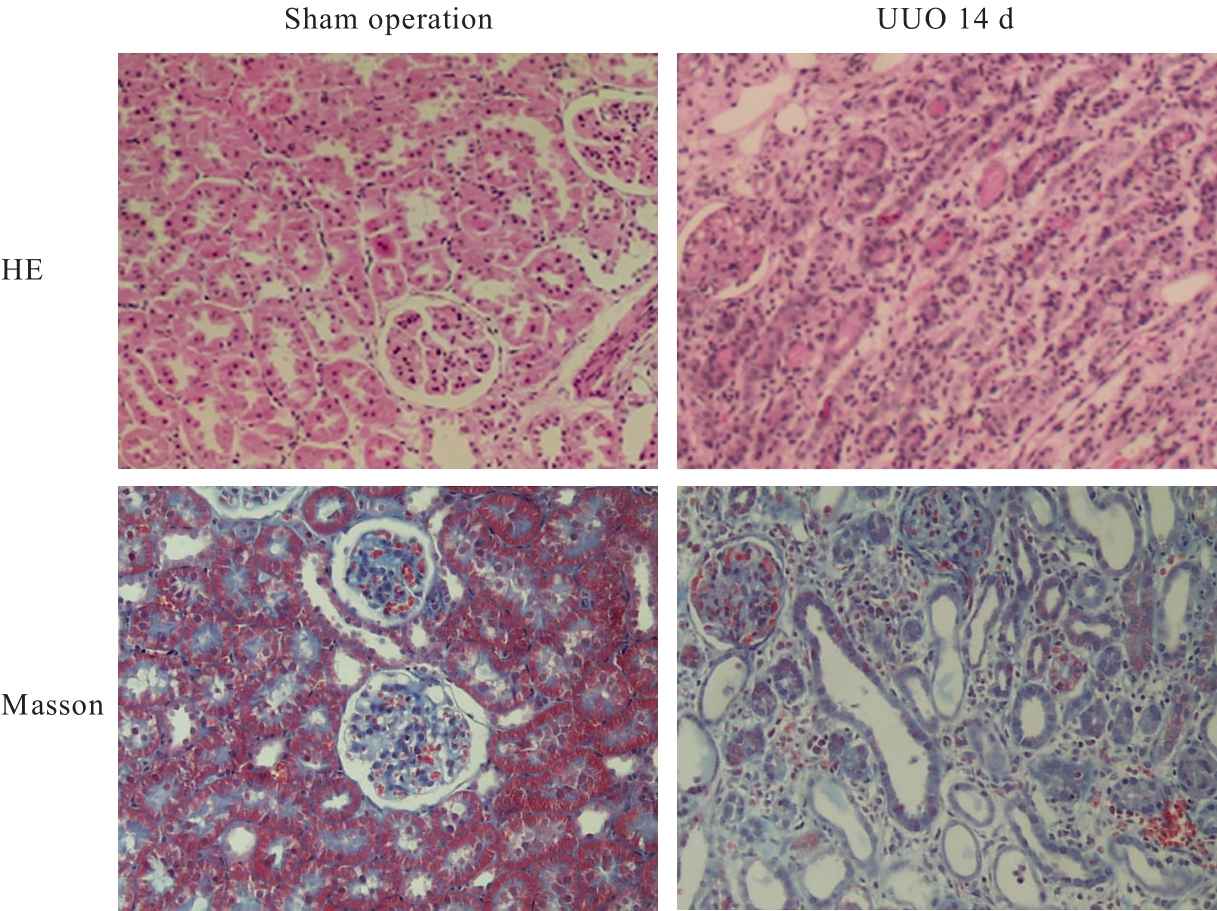
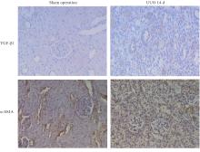
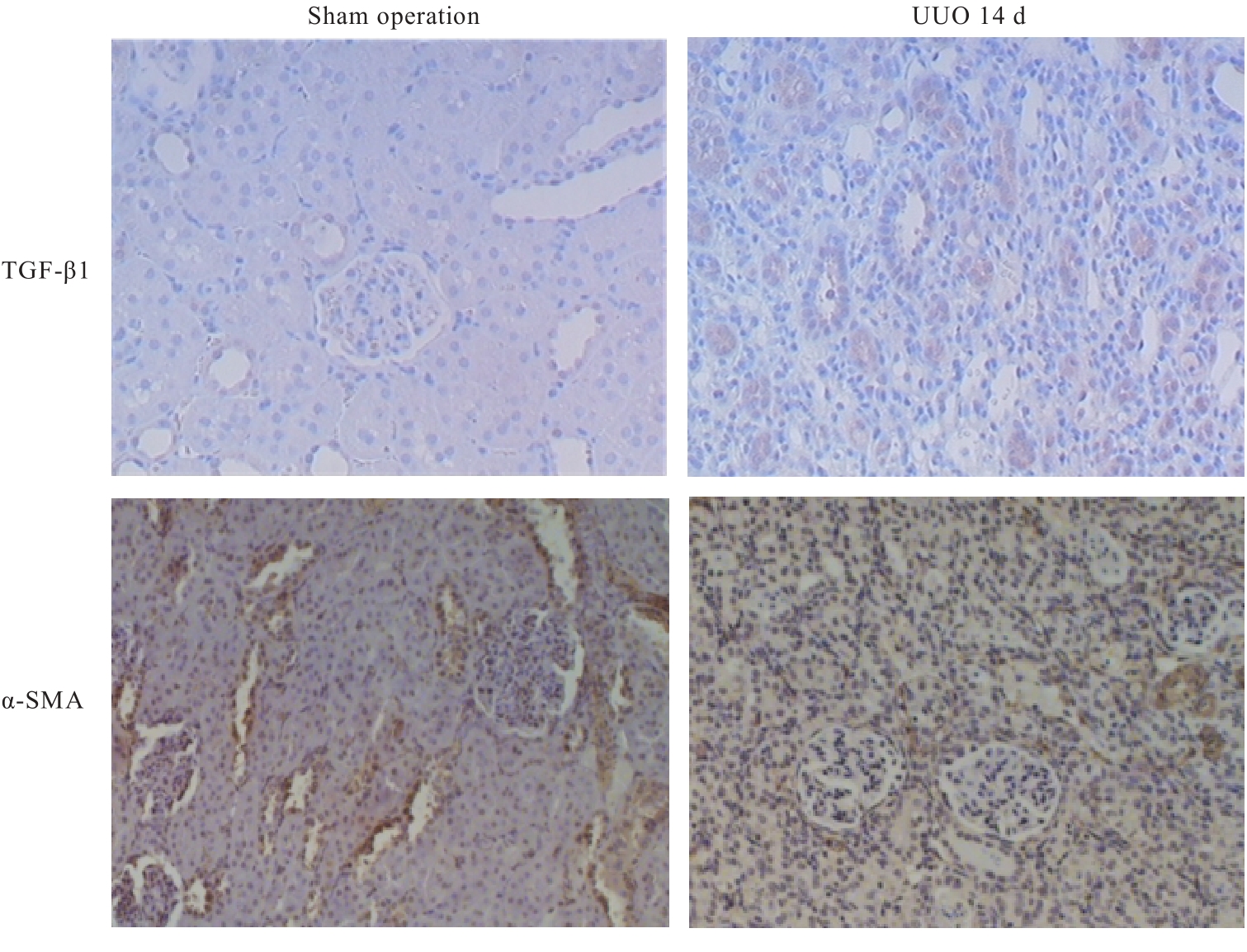
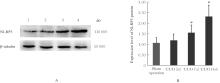
目的 探讨核苷酸结合寡聚化结构域样受体蛋白3(NLRP3)炎性小体在大鼠单侧输尿管梗阻(UUO)模型肾间质纤维化中的作用,并阐明其可能的作用机制。 方法 健康雄性Wistar大鼠30只随机分为假手术组(n=6)和UUO组(n=24),假手术组大鼠仅分离输尿管不结扎,UUO组分别于术后3、7和14 d处死大鼠,并按照处理时间分为UUO 3 d组(n=8)、UUO 7 d组(n=8)和UUO 14 d组(n=8)。HE染色和Masson染色观察各组大鼠肾组织病理形态表现,试剂盒检测各组大鼠肾组织中丙二醛(MDA)水平、超氧化物歧化酶(SOD)活性和羟脯氨酸(HYP)水平,免疫组织化学法检测各组大鼠肾组织中α-平滑肌肌动蛋白(α-SMA)和转化生长因子β1(TGF-β1)蛋白表达水平,Western blotting法检测各组大鼠肾组织中NLRP3蛋白表达水平。 结果 HE染色,UUO组大鼠出现明显肾小管扩张,肾间质水肿和增宽,可见较多炎症细胞浸润,部分肾小管腔内可见脱落的上皮细胞。与假手术组比较,UUO 3 d组、UUO 7 d组和UUO 14 d组大鼠HE染色肾间质纤维化评分均明显升高(P<0.05);与UUO 3 d组和UUO 7 d组比较,UUO 14 d组大鼠HE染色肾间质纤维化评分明显升高(P<0.05)。Masson染色,UUO组大鼠肾间质炎症细胞浸润明显,可见明显纤维组织增生;随UUO作用时间增加,大鼠部分肾小管消失,肾间质明显增宽,胶原沉积逐渐增多,皮髓交界处胶原沉积程度更加明显。与假手术组比较,UUO 3 d组、UUO 7 d组和UUO 14 d组大鼠Masson染色肾间质纤维化评分均明显升高(P<0.05);与UUO 3 d组和UUO 7 d组比较,UUO 14 d组大鼠Masson染色肾间质纤维化评分明显升高(P<0.05)。与假手术组比较,UUO 3 d组、UUO 7 d组和UUO 14 d组大鼠梗阻侧肾组织中MDA水平均明显升高(P<0.05),SOD活性明显降低(P<0.05)。与假手术组比较,UUO 3 d组、UUO 7 d组和UUO 14 d组大鼠梗阻侧肾组织中HYP水平均明显升高(P<0.05);与 UUO 3 d 组比较, UUO 14 d 组大鼠梗阻侧肾组织中 HYP 水平明显升高(P<0.05)。免疫组织化学法,与假手术组比较,UUO 3 d组、UUO 7 d组和UUO 14 d组大鼠肾组织中α-SMA蛋白表达水平明显升高(P<0.05);与UUO 3 d组和UUO 7 d组比较,UUO 14 d组大鼠肾组织中α-SMA蛋白表达水平均明显升高(P<0.05);与假手术组比较,UUO 3 d组、UUO 7 d组和UUO 14 d组大鼠肾小管上皮细胞和肾小管间质组织中TGF-β1蛋白表达水平明显升高(P<0.05);与UUO 3 d组比较,UUO 14 d组大鼠肾小管上皮细胞和肾小管间质组织中TGF-β1蛋白表达水平明显升高(P<0.05)。Western blotting法,与假手术组比较,UUO 7 d组和UUO 14 d组大鼠肾组织中NLRP3蛋白表达水平均明显升高(P<0.05)。 结论 NLRP3炎症小体在UUO大鼠肾纤维化过程中发挥重要作用,其作用机制与氧化应激增加和TGF-β1蛋白表达水平升高有关。

关键词: 核苷酸结合寡聚化结构域样受体蛋白3, 氧化应激, 转化生长因子β1, 单侧输尿管梗阻, 肾间质纤维化

Abstract:

Objective To discuss the effect of NOD-like receptor protein 3 (NLRP3) inflammasome on the renal interstitial fibrosis in the unilateral ureteral obstruction (UUO) model rats, and to clarify its potential mechanism. Methods Thirty healthy male Wistar rats were randomly divided into sham operation group (n=6) and UUO group (n=24). The rats in sham operation group underwent the dissection of the ureter without ligation, while the rats in UUO group were sacrificed on the 3rd, 7th, and 14th days after operation, and based on the treatment duration,the rats were divided into UUO 3 d group (n=8), UUO 7 d group (n=8), and UUO 14 d group (n=8). HE staining and Masson staining were used to observe the pathomorphology of kidney tissue of the rats in various groups; reagent kits were used to detect the levels of malondialdehyde (MDA), activities of superoxide dismutase (SOD), and levels of hydroxyproline (HYP) in kidney tissue of the rats in various groups;immunohistochemistry method was used to detect the expression levels of α-smooth muscle actin (α-SMA) and transforming growth factor-β1 (TGF-β1) proteins in kidney tissue of the rats in various groups; Western blotting method was used to detect the expression levels of NLRP3 protein in kidney tissue of the rats in various groups. Results The HE staining results showed significant tubular dilation, interstitial edema, and widening, with increased infiltration of inflammatory cells, and shedding of epithelial cells was seen in parts of the tubular lumina of the rats in UUO group. Compared with sham operation group, the interstitial fibrosis scores of the rats in UUO 3 d, UUO 7 d, and UUO 14 d groups were significantly increased (P<0.05); compared with UUO 3 d group and UUO 7 d group, the interstitial fibrosis score of the rats in UUO 14 d group was significantly decreased (P<0.05). The Masson staining results showed that in UUO group, there was evident infiltration of inflammatory cells in the renal interstitium and a noticeable increase in fibrotic tissue proliferation; with the increasing of duration of UUO, some tubular structures disappeared, and the interstitial widened further with gradually increasing collagen deposition, particularly at the corticomedullary junction. Compared with sham operation group, the interstitial fibrosis scores of the rats in UUO 3 d, UUO 7 d, and UUO 14 d groups were significantly increased (P<0.05); and compared with UUO 3 d and UUO 7 d groups, the interstitial fibrosis score of the rats in UUO 14 d group was significantly decreased (P<0.05). Compared with sham operation group, the levels of MDA in obstructed kidney tissue of the rats in UUO 3 d, UUO 7 d, and UUO 14 d groups were significantly increased (P<0.05), and the SOD activities were significantly decreased (P<0.05). Compared with sham operation group, the levels of HYP in obstructed kidney tissue of the rats in UUO 3 d, UUO 7 d, and UUO 14 d groups were also significantly increased (P<0.05);compared with UUO 3 d group, the level of HYP in obstructed kidney tissue of the rats in UUO 14 d group was significantly increased (P<0.05). The immunohistochemistry results showed that compared with sham operation group, the expression levels of α-SMA protein in kidney tissue of the rats in UUO 3 d, UUO 7 d, and UUO 14 d groups were significant increased (P<0.05); compared with UUO 3 d and UUO 7 d groups, the expression levels of α-SMA protein in kidney tissue of the rats in UUO 14 d group was significantly increased (P<0.05); compared with sham operation group,the expression levels of TGF-β1 protein in renal tubular epithelial cells and renal tubule interstitial tissue of the rats in UUO 3 d, UUO 7 d, and UUO 14 d groups were also significantly increased (P<0.05); compared with UUO 3 d group, the expression levels of TGF-β1 protein in the tubular epithelial cells and renal tubule interstitial tissue of the rats in UUO 14 d group were significantly decreased(P<0.05). The Western blotting results showed that compared with sham operation group, the expression levels of NLRP3 protein in kidney tissue of the rats in UUO 7 d and UUO 14 d groups were significantly increased (P<0.05). Conclusion The NLRP3 inflammasome plays a critical role in renal fibrosis of the UUO rats,and its mechanism may be related to the increasing of oxidative stress and the increasing of expression level of TGF-β1 protein.

Key words: Nucleotide combined with structure of oligomerization domain receptor protein 3, Oxidative stress, Transforming growth factor β1, Unilateral ureteral obstruction, Renal interstitial fibrosis

中图分类号: 

  • R692