吉林大学学报(医学版) ›› 2025, Vol. 51 ›› Issue (2): 341-351.doi: 10.13481/j.1671-587X.20250208

• 基础研究 • 上一篇    

IκB激酶相互作用蛋白在宫颈癌组织中的表达及其对宫颈癌细胞增殖、迁移和侵袭的影响

王妍,赵邹宇,于盼盼,杨萍()   

  1. 石河子大学第一附属医院妇科,新疆 石河子 832000
  • 收稿日期:2024-05-20 接受日期:2024-07-03 出版日期:2025-03-28 发布日期:2025-04-22
  • 通讯作者: 杨萍 E-mail:pingy2018@163.com
  • 作者简介:王 妍(1997-),女,新疆维吾尔自治区乌鲁木齐市人,在读硕士研究生,主要从事妇科肿瘤方面的研究。
  • 基金资助:
    国家自然科学基金面上项目(82072893);新疆生产建设兵团科技局兵团重点领域科技攻关计划项目(2023AB055);新疆生产建设兵团科技局天山英才科技创新领军人才-高层次领军人才项目(CZ001201)

Expression of I kappa B kinase-interacting protein in cervical cancer tissue and its effect on proliferation, migration and invasion of cervical cancer cells

Yan WANG,Zouyu ZHAO,Panpan YU,Ping YANG()   

  1. Department of Gynecology,First Affiliated Hospital,Shihezi University,Shihezi 832000,China
  • Received:2024-05-20 Accepted:2024-07-03 Online:2025-03-28 Published:2025-04-22
  • Contact: Ping YANG E-mail:pingy2018@163.com

摘要:

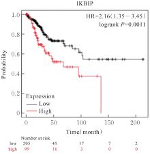
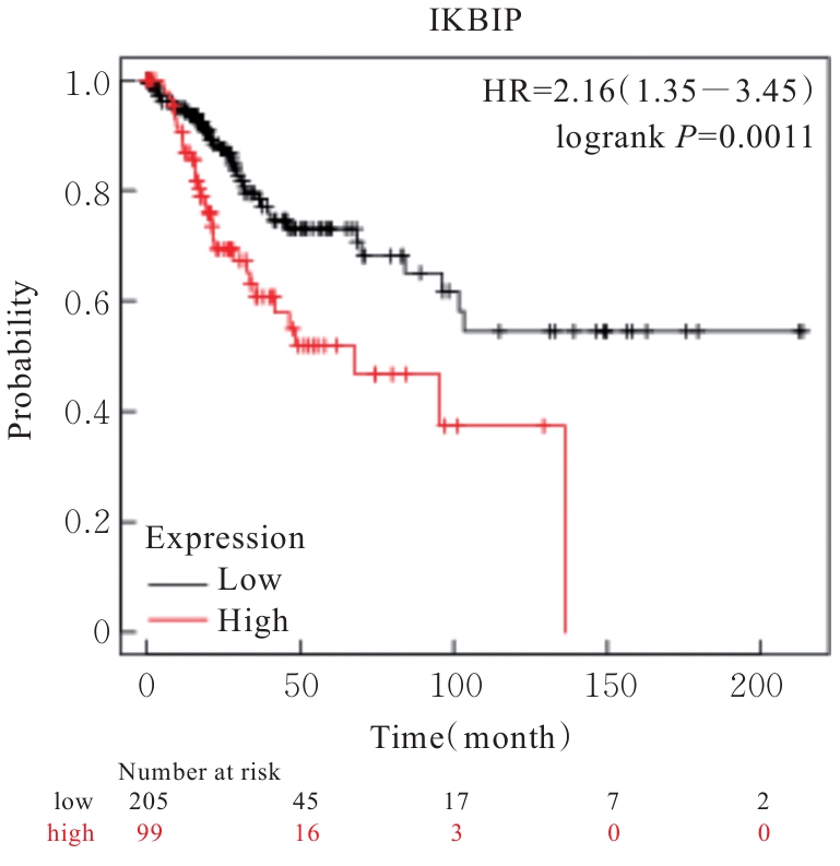
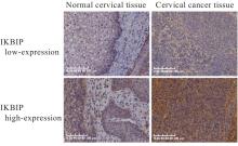
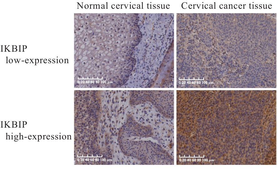
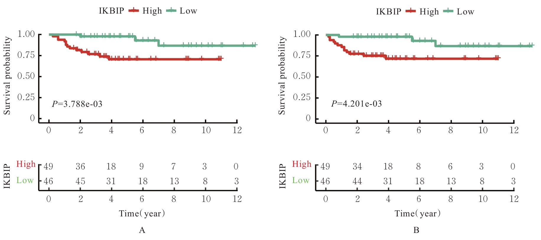
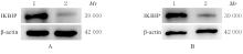
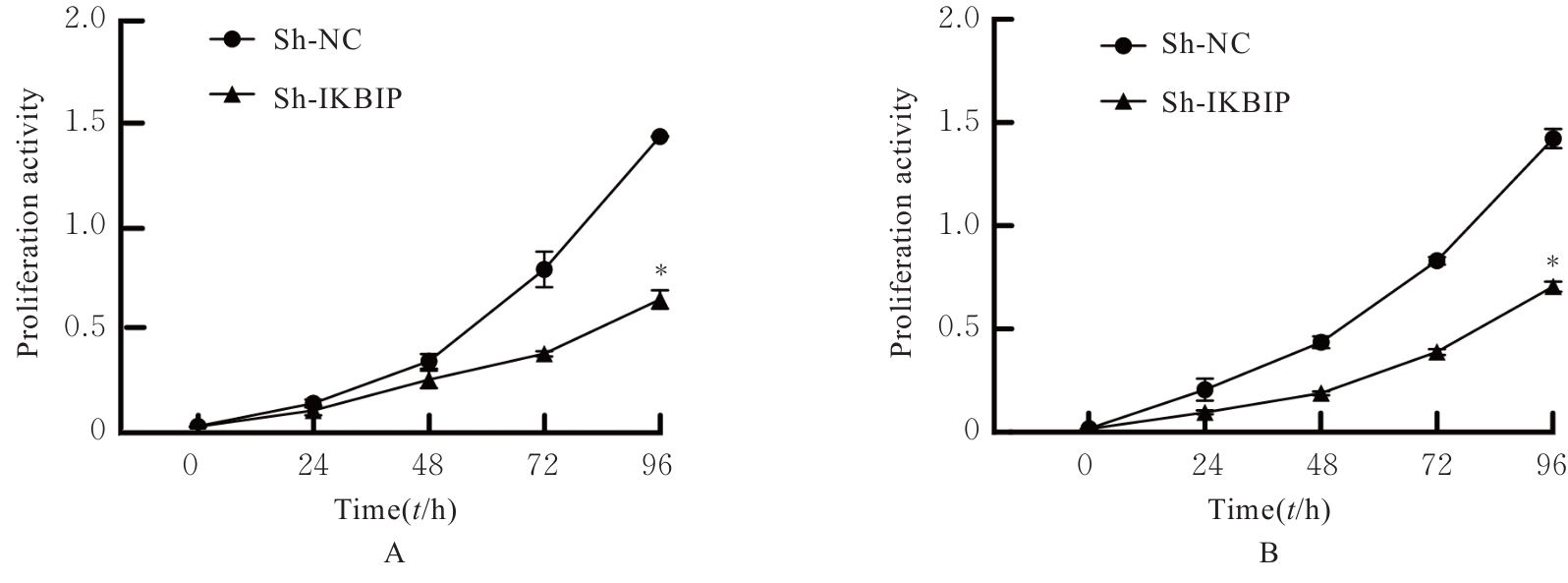
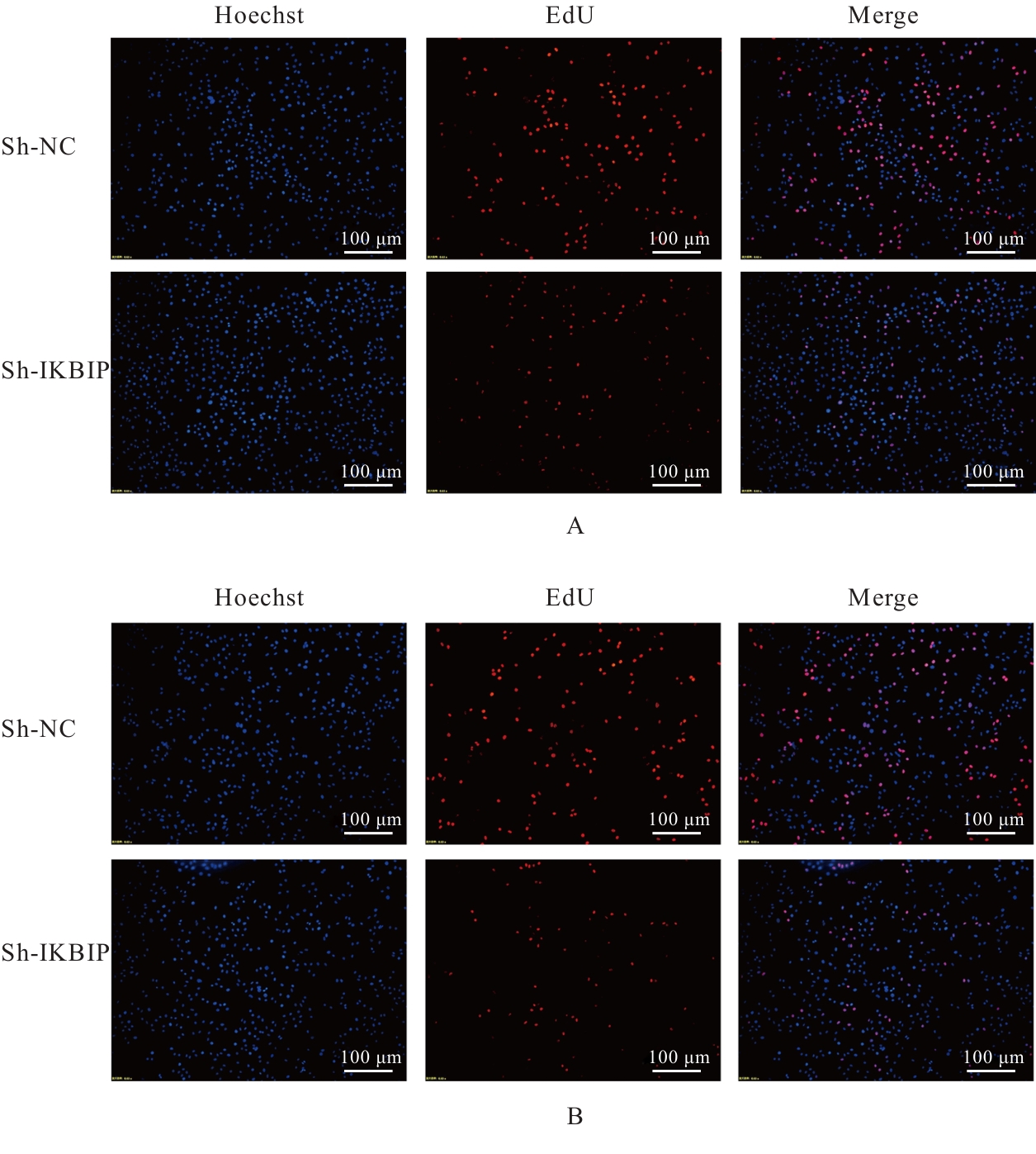
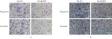
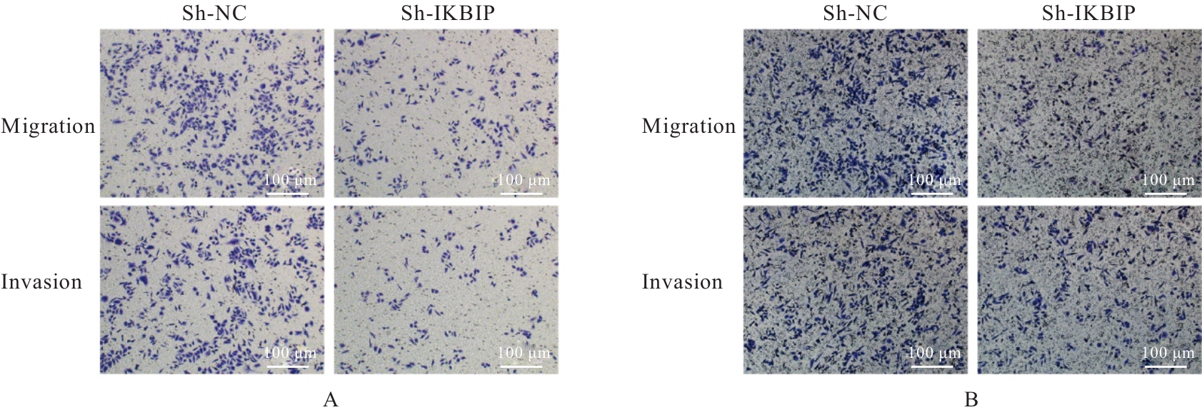
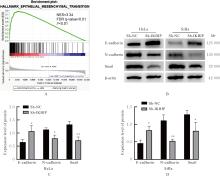
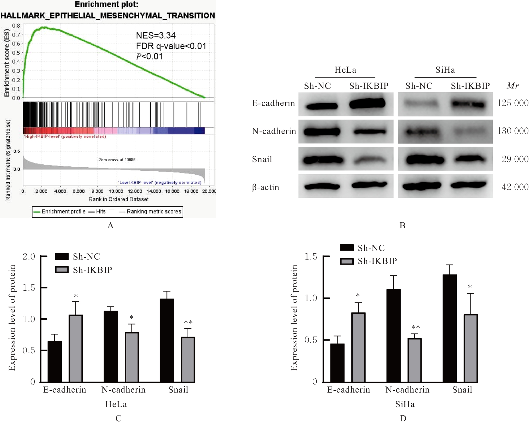
目的 探讨IκB激酶相互作用蛋白(IKBIP)在宫颈癌组织肿瘤细胞(TCCCT)中的表达与患者临床病理特征和预后的关系及其对宫颈癌(CC)HeLa和SiHa细胞生物学行为的影响,阐明其可能的作用机制。 方法 采用GENT2数据库和Kaplan-Meier(K-M)plotter数据库分析CC及正常宫颈组织中的IKBIP mRNA表达水平差异以及其表达水平与CC患者临床预后的关系。基因集富集分析(GSEA)软件中选取参考基因集为Hallmark,进行相关通路富集。采用免疫组织化学法检测IKBIP蛋白在TCCCT和正常宫颈组织上皮细胞(ECNCT)中的表达情况,分析其表达水平与CC患者临床病理特征和预后的关系;单因素和多因素Cox回归分析影响CC患者预后的危险因素。构建IKBIP敲低的CC细胞(HeLa和SiHa细胞),实验分为sh-NC组和sh-IKBIP组;采用Western blotting法评估各组细胞中IKBIP蛋白表达情况;细胞计数试剂盒8(CCK-8)和5-乙炔基-2'-脱氧尿苷(EdU)法检测各组细胞增殖活性及EdU阳性细胞率,Transwell小室实验检测各组迁移细胞数和侵袭细胞数,Western blotting法检测各组细胞中E-钙黏蛋白(E-cadherin)、N-钙黏蛋白(N-cadherin)和Snail蛋白表达水平。 结果 GENT2数据库分析,CC组织中IKBIP mRNA表达水平高于正常宫颈组织(P<0.001)。GSEA富集分析,上皮-间质转化(EMT)通路位于IKBIP高表达组的第1位。免疫组织化学法,TCCCT中IKBIP蛋白阳性表达率高于ECNCT(50.5% vs 8.0%),且其过表达与FIGO分期(2018)和肿瘤分化程度有关联(P<0.05);单因素和多因素Cox回归分析,淋巴结转移(LNM)和IKBIP高表达是影响CC患者总生存期(OS)及无进展生存期(PFS)的独立危险因素(P<0.05)。Western blotting法,与sh-NC组比较,sh-IKBIP组细胞IKBIP蛋白表达水平明显降低(P<0.05);CCK-8和EdU法,与sh-NC组比较,sh-IKBIP组细胞增殖活性和EdU阳性百分率均明显降低(P<0.05);Transwell小室实验,与sh-NC组比较,sh-IKBIP组迁移细胞数和侵袭细胞数明显减少(P<0.05)。Western blotting法,与sh-NC组比较,sh-IKBIP组细胞中E-cadherin蛋白表达水平升高(P<0.05),N-cadherin和Snail蛋白表达水平均降低(P<0.05)。 结论 IKBIP蛋白在CC细胞中高表达,且与CC患者不良预后有密切关联。下调IKBIP蛋白表达可抑制CC细胞的增殖、迁移和侵袭能力及EMT进程。

关键词: IκB激酶相互作用蛋白, 宫颈肿瘤, 细胞增殖, 细胞迁移, 上皮-间质转化

Abstract:

Objective To explore the correlation between IκB kinase-interacting protein (IKBIP) expression in tumor cells within cervical cancer tissues (TCCCT) and the clinical pathological characteristics and prognosis of patients, as well as its impact on the biological behaviors of cervical cancer (CC) cells HeLa and SiHa, and to elucidate its potential mechanism. Methods GENT2 and Kaplan-Meier plotter databases were utilized to analyze the differential expression of IKBIP mRNA in CC and normal cervical tissues, as well as its correlation with the clinical prognosis of CC patients. The Hallmark reference gene set was chosen for pathway enrichment analysis using the Gene Set Enrichment Analysis(GSEA) software. Immunohistochemistry method was used to detect the IKBIP protein expression in TCCCT and epithelial cells in normal cervical tissue (ECNCT), and analyze the correlations between its expression level and clinicopathological characteristics and prognosis of CC patients. Furthermore, univariate and multivariate Cox regression analyses were performed to identify the risk factors impacting the prognosis of CC patients. The stably transfected cells of CC (HeLa cells and SiHa cells) with IKBIP knockdown were established for the experiment, which were divided into sh-NC group and sh-IKBIP group. The expression of IKBIP protein in various groups was assessed using Western blotting method. The cell proliferation activity and the percentage of 5-ethynyl-2'-deoxyuridine (EdU) positive cells were measured using cell counting kit-8 (CCK-8) and EdU methods, while Transwell chamber assay was employed to determine the numbers of migration and invasion cells in various groups. Additionally, the expression levels of E-cadherin, N-cadherin, and Snail proteins in the cells in various groups were analyzed by Western blotting method. Results The GENT2 database analysis revealed that the expression level of IKBIP mRNA in CC tissue was higher than that in normal cervical tissue (P<0.001). The GSEA enrichment analysis identified the epithelial-mesenchymal transition (EMT) pathway as the top-ranked pathway in IKBIP high-expression group. The immunohistochemistry results indicated the positive expression rate of IKBIP protein in TCCCT was higher than that in ECNCT (50.5% vs 8.0%), and its over-expression was associated with FIGO stage (2018) and differentiation grade of tumor (P<0.05). The univariate and multivariate Cox regression analyses suggested that lymph node metastasis (LNM) and high expression of IKBIP were the risk factors affecting the overall survival (OS) and progression-free survival (PFS) of CC patients (P<0.05). The Western blotting method results showed that compared with sh-NC group, the expression level of IKBIP protein in the cells in sh-IKBIP group was decreased (P<0.05). The CCK-8 and EdU assay results showed that compared with sh-NC group, the proliferation activity and the percentage of EdU positive cells in sh-IKBIP group were decreased (P<0.05). The Transwell chamber assay results showed that compared with sh-NC group, the numbers of migration and invasion cells in sh-IKBIP group were significantly decreased (P<0.05). Additionally, the Western blotting method results indicated that compared with sh-NC group, the expression level of E-cadherin protein in sh-IKBIP group in CC cells was increased (P<0.05), while the expression levels of N-cadherin and Snail protein were decreased (P<0.05). Conclusion The expression of IKBIP protein is significantly up-regulated in HeLa and SiHa cells derived from CC, and it is closely correlated with poor prognosis in CC patients. Suppression of IKBIP protein expression can effectively inhibit the proliferation, invasion and migration capabilities as well as EMT process of CC cells.

Key words: I kappa B kinase-interacting protein, Cervical neoplasms, Cell proliferation, Cell invasion, Epithelial-mesenchymal transition

中图分类号: 

  • R711.74